DOI: 10.5935/2359-4802.20170038
Introduction
Studies have shown that calcium deposits in the cardiovascular system are associated with atherosclerotic disease.1-4 Similar to computed tomography (CT), bidimensional transthoracic echocardiography (TTE) is capable of detecting cardiac calcium deposits, and is a portable, noninvasive, nonradioactive, and low-cost method.5 Coronary angiotomography is known to predict individual risk of coronary events, evaluating the calcium
score of the coronary arteries and the semiquantitative scores of valvular calcium.5
A study by Gaibazzi et al.6 has established a cardiac calcification index by TTE and predicted the presence of important coronary calcification, with results comparable to those of coronary angiotomography.6 In patients with coronary artery disease (CAD), mitral annular calcification and aortic valve sclerosis on TTE are independent risk factors for cardiovascular events.7
ORIGINAL ARTICLE
Mailing Address: Cintia Rocha Fortes de Sá
Rua José Brusamolin, 658, Casa 2. Postal Code: 82210-280. São Lourenço, Curitiba, PR – Brazil E-mail: [email protected]; [email protected]
Correlation Between Cardiac Calcium Index and Coronary Artery Disease
Cintia Rocha Fortes de Sá,1 Ana Cristina Camarozano Wermelinger,1 Daniane Rafael,1 Rubens Zenóbio Darwich,2
Jerônimo Antonio Fortunato Junior,2 Daniela de Castro Carmo,1 Liz Andréa Villela Baroncini1
Instituto Saber e Aprender;1 Hospital da Cruz Vermelha2 – Cruz Vermelha Brasileira- Filial do Estado do Paraná, Curitiba, PR – Brazil
Manuscript received October 23, 2016; revised manuscript November 10, 2016; accepted December 1, 2016.
Resumo
Introduction: Valvular deposits of calcium quantified by transthoracic echocardiography are associated with the
occurrence of systemic atherosclerotic disease, but its prognostic value and influence of associated cardiovascular risk factors have not been defined yet.
Objectives: To correlate the valvular cardiac calcium index by transthoracic echocardiography with cardiovascular
risk factors and presence of coronary artery disease (CAD).
Methods: We selected 203 patients (61.2 ± 14.3 years; 57.6% females) who underwent transthoracic echocardiography
with cardiac calcium index quantification. The presence or absence of CAD, hypertension, diabetes mellitus (DM), dyslipidemia, and smoking was assessed.
Results: Age above 65 years (p < 0.001) and the presence of hypertension (p < 0.001) showed a significant correlation with the presence of cardiac calcification, whereas DM (p = 0.056) and CAD (p = 0.08) showed only a trend toward a correlation with calcification. Mitral valve calcification alone correlated significantly with age above 65 years (p < 0.001), presence of CAD (p = 0.004), hypertension (p = 0.054), and DM (p = 0.07). On multivariate analysis, CAD (odds ratio [OR] 3.39, 95% confidence interval [95%CI] 1.58-7.29, p = 0.002) and age > 65 years (OR 1.05, 95%CI 1.02-1.08, p = 0.003) correlated significantly and independently with mitral valve calcification. Aortic valve calcification alone showed no correlation with the presence of CAD (p = 0.435), but correlated significantly with age above 65 years (p < 0.001) and hypertension (p < 0.001). On multivariate analysis, only age (OR 1.1, 95%CI 1.06-1.14, p < 0.001) remained independently and significantly correlated with aortic calcification.
Conclusion: Age above 65 years and hypertension were independent risk factors for the presence of valvular
cardiac calcification, with mitral valve calcification alone emerging as significantly and independently associated with the presence of CAD. (Int J Cardiovasc Sci. 2017;30(2):136-144)
Some studies have shown that calcium deposition in the aortic leaflets and fibrous skeleton at the base of the heart is related to aging. In the elderly, up to 55% of the bidimensional TTEs demonstrate calcifications.8,9 Other studies have shown that the absence of cardiac calcification is a stronger predictor of absence of coronary disease than the absence of traditional risk factors, with the exception of diabetes mellitus (DM).10 Mitral annular calcification is associated with calcification of other cardiac structures, such as the aortic root, papillary muscles, chordae tendineae, and aortic valves, suggesting that it may be a result of a degenerative process that increases with age, or secondary to increased valvular stress.11 As a result of the increased turbulence and mechanical stress, the aortic valve serves as a focus of deposition of lipids involved in the process of atherosclerosis. Histopathological studies of aortic valves with valve sclerosis show accumulation of atherogenic lipoproteins, infiltration of inflammatory cells, extracellular matrix, and microscopic calcifications.12 Finally, aortic valve sclerosis may be related to systemic atherosclerosis and cardiovascular events, but its usefulness in predicting CAD is questionable.13
The aim of this study was to correlate the cardiac calcification index on TTE with cardiovascular risk factors and the presence of CAD.
Methods
This observational, cross-sectional study evaluated consecutive outpatients users of the public health system who underwent TTE with quantification of the cardiac calcification index from May 2015 to February 2016.
Patients
Among the outpatient population referred by the attending physician for TTE due to any clinical indication, we selected patients of both genders, older than 18 years of age, and of any race. The exclusion criteria were patients with chronic kidney disease undergoing dialysis, since these patients show changes in calcium metabolism and ectopic calcification associated with the renal dysfunction, patients with prosthetic heart valves, and patients with valvular stenosis, due to calcification of the valves being due primarily to mechanical stress. The following
clinical data were evaluated: age, gender, body mass index (BMI), hypertension, DM, CAD, smoking (current or previous), dyslipidemia, and use of medications (statins, angiotensin-converting enzyme inhibitors [ACEIs], angiotensin receptor blockers [ARBs], aspirin, and beta-blockers). The diagnoses of hypertension, DM, dyslipidemia, and smoking were retrieved from medical records and/or reported by the patients. The presence of CAD was confirmed by data from medical records and included the occurrence of non-fatal myocardial infarction and surgical or percutaneous myocardial revascularization.
All patients signed two copies of a free and informed consent form and retained one of the copies. The study was approved by the local Research Ethics Committee (CAAE 58034716.9.0000.0093).
Echocardiographic equipment
Table 1 – Echocardiographic grading system of cardiac and aortic root calcium (adapted from Gaibazzi et al.6)
Degree Papillary muscle calcium Mitral annular calcium Aortic valve sclerosis Aortic root calcium
0 Absent Absent Absent (leaflet thickness < 2 mm) Absent
1 Present Mild (< 5 mm) Mild (leaflet thickness > 2 mm or focal or
diffuse increased reflectivity) Present
2 - Moderate (5-10 mm)
Moderate (leaflet thickness > 4 mm and/
or marked hyperreflectivity)
-3 - Severe (> 10 mm) Severe (leaflet thickness > 6 mm and/or
marked hyperreflectivity)
-Statistical analysis
We described quantitative variables as mean, median, minimum and maximum values, and standard deviation. To summarize the qualitative variables, we used frequencies and percentages. We compared two classifications of a single variable in relation to a quantitative variable with Student's t test for independent samples. To evaluate the association between two qualitative variables, we used the chi-square test and Fisher's exact test. To evaluate the combined association of variables of interest with the presence of CAD, we fitted a logistic regression model using Wald’s test to evaluate the statistical significance of each variable included in the model. P values lower than 0.05 indicated statistical significance.
Results
Population characteristics
In all, we evaluated 203 patients with a mean age of 61.2 ± 14.3 years, 117 (57.6%) of whom were female. The most prevalent risk factors were hypertension, dyslipidemia, and smoking (Table 2). The medications mostly used by the patients were ACEI/ARB (65.5%) and beta-blockers (45.3%).
Calcification indices
With respect to the presence of valvular calcifications, 41% of the study population presented visible calcifications on TTE, while this index increased to 58.8% in patients older than 65 years (n = 90). In the overall sample, 16% of the calcifications observed affected both the valves, 15% affected only the aortic valve, and 9% affected only the mitral valve (Table 3).
As for the distribution of the indices, 59% of the patients presented a valvular cardiac calcium index of 0 (no calcification), 29.5% presented an index of 1 or 2, and 11.3% had an index of 3, 4 or 5. None of the patients had calcification indices between 6 and 8.
With respect to the calcium index and patients' age, there was a statistically significant difference (p < 0.001) between patients with a calcium index of 0 (mean age 56.9 years) and those with calcium indices of 1 or 2 (mean age 65.6 years) and 3, 4 or 5 (mean age of 72.1 years).
With respect to the calcium index versus presence of hypertension, we observed that 50.7% of the hypertensive patients (versus 81.8% of the non-hypertensive ones) presented a calcium index of 0; 35.8% (versus 12.7% of non-hypertensive ones) had indices between 1 and 2; and 13.5% (versus 5.5% of non-hypertensive ones) had indices of 3, 4 or 5. From these data, we conclude that hypertension is associated with the presence of calcification (p < 0.001).
Risk factors and combined mitral and aortic valvular cardiac calcification
Table 2 – Baseline characteristics of the study population
Variable Number of patients (n) Percentage
Female sex 117 57.6%
Male sex 86 42.4%
Age < 65 years 113 56.2%
Age ≥ 65 years 90 44.3%
BMI < 25 kg/m2 58 28.6%
BMI ≥ 25 and < 30 kg/m2 89 43.8%
BMI ≥ 30 kg/m2 56 27.6%
Hypertension 149 73.4%
Dyslipidemia 77 37.9%
Smoking 71 35%
Diabetes 54 26.6%
CAD 44 21.7%
Family history of CAD 12 6%
Use of ACEI/ARB 133 65.5%
Aspirin use 93 45.8%
Beta-blocker use 92 45.3%
Statin use 36 17.7%
Abbreviations: BMI: body mass index; CAD: coronary artery disease; ACEI/ARB: angiotensin-converting enzyme inhibitors/angiotensin receptor blockers.
Table 3 – Distribution of valvular cardiac calcification on transthoracic echocardiography
Absence of calcification 120 (59%)
Mitral and aortic valvular calcification 33 (16.3%)
Mitral calcification (of the papillary muscle) 3 (1.5%)
Mitral calcification (annular) 51 (25%)
Aortic valve calcification (aortic valve sclerosis) 48 (23.6%)
Aortic calcification (aortic root) 33 (16.3%)
Calcification index 0 120 (59.1%)
Calcification index 1 or 2 60 (29.5%)
80.00% 70.00% 60.00% 50.00% 40.00% 30.00% 20.00% 10.00% 0.00%
Age < 65 years Age ≥ 65 years Absence of cardiac calcification Presence of cardiac calcification
Figure 1 – Association between cardiac calcification and age.
90.00% 80.00% 70.00% 60.00% 50.00% 40.00% 30.00% 20.00% 10.00% 0.00%
Hypertensive Non-hypertensive
Absence of cardiac calcification Presence of cardiac calcification
Figure 2 – Association between cardiac calcification and hypertension.
Risk factors and mitral valve calcification
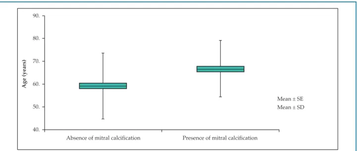
The risk factors associated with mitral annular or papillary muscle calcification were age over 65 years (p < 0.001), presence of hypertension (p = 0.05), and presence of CAD (p = 0.004). The presence of DM showed only a trend toward an association with mitral valve calcification (p = 0.076). On multivariate analysis, the presence of CAD (p = 0.002, odds ratio [OR] 3.39, 95% confidence interval [CI] 1.58-7.29) was an independent
90.
80.
70.
60.
50.
40.
Age (years)
Absence of mitral calcification Presence of mitral calcification
Mean ± SE Mean ± SD
Figure 3 – Association between mitral calcification and age. EP: standard error; SD: standard deviation.
90.00% 80.00% 70.00% 60.00% 50.00% 40.00% 30.00% 20.00% 10.00% 0.00%
Absence of CAD Presence of CAD Absence of mitral annular or papillary muscle calcification Presence of mitral annular or papillary muscle calcification
Figure 4 – Combined association between mitral calcification and coronary artery disease. CAD: coronary artery disease.
Risk factors and aortic valve calcification
Regarding the presence of aortic root calcification and/or aortic valve sclerosis, the risk factors associated were age over 65 years (p < 0.001) and presence of hypertension (p < 0.001). The presence of CAD did not correlate with aortic calcification (p = 0.435). On multivariate analysis, only age above 65 years
90.0
80.0
70.0
60.0
50.0
40.0
Mean ± SE Mean ± SD
Absence of aortic valvular sclerosis or aortic root calcification
Presence of aortic valvular sclerosis or aortic root calcification
Age (years)
Figure 5 – Association between aortic calcification and age. EP: standard error; SD: standard deviation.
Discussion
The present study identified the occurrence of mitral valve annular calcification as a risk factor independently and significantly associated with CAD. This fact has been already demonstrated in necropsy1 and population studies, which have shown the occurrence of calcium deposits in the cardiovascular system of individuals with atherosclerotic disease.2-4
TTE is a practical tool to assess cardiac calcium and its efficacy is comparable to that of angiotomography, as demonstrated in the Heinz Nixdorf Recall study, published in JAMA in 2010.5 There is no standardization in regards to the quantification of cardiac calcium on TTE, and the present study used the Gaibazzi6 score for this purpose.
Numerous studies have correlated cardiac calcium to established CAD. A 2015 study published in the American Journal of Cardiology7 including 595 outpatients with stable CAD undergoing TTE has shown that the risk of death was higher in patients with a
calcium score ≥ 2, even when adjusted for age, previous
revascularization, DM, diastolic blood pressure, and glomerular filtration rate. However, the presence of cardiac calcification did not emerge as an independent risk factor in our study patients with previous CAD.
A 2006 study published in Circulation12 attempted to determine whether atherosclerotic calcification of multiple vascular areas would be significantly associated with mitral and aortic annular calcification, independently of traditional risk factors. To determine
that, the study evaluated the extension of the atherosclerotic calcification by CT of five vascular beds and mitral and aortic annular calcification in 1,242 patients. It observed that 24% of the patients had aortic annular calcification and 8% had mitral annular calcification, and that age and hypertension were the only cardiovascular risk factors independently associated with the prevalence of these calcifications. We found similar results in the present study: 41% of the study population showed some degree of valvular cardiac calcification on TTE, whereas 15% had calcification only in the aortic valve and 9% only in the mitral valve. Similarly, only age and hypertension emerged as risk factors independently associated with valvular calcification.
A study has shown that patients with mitral annular calcification have cardiovascular, renal, metabolic, and functional profiles worse than those with aortic ring calcification or aortic valve sclerosis. The same study also demonstrated that the more intense the calcification, the greater is the relationship with cardiovascular disease. When the risk was adjusted for the presence of DM, hypertension, previous CAD, ankle-brachial index (ABI)
≤ 0.9, serum creatinine ≥ 1.5 mg/dL, carotid stenosis ≥ 25%, left ventricular ejection fraction (LVEF), and
myocardial mass (g), only mitral annular calcification was associated with cardiovascular disease.14 Similarly, the present study revealed in the multivariate analysis that the presence of CAD and age above 65 years are independent risk factors for the presence of mitral annular and/or papillary muscle calcification.
Although several studies have shown a connection between aortic valve sclerosis and risk factors for atherosclerosis and cardiovascular morbidity, the data are still controversial. A 2015 study by Bhatt et al.13 attempted to determine the association between the presence or absence of aortic valve sclerosis and cardiovascular risk factors, CAD extent, and severity of coronary lesions. After allocating 482 patients evaluated with coronary cineangiography into two groups, with and without valvular sclerosis, age was the only independent predictor of aortic valve sclerosis. Aortic valve sclerosis was also not independently associated with the number of obstructed vessels or degree of obstruction, showing that it is probably a benign marker of senile degenerative cardiac changes, regardless of the severity and complexity of the CAD. Finally, aortic valve sclerosis may be related to systemic atherosclerosis and cardiovascular events, but its usefulness in predicting CAD is questionable. In the present study, multivariate analysis revealed that only age was an independent risk factor for the presence of aortic valve calcification. In contrast, some studies have considered the absence of aortic root calcification and mitral annular calcification as a stronger predictor of absence of coronary disease than the absence of traditional risk factors, with the exception of DM.15
The use of ACEIs, aspirin, beta-blockers, and statins was frequent among the patients in this study due to the prevalence of hypertension and dyslipidemia in these individuals. There is no treatment based on evidence to reduce or prevent mitral annular calcification, and it unknown whether commonly used medications such as aspirin, beta-blockers, and statins may prevent its pathogenesis. Several studies suggest that warfarin is associated with increased valvular calcification.11
Although Bhatt et al. found no association between hypercholesterolemia and mitral annular calcification, further studies are needed to determine whether the use of statins would affect prognosis in these patients. Although many patients in the present study used drugs such as ACEIs (65.5% of the patients), aspirin (45.8%), and statin (17.7%), the study was not delineated to correlate the use of drugs and the presence of cardiac calcification.
Some factors have limited this study. First, the study population consisted mainly of elderly and hypertensive patients, since these patients were referred to TTE from an outpatient cardiology clinic. Second, the clinical diagnoses of the risk factors were established by self-report by the patients or through information obtained by review of their medical records. Another limiting factor of the study was the relative subjectivity in the quantification of cardiac calcification. Finally, the medications used by the patients were not included in the multivariate analysis.
Conclusion
The present study concluded that age above 65 years and hypertension were independent risk factors for the presence of valvular cardiac calcification, with mitral valvular calcification alone significantly and independently associated with the presence of CAD.
Author contributions
Conception and design of the research and Critical revision of the manuscript for intellectual content: Sá
CRF, Baroncini LAV, Wermelinger ACC; Acquisition of
data: Sá CRF, Rafael D, Darwich RZ, Fortunato Junior JA, Carmo DC; Analysis and interpretation of the data and
Statistical analysis: Sá CRF, Baroncini LAV; Obtaining
funding and Writing of the manuscript: Sá CRF.
Potential Conflict of Interest
No potential conflict of interest relevant to this article was reported.
Sources of Funding
There were no external funding sources for this study.
Study Association
1. Roberts WC. Morphologic features of the normal and abnormal mitral valve. Am J Cardiol. 1983;51(6):1005-28.
2. Rossi A, Faggiano P, Amado AE, Cicoira M, Bonapace S, Franceschini
L, et al. Mitral and aortic valve sclerosis/calcification and
carotid atherosclerosis: results from 1065 patients. Heart Vessels. 2014;29(6):776-83.
3. Agno FS, Chinali M, Bella JN, Liu JE, Arnett DK, Kitzman DW, et al.
Aortic valve sclerosis is associated with preclinical cardiovascular disease in hypertensive adults: the Hypertension Genetic Epidemiology Network study. J Hypertens. 2005;23(4):867-73.
4. Agmon Y, Khandheria BK, Meissner I, Sicks JR, O Fallon WM, Wiebers
DO, et al. Aortic valve sclerosis and aortic atherosclerosis different manifestations of the same disease?Insights from a population-based study. J Am Coll Cardiol. 2001;38(3):827-34.
5. Erbel R, Möhlenkamp S, Moebus S, Schmermund A, Lehmann N, Stang
A, et al; Heinz Nixdorf Recall Study Investigative Group. Coronary risk stratification, distribution, and reclassification improvement based on quantification of subclinical coronary atherosclerosis: the Heinz Nixdorf recall study. J Am Coll Cardiol. 2010;56(17):1397-406.
6. Gaibazzi N, Baldari C, Faggiano P, Albertini L, Faden G, Pigazzani F,
et al. Cardiac calcium score on 2D echo: correlations with cardiac and coronary calcium at multi-detector computed tomography. Cardiovasc Ultrasound. 2014;12:43.
7. Saha AS, Beatty AL, Mishra RK, Whooley MA, Schiller NB. Usefulness of
an echocardiographic composite cardiac calcium score to predict death in patients with stable coronary artery disease (from the Heart and Soul Study) Am J Cardiol. 2015;116(1):50-8.
8. Koulaouzidis G, Nicoll R, MacArthur T, Jenkins PJ, Henein MY. Coronary
artery calcification correlates with the presence and severity of valve calcification. Int J Cardiol. 2013;168(6):5263-6.
9. Utsunomiya H, Yamamoto H, Kunita E, Kitagawa T, Ohashi N, Oka T,
et al. Combined presence of aortic valve calcification and mitral anular calcification as a marker of the extent and vulnerable characteristics of coronary artery plaque assessed by 64 multidetector computed tomography. Atherosclerosis. 2010;213(1):166-72.
10. Acartürk E, Bozkurt A, Cayli M, Demir M. Mitral annular calcification and aortic valve calcification may help in predicting significant coronary artery disease. Angiology. 2003;54(5):561-567.
11. Sherif HM. Calcification of left-sided valvular structures: evidence of a proinflammatory milieu. J Heart Valve Dis. 2009;18(1):52-60.
12. Freeman RV, Otto CM. Spectrum of calcific aortic valve disease. Pathogenesis, disease progression, and treatment strategies. Circulation. 2005;111(24):3316-26.
13. Bhatt H, Sanghani D, Julliar K, Fernaine G. Does aortic valve sclerosis
predicts the severity and complexity of coronary artery disease? Indian Heart J. 2015;67(3):239-44.
14. Jeon DS, Atar S, Brasch AV, Luo H, Mirocha J, Navqi TZ, et al. Association
of mitral annulus calcification, aortic valve sclerosis and aortic root calcification with abnormal myocardial perfusion single photon emission tomography in subjects age < or =65 years old. J Am Coll Cardiol. 2001;38(7):1988-93.
15. Acartürk E, Bozkurt A, Cayli M, Demir M. Mitral annular calcification and aortic valve calcification may help in predicting significant coronary artery disease. Angiology. 2003;54(5):561-7.