241 Sonographic evaluation of swallowing biomechanics: a preliminary study
Radiol Bras. 2008 Jul/Ago;41(4):241–244 Original Article • Artigo Original
Sonographic evaluation of swallowing biomechanics:
a preliminary study*
Biomecânica ultra-sonográfica da deglutição: estudo preliminar
Cinthya da Silva Lynch1, Maria Cristina Chammas2, Letícia Lessa Mansur3, Giovanni Guido Cerri4
OBJECTIVE: To evaluate, by means of ultrasonography, the spatial parameters of the hyolaryngeal complex in the deglutition dynamics as well the correlation between age and effects. MATERIALS AND METHODS: The present prospective and quantitative study included 39 healthy men and women in the age range between 20 and 70 years (mean, 45.56; standard deviation, 14.53). The swallowing biomechanics corresponding to the measurement of the distance between the upper portion of the hyoid bone and the upper rim of the thyroid cartilage at the moment of maximum laryngeal elevation was evaluated. Measurements were performed with the ingestion of pasty or liquid food. RESULTS: The variation of the distance between the hyoid bone and the larynx presented a positive association with aging only in the swallowing of pasty food that requires a higher lingual propulsive activity than liquid food does. CONCLUSION: An increase in the distance between the hyoid bone and the larynx, corresponding to a poorer laryngeal elevation, may occur during the deglutition of pasty food as a result of aging, a process where a decrease in functional reserves is observed even in healthy individuals. Ultrasonography can detect the swallowing behavior related to foods consistency, demonstrating the diagnostic potentiality of this method in the evaluation of the deglutition. Keywords: Ultrasonography; Swallowing; Cervical; Dysphagia; Biomechanics; Larynx.
OBJETIVO: Verificar, por meio da ultra-sonografia, os parâmetros espaciais do complexo hiolaríngeo na di-nâmica da deglutição e a associação entre idade e efeitos. MATERIAIS E MÉTODOS: Neste estudo quanti-tativo e prospectivo foram incluídos 39 indivíduos sadios, de ambos os gêneros, na faixa etária de 20 a 70 anos (média, 45,56; desvio-padrão, 14,53). Avaliou-se a biomecânica da deglutição correspondente à me-dida da distância entre a porção superior do osso hióide e a borda superior da cartilagem tireóide, no mo-mento de máxima elevação laríngea. As medidas foram realizadas com a ingestão de alimo-mentos de consis-tências líquida e pastosa. RESULTADOS: A variação da distância da laringe ao hióide apresentou associação positiva com a idade, somente na deglutição de alimento pastoso, consistência na qual é solicitada maior atividade de propulsão lingual do que nos líquidos. CONCLUSÃO: É possível que o aumento da distância entre o hióide e a laringe, que representa menor elevação laríngea, na deglutição de pastosos, ocorra por efeito do envelhecimento, processo no qual se verifica diminuição de reservas funcionais, mesmo em indi-víduos sadios. A ultra-sonografia pode detectar comportamentos relacionados a consistências alimentares, o que mostra sua possível potencialidade diagnóstica na avaliação da deglutição.
Unitermos: Ultra-sonografia; Deglutição; Cervical; Disfagia; Biodinâmica; Laringe. Abstract
Resumo
* Study developed at Instituto de Radiologia do Hospital das Clínicas da Faculdade de Medicina da Universidade de São Paulo (InRad/HC-FMUSP), São Paulo, SP, Brazil. Financial support: Universidade da Amazônia (Unama) – Fundação Instituto para o Desenvolvimento da Amazônia (Fidesa).
1. Fellow PhD degree, Department of Radiology, Faculdade de Medicina da Universidade de São Paulo (FMUSP), São Paulo, SP, Assistant Professor I, Universidade da Amazônia (Unama), Ma-naus, AM, Brazil.
2. PhD, Director for the Unit of Ultrasonography, Instituto de Radiologia do Hospital das Clínicas da Faculdade de Medicina da Universidade de São Paulo (InRad/HC-FMUSP), São Paulo, SP, Brazil.
3. PhD, Assistant Professor for the Course of Phonoaudiology, Department of Physiotherapy, Phonoaudiology and Occupational Therapy, Universidade de São Paulo (USP), São Paulo, SP, Brasil. 4. PhD, Titular Professor, Department of Radiology, Faculdade de Medicina da Universidade de São Paulo (FMUSP), São Paulo, SP, Brazil.
Mailing address: Dra. Cinthya da Silva Lynch. Avenida Alcindo Cacela, 287, Bairro Umarizal. Belém, PA, Brazil, 66060-902. E-mail: [email protected]
phase), pharyngeal and esophageal (con-sidered as reflex phases)(2).
According to the American Speech-Language-Hearing Association(ASHA)(3),
any alteration in this alimentary transit can be defined as dysphagia. Dysphagia corre-sponds to a set of symptoms characterized by difficulty in propelling liquid or solid foods from the oral cavity through the esophagus(2,3). Dysphagic problems and
their secondary complications are associ-ated with high rates of mortality and mor-bidity.
Deglutition difficulties may occur at any age and affect health and social aspects of Lynch CS, Chammas MC, Mansur LL, Cerri GG. Sonographic evaluation of swallowing biomechanics: a preliminary study. Radiol Bras. 2008;41(4):241–244.
INTRODUCTION
Deglutition consists of a sequence of physiological events resulting, after all, in the movement of the food from the mouth into the stomach.
According to Bass & Morrell(1), the
de-glutition process depends on the integrity of a complex neuromotor mechanism and involves a coordinated process that is typi-cally divided into the following phases: oral preparatory (considered as a voluntary
0100-3984 © Colégio Brasileiro de Radiologia e Diagnóstico por Imagem
242
Lynch CS et al.
Radiol Bras. 2008 Jul/Ago;41(4):241–244 feeding. During the deglutition, it is
nec-essary to protect the airways for preventing rinks for aspiration pneumonia(2,4).
The baseline laryngeal protective mechanism consists of reflex inhibition of the respiration, closure of the glottic sphincter, anterior laryngeal displacement and elevation, remaining under the tongue protection(4). This displacement contributes
to the airways closure and opening of the pharyngoesophageal transition necessary to the protection of the lower airways(4–6).
Healthy aging does not cause dysph-agia, but the deglutition performance in the elderly is differentiated. Generally, the eld-erly experiment a decrease in functional reserves of different organs and systems, added of changes in all the deglutition phases(7). In the absence of morbid
condi-tions, the elderly resort to several compen-satory strategies such as exerting strength in the deglutition to increase the pressure of the tongue inside the oral cavity, aiding in the food propulsion(8).
Imaging methods have attracted the at-tention of health professionals in areas such as radiology and phonoaudiology, consid-ering the usefulness of these methods in the recording, description and quantification of relevant parameters of the structures in-volved in the normal deglutition process, as well as in deglutition disorders. Tradi-tionally, videofluoroscopy has been the method of choice for investigating degluti-tion disorders(5,9).
The evaluation of the deglutition by ultrasonography (US) has been utilized for investigating the oral phase(10). The
appli-cation of this method for investigating the pharyngeal phase was experimented early in the decade of 1990 with the publication of the first studies demonstrating the pos-sibility of visualizing anatomical structures and the movements temporal relationship during the oral and pharyngeal phases of deglutition, such as in the study developed by Miller et al.(11) investigating the
appli-cation of M-mode ultrasonography during the swallow of two different boluses of food and three deglutition maneuvers. Sig-nificant differences were found between duration times in lateral pharyngeal views in the different maneuvers performed.
Ultrasonography presents some advan-tages in the assessment of dysphagia when
compared with other traditional diagnostic methods: optimum spatial and temporal resolution in the evaluation of small seg-ments, multiplanar study capability, low cost, equipment portability, besides not requiring contrast enhancement and ioniz-ing radiation(6,12,13).
On the other hand, this method presents limitations as compared with videofluoro-scopy, considering that a panoramic visu-alization of the deglutition process cannot be achieved and the access to some pharyn-geal structures is restricted(6). It is a role of
the specialist to identify the possible clues and the process mechanisms on diagnostic sonographic images.
The present study is aimed at investigat-ing the feasibility of ultrasonography in evaluating the deglutition process.
MATERIALS AND METHODS
The present study included 39 healthy volunteers (8 men and 31 women) in the age range between 20 and 70 years. The individuals were distributed according to their age range as shown on Table 1. Mean age was 45.56, and the median, 46 years, standard deviation 14.53. All the individu-als participating in the present study met the normality criterion required for deglutition function by the test proposed by Logemann for clinical evaluation of dysphagia(2) and
by the adapted Cotœs protocol(14).
The following aspects were evaluated in the clinical deglutition test: recent changes in feeding (weight loss, appetite decrease, duration of meals).
The following aspects were included in the physical evaluation: muscular tonus, mobility and amplitude of the orofacial musculature, and dental status.
Previous history of neurological or neurodegenerative diseases and presence
of other direct or indirect factors affecting the deglutition constituted exclusion crite-ria. A term of free and informed consent was signed by all the individuals. The re-search protocol was approved by the Com-mittee for Ethics in Research of Hospital das Clínicas da Faculdade de Medicina da Universidade de São Paulo, São Paulo, SP, Brazil.
Studies were performed with an ATL HDI 5000 (Philips Medical Systems; Phila-delphia, USA) with a multifrequency, con-vex 2–5 MHz transducer, and a thick layer of hydrosoluble contact gel.
During the whole examination, the pa-tient remained in the sitting position, form-ing an angle of 90º between the buccal floor and the neck, in an attempt to simulate the usual positioning during a meal. The pa-tient was asked to swallow foods of two different consistencies liquid and pasty -represented respectively by water and yo-gurt, offered by 10 ml disposable spoons. The food bolus was maintained in the mouth until the patients was verbally asked to start swallowing as usual. The study se-quence was recorded in real-time on VHS videotapes. No complication related to the administration of food was observed.
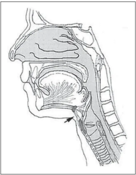
The convex transducer was positioned on the cervical region, at the level of the hyoid-larynx complex (Figure 1). Longitu-dinal images were acquired for measuring the amplitude of movements related to the
Figure 1. Transducer positioning for images record-ing in the cervical region, particularly of the hyoid-larynx complex.
Table 1 Distribution of patients according to age range.
Age range (years)
20–30
31–40
41–50
51–60
61–70
Total
No. of individuals
4
12
7
7
243 Sonographic evaluation of swallowing biomechanics: a preliminary study
Radiol Bras. 2008 Jul/Ago;41(4):241–244
Figure 3. Distance in centimeters of the hyoid-larynx segment during degluti-tion of liquid food (water).
Figure 2. Distance in centimeters of the hyoid-larynx segment at rest.
Figure 4. Distance in centimeters of the hyoid-larynx segment during deglutition of pasty food (yogurt).
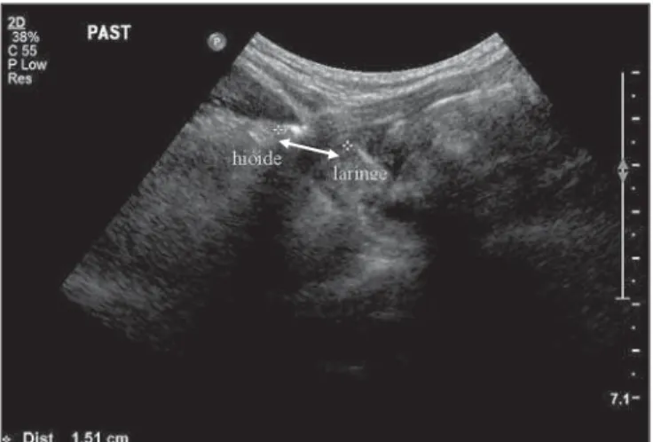
distance between the upper segment of the hyoid bone and the upper edge of the lar-ynx (thyroid cartilage). At the moment of maximum laryngeal elevation, the mea-surement of this segment (hyoid-larynx) was performed in centimeters (Figures 2, 3, and 4).
The version 13.0 of the Statistical Pack-age for Social Sciences (SPSS) software was utilized for statistical analysis, consid-ering p = 0.05 as significance level. The Student’s t test was utilized for evaluating possible differences between values for male and female individuals, and the Levene’s test, for testing the homogeneity of variances. Pearson´s correlation coeffi-cient was applied to evaluate possible cor-relation between paired measurements.
RESULTS
No statistically significant difference was found among mean ages between men and women.
Sonographic evaluation of deglutition
Comparison between genders in the swallowing of liquid and pasty foods – The analysis of the values obtained by the Student’s t test controlled by the Lavene´s test for homogeneity of variances demon-strated a similar behavior both for men and women in the swallowing of liquid and pasty foods. Considering that no statisti-cally significant intergroup difference was found (liquid food, p = 0.785; pasty food, p = 0.460), the authors decided to unify the gender variable.
Measurement of the hyoid-thyroid cartilage distance – The measurements of the distance between the hyoid bone and the thyroid cartilage and their correlation with age provided statistically significant results (liquid food, p = 0.175; pasty food, p = 0.011).
DISCUSSION
The different physiological aspects of the normal deglutition and dysphagic dis-orders represent a recent area of interest shared by several health professionals.
The management of dysphagia requires the investigation of the deglutition physi-ology by means of diagnostic imaging methods.
The utilization of US as a tool for evalu-ating the deglutition with emphasis on the easy equipment operation for specific re-quirements in the investigation of
dysph-agic disorders, corroborates the results re-ported by Kuhl et al.(6), who demonstrated
significant differences among laryngeal displacements, allowing the utilization of this method.
Despite the imbalance in the number of male and female individuals included in the sample of the present study, the US evalu-ation demonstrated a small standard devia-tion.
The previous clinical examination dem-onstrated normality in anatomical and func-tional aspects of the organs and systems involved in the deglutition. Specific age-related signs, such as decrease in laryngeal elevation, could not be detected during the clinical examination, requiring an objective evaluation by US.
244
Lynch CS et al.
Radiol Bras. 2008 Jul/Ago;41(4):241–244 (voluntary deglutition), including the
in-crease in the strength of food propulsion among other mechanisms, similarly to re-sults reported by Logemann et al.(13) and
Kays & Robbins(7).
Even with the attention level under con-trol, the individuals presented a perfor-mance in pasty food deglutition demon-strating a significant positive correlation with age. Despite their satisfactory func-tional levels, the elders present different performance levels as compared with the levels achieved by the other individuals in de sample, in agreement with some au-thors(6–8). This information should be taken
as a relevant warning to health profession-als, considering that, frequently, the utili-zation of pasty foods is proposed in the adaptation of food consistency for the eld-erly with deglutition difficulties, because of the generally diffused idea that this consis-tency is more easily processed in the oral phase.
There are still few studies approaching the utilization of US in the deglutition evaluation(15). The present study is a pilot
for future initiatives involving comparison with other results obtained with video-fluoroscopy.
Promising suggestions for utilization of US in deglutition studies include temporal measurements in order to allow the obser-vation of structures temporal and spatial relationships, and inference of integrity or impairment of airways protective mecha-nisms, as well as the utilization of the
equipment as a monitoring resource in speech and language therapies.
Images suggesting the presence of food remainders, penetration, food aspiration, as well as the presence of protuberant thyroid cartilage impairing the acquisition of im-ages constitute limiting factors in the US evaluation of the deglutition.
CONCLUSION
Ultrasonography has allowed the evalu-ation of the deglutition functionality as well as its variation with aging, representing a complementary resource in the evaluation of possible alterations in the swallowing process. Sonographic studies can be useful in the detection of direct effects on the re-habilitation process with the adoption of specific postural and protective maneuvers, as well as changes in foods consistency, allowing a better management of dysph-agia.
REFERENCES
1. Bass NH, Morrell RM. The neurology of swallow-ing. In: Groher ME, editor. Dysphagia: diagnosis and management. Boston: Butterworth-Heine-mann; 1992. p. 1–31.
2. Logemann JA. Evaluation and treatment of swal-lowing disorders. 2nd ed. Austin: Pro-Ed; 1998. 3. American Speech-Language-Hearing Associa-tion. Instrumental diagnostic procedures for swal-lowing. Ad Hoc Committee on Advances in Clini-cal Practice. ASHA Suppl. 1992;34(Suppl. 7):25– 33.
4. Costa MMB. Como proteger fisiologicamente as vias aéreas durante a deglutição. In: Castro LP, Savassi-Rocha PR, Melo JRC, et al. Tópicos em
gastroenterologia 10 – deglutição e disfagia. Rio de Janeiro: Medsi; 2000. p. 37–48.
5. Leonard RJ, Kendall KA, McKenzie S, et al. Structural displacements in normal swallowing: a videofluoroscopic study. Dysphagia. 2000;15: 146–52.
6. Kuhl V, Eicke BM, Dieterich M, et al. Sono-graphic analysis of laryngeal elevation during swallowing. J Neurol. 2003;250:333–7. 7. Kays S, Robbins J. Effects of sensorimotor
exer-cise on swallowing outcomes relative to age and age-related disease. Semin Speech Lang. 2006; 27:245–59.
8. Hind JA, Nicosia MA, Roecker EB, et al. Com-parison of effortful and noneffortful swallows in healthy middle-aged and older adults. Arch Phys Med Rehabil. 2001;82:1661–5.
9. Bilton TL. Estudo da dinâmica da deglutição e das suas variações associadas ao envelhecimento, avaliadas por videodeglutoesofagograma, em adultos assintomáticos de 20 a 86 anos [Tese de Doutorado]. São Paulo: Universidade Federal de São Paulo; 2000.
10. Chi-Fishman G. Quantitative lingual, pharyngeal and laryngeal ultrasonography in swallowing re-search: a technical review. Clin Linguist Phon. 2005;19:589–604.
11. Miller JL, Watkin KL. Lateral pharyngeal wall motion during swallowing using real time ultra-sound. Dysphagia. 1997;12:125–32.
12. Huckabee M, Hiss S, Barclay M, et al. The rela-tionship between submental SEMG measurement and pharyngeal pressures during normal and effortful swallowing. Dysphagia. 2005;20:73. 13. Logemann JA, Pauloski BR, Rademaker AW, et
al. Temporal and biomechanical characteristics of oropharyngeal swallow in younger and older men. J Speech Lang Hear Res. 2000;43:1264–74. 14. Cot F. Évaluation de la déglutition. In: Cot F,
editor. La dysphagie oro-pharingée chez l’adulte. Québéc: Maloine-Edisem; 1996. p. 79–99. 15. Santos RS, Macedo Filho ED. Sonar Doppler