Cephalometric evaluation of the airway space and hyoid
bone in children with normal and atypical deglutition:
correlation study
Avaliação cefalométrica de via aérea e do osso hioide em crianças com deglutição
normal e atípica: estudo de correlações
Almiro José Machado Júnior
I, Agrício Nubiato Crespo
IIDiscipline of Otorhinolaryngology, Faculdade de Ciências Médicas da Universidade Estadual de Campinas (FCM-Unicamp), Campinas,
São Paulo, Brazil
ABSTRACT
CONTEXT AND OBJECTIVE: Although there is a close relationship between swallowing and breathing, there are no studies evaluating the radiographic anatomy of the airway and its possible correlation with the radiographic position of the hyoid bone. The aim of this study was to evaluate the possible correlation of the radiographic position of the hyoid bone and airway space (PAS) in lateral radiographs on children with atypical deglutition, in comparison with those with normal swallowing.
DESIGN AND SETTING: Cross-sectional analytical study with control group in a public university.
METHODS: Using cephalometric analysis on lateral teleradiographs, the distance from the hyoid bone to the mandibular plane (MP-H) and the distance from the hyoid bone to the tuber (T-H) were correlated with the PAS measurement (airway) in two groups: 55 teleradiographs in the experimental group (with atypical deglutition) and 55 teleradiographs in the control group (normal deglutition). Both groups in-cluded subjects at the mixed dentition stage.
RESULTS: The variable T-H presented a statistically signiicant correlation with PAS (0.0286) and the vari-able MP-H had a signiicant correlation with the varivari-able PAS (0.0053). This positive correlation was signii-cant only in the control group and not in the group with atypical swallowing.
CONCLUSIONS: There was a positive correlation between the MP-H and PAS measurements and between the T-H and PAS measurements only in the group with normal swallowing. These correlations were not observed in the group with atypical swallowing.
RESUMO
CONTEXTO E OBJETIVO: Embora haja estreita relação entre respiração e deglutição, não existem estudos que avaliem a anatomia radiográica de via aérea e sua possível correlação com a posição radiográica do osso hioide. O objetivo deste estudo foi avaliar possível correlação da posição radiográica do osso hioide e do espaço aéreo na radiograia lateral de crianças com deglutição atípica quando comparada com aquelas com deglutição normal.
TIPO DE ESTUDO E LOCAL: Estudo transversal analítico com grupo controle em universidade pública.
MÉTODOS: Por meio de análise cefalométrica em telerradiograias laterais, foi correlacionada a distância do osso hioide ao plano mandibular (MP-H) e do túber ao osso hioide (T-H) com a medida do espaço da via aérea (PAS) em dois grupos: 55 telerradiograias do grupo experimental (com deglutição atípica) e 55 telerradiograias do grupo controle (deglutição normal). Ambos os grupos incluíram indivíduos em fase de dentição mista.
RESULTADOS: A variável T-H apresentou correlação estatisticamente signiicativa com PAS (0,0286) e a variável MP-H teve correlação signiicativa com a variável PAS (0,0053). Esta correlação positiva foi signiica-tiva apenas no grupo controle e não no grupo de deglutição atípica.
CONCLUSÕES: Há correlação positiva entre as medidas MP-H e PAS e entre as medidas T-H e PAS somente no grupo de deglutição normal. Estas correlações não foram observadas no grupo de deglutição atípica.
IDDS. Researcher, Discipline of
Otorhinolaryngology, Faculdade de Ciências Médicas da Universidade Estadual de Campinas (FCM-Unicamp), Campinas, São Paulo, Brazil.
IIPhD. Otorhinolaryngologist, Discipline of
Otorhinolaryngology, Faculdade de Ciências Médicas da Universidade Estadual de Campinas (FCM-Unicamp), Campinas, São Paulo, Brazil.
KEY WORDS:
Cephalometry. Deglutition. Hyoid bone. Oropharynx. Mouth.
PALAVRAS-CHAVE:
Circunferência craniana. Deglutição.
Osso hióide. Orofaringe. Boca.
INTRODUCTION
beyond the fourth year of life. However, it is then considered to be a dysfunction or abnormality because of its association with cer-tain dental malocclusions and facial growing abnormalities.1,2 Such deglutition is classiied as atypical.3-5
Recent studies have investigated the swallowing pattern in relation to child development and have concluded that atypical swallowing is present in half of the children examined at the age of three years, but changes signiicantly ater the age of six years. Nevertheless, atypical swallowing is still present in 25 percent at the age of 12 years.4 he movements of the tongue during swallow-ing may be clinically assessed by askswallow-ing the child to swallow liq-uids, semi-solids or solids, or even only saliva, to observe the pro-trusion of the tongue with the lips half-open or, if necessary, with lips opened with the ingers (forced opening method).1,3 By placing the hands on the masseters, it is possible to observe the presence or absence of contraction and to observe the ascendant movement of the hyoid bone under the thyroid cartilage. he participation of the perioral muscles is also observed, as well as whether the swal-lowing is loud, whether there is any retraction movement with the head, or whether any sign characterizing childlike swallowing is present.1,3-5 For a variety of reasons that so far remain incom-pletely explained, “infantile swallowing” may continue beyond the replacement of the deciduous teeth. Atypical deglutition has been attributed to sucking without nutritive purposes, use of feeding bottles, oral respiration, abnormalities of the central nervous sys-tem and anatomical abnormalities.5-7 However, there is no consen-sus regarding the etiology of atypical deglutition.8-10
Synchronization of sucking and swallowing is achieved through a close relationship between the muscles of the oral region, in order to generate suction pressure for opening and closing the mandible and for using the tongue for bolus forma-tion and peristaltic transportaforma-tion to the pharynx.10 During oral feeding, mechanical respiration involves appropriate activation of the diaphragm, intercostal muscles and muscles of the upper airways from the nose to the glottis.10 Among the likely anatomi-cal abnormalities in cases of atypianatomi-cal deglutition is the position-ing of the hyoid bone, since this is the origin or insertion point of several muscles relating to deglutition.11-13
Recent studies have evaluated the airway space and hyoid bone position in mouth breathing and obstructive sleep apnea (OSA).14-17 Although there is a close relationship between swallowing and breathing, there are no studies relating to the radiographic anatomy of the airway space in cases of atypical swallowing and its possible correlation with the radiographic position of the hyoid bone.
OBJECTIVE
he objective of this study was to evaluate the possible correla-tion between the radiographic posicorrela-tion of the hyoid bone and the airway space on lateral radiographs in children with atypical deglutition, in comparison with those with normal swallowing.
METHODS
he research protocol for this study received unrestricted prior approval from the Research Ethics Committee of Faculdade de Medicina da Universidade Estadual de Campinas (FCM-Unicamp) (# 619/2005). his was a cross-sectional analytical study with a con-trol group in which lateral teleradiographs from children of both genders at the phase of mixed dentition were evaluated. he whole study sample consisted of 110 teleradiographs in lateral view, from 52 female and 58 male subjects. he two groups were similar with regard to gender distribution. he mean ages of the control group (normal deglutition) and the experimental group were 9.46 years and 10.05 years, respectively. To deine the control and experimen-tal groups, an initial test using the forced opening method was con-ducted1-3 by three senior orthodontists simultaneously. he group to which the child’s teleradiograph should be allocated was deined by consensus.
Twenty lateral teleradiographs on 20 patients with a clinical diagnosis of atypical deglutition and another 20 lateral teleradio-graphs on 20 subjects with normal deglutition were selected for a pilot study in order to calculate the sample size. For this, the stan-dard deviation of the control group and the diference between the means of the control and experimental groups were calculated. he alpha and beta values were 0.05 and 0.10 respectively. he size of the sample obtained through the calculation was 12 teleradiographs for measurements on the distance from the hyoid bone to the tuber (T-H), 35 for measurements on the distance from the hyoid bone to the mandibular plane (MP-H) and 26 for measurements on the air-way space (PAS) in each group. A total of 55 teleradiographs were used, in order to also evaluate other variables within this research protocol that are not presented here.
At a signiicance level of 0.05, 110 teleradiographs (i.e. 55 in each group) were required to achieve a test power of 0.10. Ater sample size estimation, the whole sample was selected using the same criteria as in the pilot study, as described above.
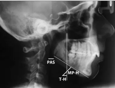
All the lateral view teleradiographs selected for the pres-ent study were of dimensions 18 cm x 24 cm, and were obtained using the same Siemens apparatus for one second at 6 kVp, with a focal length of 1.5 meters. he examinations were performed with the patient’s head in a natural position (mirror position), and were performed by the same examiner. Using the selected lat-eral teleradiographs, cephalometric examination was performed in a darkened room with a negatoscope. An acetate sheet was laid over the teleradiograph and the following anatomoradiographic points and planes were marked on the sheet (Figure 1):
T-H: tuber (line of intersection between the center of the pterygomaxillary issure and the posterior nasal spine) to hyoid (most anterosuperior point of the body of the hyoid bone);
PAS: frontal wall of pharyngeal airway to posterior wall of pharyngeal airway.
Lateral teleradiographs that did not provide a good view of the anatomical structures used in the cephalometric examination were excluded from the study sample. Patients with dental agenesis, con-genital poor orofacial formation or orthodontic and/or functional orthopedic treatment prior to the study, and those for whom there were doubts and imprecision regarding the diagnosis of deglutition, were also excluded. Lack of unanimity among the examiners regard-ing the clinical diagnosis was also a factor for exclusion from the sample. he patients’ skeletal pattern and any occurrences of maloc-clusion were not taken into consideration in this study.
he lateral teleradiographs from the experimental group and the control group were randomly put aside and numbered sequen-tially. he examiner performing the manual measurements was
Table 1. Comparative analysis on the variable of male and female subjects
Deglutition Male Female P-value from chi-square test
Normal 33 22
0.1266 Atypical 25 30
Table 2. Comparative analysis of the age variable (years)
Deglutition n Mean Standard deviation Minimum Median Maximum P-value from Mann-Whitney test
Normal 55 9.32 1.83 6.41 9.25 11.66
0.6345
Atypical 55 9.58 2.13 6.41 9.08 11.91
Table 3. Comparative analysis on the MP-H variable (mm)
Deglutition n Mean Standard deviation Minimum Median Maximum P-value from Mann-Whitney test
Normal 55 11.69 5.13 3.00 12.00 21.00
0.016 Atypical 55 16.14 4.86 7.00 16.00 27.00
Figure 1. Cephalometric measurements: TH = tuber and MP-H = madibular plane.
blinded to the patient data. he sequentially numbered teleradio-graphs were handed over to the examiner for the abovementioned standardized measurements to be made, and the results were recorded on a data-gathering instrument. To minimize systematic errors, the same examiner carried out data gathering on the whole sample on two occasions separated by a 20-day interval. Ater col-lection of radiographic data, age and sex data were added, along with whether atypical deglutition was present or not. On the other hand, all appropriate measures were taken to ensure conidential-ity of the subjects’ personal data. Only the initials were recorded on the data-gathering instrument. here was no way in which anyone other than the investigator would be able to identify the individual to whom each teleradiograph belonged.
To investigate possible linear associations (correlations) between the MP-H, T-H and PAS variables, Spearman’s correla-tion analysis was performed. To investigate the intra-examiner consistency, the Wilcoxon test for related samples was used to detect possible diferences between measurements obtained on two diferent occasions. he signiicance level used in the statisti-cal tests was P = 0.05.
RESULTS
he whole study sample consisted of 110 teleradiographs in lat-eral view, from 52 female and 58 male subjects. Only four telera-diographs were discarded because of the exclusion criteria. he two groups were similar regarding gender distribution (Table 1). he mean ages of the control group (normal swallowing) and the experimental group were 9.46 years and 10.05 years, respectively, without any signiicant diference between the groups (Table 2).
he average distance of the PAS variable was 7 mm in the experimental group and 10 mm in the control group, with a statistically signiicant diference (P < 0.001) (Table 5).18 here were positive correlations between MP-H and PAS (P = 0.0053) and T-H and PAS (P = 0.0286), but these correlations were only observed in the control group (Table 6).
DISCUSSION
he study presented here shows that there was a signiicant dif-ference in the radiographic size of the PAS measurement between the study groups, such that it was smaller in the group with atypi-cal swallowing. here were also signiicant diferences in the T-H and MP-H measurements, such that in the group with normal swallowing, the hyoid bone was radiographically closer to the mandibular plane and to the T line (tuber). In assessing the pos-sibility of a correlation between the PAS and MP-H variables and between the PAS and T-H variables, it could be seen that there was a positive correlation in radiographic position between the hyoid bone and the PAS measurement only in the group with normal swallowing.
he PAS measurement has been described as the distance between the posterior and anterior parts of the pharynx, where the base of the tongue is located.12,18 his leads us to believe that the shortening of the airways in patients with atypical swal-lowing might cause changes in tongue positioning, which would lead to changes in the position of the hyoid bone.
Craniofacial abnormalities in children with respiratory obstruction have been studied over recent years. However, the absence of a direct relationship between the cause of respira-tory obstruction and its efect on craniofacial growth has led to considerable controversy in the literature.11-15 he most widely accepted theory is that tonsil hypertrophy, which leads to pha-ryngeal obstruction, causes mouth breathing11 and changes in
the child’s way of positioning the orofacial muscles and mandible. hese changes, in turn, inluence mastication, swallowing and phonation, and lead to occlusal and skeletal abnormalities.11,16
he MP-H variable has been used in cephalometric stud-ies in relation to obstructive sleep apnea and hypopnea syn-drome (OSAHS).13-14 he data from these studies are similar to the results from our study, in that they show that in OSAHS, the hyoid bone is more distant from the mandibular plane, which was also observed in our study on atypical swallowing. his observa-tion leads us to believe that the hyoid bone is perhaps related to maintaining and stabilizing the airway. We also believe that the lower position of the hyoid bone in the group with atypical swal-lowing may have been caused by a change in the suprahyoid and infrahyoid muscles, and possibly hypertonia of the infrahyoid and hypotonia of the suprahyoid muscles. hese changes to trac-tion may have been responsible for the abnormal positrac-tion of the tongue in cases of atypical swallowing,1 which is a lower position than in cases of normal swallowing.
he T-H variable was originally used in this study with the intention of observing the anterior-posterior positioning of the hyoid bone in relation to the face. We took the descendant line of the pterygomaxillary issure to the level of the hyoid bone as mark zero. Distances to its right were measured as positive values and distances to its let were taken to be negative values. here-fore, the negative value of the T-H variable found in the group with atypical swallowing refers to the more posterior position of the hyoid bone, in relation to the descendant line of the pterygo-maxillary issure. One hypothesis that has already been studied is that the radiographic position of the hyoid bone is dependent on facial type and is associated with factors such as age, obesity, breathing and apnea,1,3,17 Our results demonstrated that func-tional alterations such as atypical swallowing may also be among the adjunct factors that alter the position of the hyoid bone.
Table 4. Comparative analysis on the T-H variable (mm)
Deglutition n Mean Standard deviation Minimum Median Maximum P-value from Mann-Whitney test
Normal 55 2.26 1.79 0.00 2.00 6.00
< 0.001 Atypical 55 -5.89 4.77 -16.00 -5.00 4.00
Table 5. Comparative analysis on the PAS (airway) variable (mm)
Deglutition n Mean Standard deviation Minimum Median Maximum P-value from Mann-Whitney test
Normal 55 10.53 2.43 5.00 10.00 15.00
< 0.001
Atypical 55 7.82 2.93 3.00 7.00 13.00
Table 6. Spearman’s linear correlation coeicients between the variables in each group
Normal deglutition Normal deglutition Atypical deglutition Atypical deglutition
PAS
T-H MP-H T-H MP-H
0.29536 0.37103 -0.19513 0.01967
0.0286* 0.0053* 0.1534 0.8867
Recent studies7,10 have correlated the pharynx measure-ment with mandibular position and have suggested that with mandibular advancement there is an increase in PAS mea-sure. New knowledge of the correlation between the measure-ments studied in the normal swallowing group may corrob-orate this hypothesis. Because there are muscles connecting the hyoid bone to the mandible and because the hyoid is a mobile bone, the position of the hyoid may be altered because of the position of the mandible. This may be the reason why we only found a correlation between the measurements stud-ied in the normal swallowing group. In the atypical swallow-ing group, the hyoid bone was in a more inferior and poste-rior position, which would indicate mandible positioning that was more posterior, with consequently decreased airway size. We believe that the decrease in the PAS measurement may be related to a more posterior position for the mandible, caused primarily by the lower positioning of the tongue, thereby causing a decrease in the PAS measurement. In the group with atypical swallowing, we did not observe this correlation, since the hyoid bone is more distant from the mandibular plane and from the pterygomaxillary line.
Clinical tests used for defining atypical swallowing have limitations, and the final diagnosis of the type of swallow-ing is based on each examiner’s experience, through assessswallow-ing the involvement of the orbicular muscles and other compen-satory components, in swallowing. Perfect sealing of the oral cavity and contraction of the masseter muscle, which helps the dental occlusion and is necessary for swallowing, was observed in individuals with normal swallowing. Lip incom-petence and perioral muscle exertion to help in swallowing, and, in some cases, spilling of content from the labial com-missure and interposition of the tongue to help in relation to incompetent lip sealing have been observed in cases of atypi-cal swallowing.1,3,5,14
his radiographic study evaluated the relationships between known parameters, but these relationships were used in a novel manner, in that they were correlated with normal and abnormal deglutition. In addition to studying a normal group, this study also evaluated patients with atypical deglutition, which is a moderately prevalent clinical condition that can have an impact on orofacial, nutritional, esthetic and psychosocial develop-ment.13 Since deglutition is a highly complex and coordinated function, it requires activation of many anatomical structures relating to the tongue. Insuicient functional stimulation of the stomatognathic system, especially the tongue, might be the main factor in persistence of childlike deglutition.1 herefore, because pediatricians are the irst professionals to have contact with such children, they should always be aware of the degree of maturation of swallowing function and refer such children to a dentist when maxillary-mandibular changes are noticed.
CONCLUSION
here are positive correlations between the MP-H and PAS mea-surements and between the T-H and PAS meamea-surements only in the group with normal swallowing. hese correlations were not observed in the group with atypical swallowing.
REFERENCES
1. Peng CL, Jost-Brinkmann PG, Yoshida N, Miethke RR, Lin CT.
Diferential diagnosis between infantile and mature swallowing with
ultrasonography. Eur J Orthod. 2003;25(5):451-6.
2. Graber TM, Rakosi T, Petrovic AG. Dentofacial orthopedics with
functional appliances. St. Louis: Mosby Company; 1985.
3. Peng CL, Jost-Brinkmann PG, Yoshida N, Chou HH, Lin CT. Comparison
of tongue functions between mature and tongue-thrust
swallowing--an ultrasound investigation. Am J Orthod Dentofacial Orthop.
2004;125(5):562-70.
4. Ovsenik M, Farcnik FM, Korpar M, Verdenik I. Follow-up study of
functional and morphological malocclusion trait changes from 3 to
12 years of age. Eur J Orthod. 2007;29(5):523-9.
5. Bertolini MM, Vilhegas S, Norato DY, Paschoal JR. Cephalometric
evaluation in children presenting adapted swallowing during mixed
dentition. Int J Orofacial Myology. 2003;29:29-41.
6. Cayley AS, Tindall AP, Sampson WJ, Butcher AR. Electropalatographic
and cephalometric assessment of myofunctional therapy in
open-bite subjects. Aust Orthod J. 2000;16(1):23-33.
7. Cheng CF, Peng CL, Chiou HY, Tsai CY. Dentofacial morphology
and tongue function during swallowing. Am J Orthod Dentofacial
Orthop. 2002;122(5):491-9.
8. Machado Júnior AJ, Crespo AN. Radiographic position of the
hyoid bone in children with atypical deglutition. Eur J Orthod.
2012;34(1):83-7.
9. de Felício CM, Folha GA, Ferreira CL, Medeiros AP. Expanded
protocol of orofacial myofunctional evaluation with scores:
Validity and reliability. Int J Pediatr Otorhinolaryngol.
2010;74(11):1230-9.
10. Valera FC, Travitzki LV, Mattar SE, et al. Muscular, functional and
orthodontic changes in pre school children with enlarged adenoids
and tonsils. Int J Pediatr Otorhinolaryngol. 2003;67(7):761-70.
11. Paskay LC. Instrumentation and measurement procedures in orofacial
myology. Int J Orofacial Myology. 2006;32:37-57.
12. Adamidis IP, Spyropoulos MN. The efects of lymphadenoid
hypertrophy on the position of the tongue, the mandible and the
hyoid bone. Eur J Orthod. 1983;5(4):287-94.
13. Machado Júnior AJ, Crespo AN. Postural evaluation in children with
atypical swallowing: radiographic study. J Soc Bras ‘Fonoaudiol.
2012;24(2):125-9.
14. Malkoc S, Usumez S, Nur M, Donaghyd CE. Reproducibility
of airway dimensions and tongue and hyoid positions on
lateral cephalograms. Am J Orthod Dentofacial Orthop.
15. Machado Júnior AJ, Crespo AN. A lateral cephalometric X-ray study
of selected vertical dimensions in children with atypical deglutition.
The International Journal of Orofacial Myology. 2010;36:17-26.
16. Kikyo T, Saito M, Ishikawa M. A study comparing ultrasound images
of tongue movements between open bite children and normal
children in the early mixed dentition period. J Med Dent Sci.
1999;46(3):127-37.
17. Bibby RE. The hyoid bone position in mouth breathers and
tongue-thrusters. Am J Orthod. 1984;85(5):431-3.
18. Machado AJ Jr, Crespo AN. Cephalometric evaluation of the
oropharyngeal space in children with atypical deglutition. Braz J
Otorhinolaryngol. 2012;78(1):120-5.
Acknowledgements: The authors wish to thank the Foundation for Coordination of Advancement of University-level Personnel (Fundação
Coordenação de Aperfeiçoamento de Pessoal de Nível Superior, Capes),
Brazil, for inancial support
This paper formed part of a doctoral thesis produced within the
Discipline of Otorhinolaryngology, Faculdade de Ciências Médicas da
Universidade Estadual de Campinas (FCM-Unicamp), Campinas, São
Paulo, Brazil
Sources of funding: Foundation for Coordination of Advancement of University-level Personnel (Coordenação de Aperfeiçoamento de Pessoal
de Nível Superior, Capes) – grant number 2008/995978
Conlict of interest: None
Date of irst submission: June 1, 2011
Last received: December 21, 2011
Accepted: December 27, 2011
Address for correspondence:
Almiro José Machado Júnior
Rua Maria Monteiro, 841 — apto 11
Cambuí — Campinas (SP) — Brasil
CEP 13025-151
Tel. (+55 19) 3253-5472