Structural characterization of unusually stable polycyclic ozonides
R.C. Cusati
a, U.A. Pereira
a, L.C.A. Barbosa
a,b,⇑
, C.R.A. Maltha
a, José W.M. Carneiro
c, R.S. Corrêa
d,
A.C. Doriguetto
eaDepartment of Chemistry, Federal University of Viçosa, Av. P.H. Rolfs, CEP 36570-000 Viçosa, MG, Brazil
bDepartment of Chemistry, ICEx, Universidade Federal de Minas Gerais, Av. Pres. Antônio Carlos, 6627, Campus Pampulha, CEP 31270-901 Belo Horizonte, MG, Brazil cDepartment of Inorganic Chemistry, Fluminense Federal University, Outeiro de São João Batista, s/n, Centro, CEP 24020-141 Niterói, RJ, Brazil
dSão Carlos Physics Institute – USP, Cx. Postal 369, CEP 13560-970 São Carlos, SP, Brazil
eChemistry Institute, Federal University of Alfenas, Rua Gabriel Monteiro da Silva, 700, CEP 37130-000, Alfenas, MG, Brazil
h i g h l i g h t s
Compound3crystallizes in the unusual space group R3m. Most ozonides crystallize in the
monoclinic system with P21/c space
group as racemic crystal.
Compound3space group represents only0.04% of small-molecule crystal
structures.
X-ray crystallographic studies of stable polycyclic ozonides were carried out.
g r a p h i c a l
a b s t r a c t
O O -+ + O O O O O O 1 2 3 4 5 6 7 8 9 11 12 13 O 3 O O O
R3m space group
_
a r t i c l e
i n f o
Article history:
Received 17 September 2014
Received in revised form 30 October 2014 Accepted 30 October 2014
Available online 8 November 2014
Keywords:
8-Oxabicycle[3.2.1]oct-6-en-3-one Ozonides
Herbicides
Single crystal X-ray structure
a b s t r a c t
The single crystal structure of seven tri- and tetracyclic ozonides derived from 8-oxabicycle[3.2.1]oct-6-en-3-ones have been characterized by X-ray diffraction method. Five ozonides (4,5,6,7and8) crystallize in the monoclinic crystal system with P21/c space group. Compound3crystallize in the unusual
centro-symmetric space group R3m, which represents 0.04% of the total number of structures know. The supramolecular structure of3 forms infinite channels in a hexagram fashion, resulting in a honey-comb-like structure. Semi-empirical (PM6) and density functional theory methods (DFT) with the B3LYP functional and the 6-31G(d) basis set were used to optimize the geometries and compute struc-tural parameters (bond lengths, angles and dihedral angles) that could be compared to the refined crystal structure. The theoretical results show good agreements with the experimental structure.
Ó2014 Elsevier B.V. All rights reserved.
Introduction
Chemical crop protection plays a vital role in ensuring sufficient
food supply to a growing world population, since the expansion of
cultivated areas is not enough to allow a harvest increase at the
required rate
[1,2]
. To improve the world agriculture performance
it is necessary to ensure an intensive control of pests, diseases, and
specially selected weeds, always taking into account
environmen-tal issues. In the face of ever more stringent demands, the
discov-ery of new agrochemicals has become an important research area
since it contributes to reduce the losses in the production and
stor-age of crops, reducing the costs of agricultural products
[3–5]
.
Natural products can be used as models to inspire the synthesis
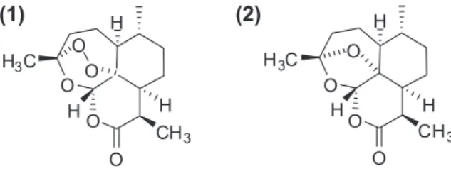
of new bioactive molecules. In this context, artemisinin (
1
,
Fig. 1
), a
http://dx.doi.org/10.1016/j.molstruc.2014.10.082
0022-2860/Ó2014 Elsevier B.V. All rights reserved.
⇑
Corresponding author at: Department of Chemistry, Federal University of Viçosa, Av. P.H. Rolfs, CEP 36570-000 Viçosa, MG, Brazil.E-mail address:[email protected](L.C.A. Barbosa).
Journal of Molecular Structure 1082 (2015) 151–161
Contents lists available at
ScienceDirect
Journal of Molecular Structure
secondary metabolite isolated from the shoots of sweet
worm-wood (
Artemisia annua
L.), and its semi synthetic analogs have a
powerful inhibitory effect on plant growth
[6–9]
. Artemisinin and
selected derivatives are also known for their antimalarial activity
against multidrug-resistant forms of the parasite
Plasmodium
falci-parum
. In fact such compounds are widely used as antimalarial
drugs as part of the Artemisinin Combination Therapy (ACT)
proto-cols recommended by WHO
[10]
.
The phytotoxic and antimalarial activities of these compounds
are dependent upon the presence of the peroxide moiety, since
deoxyartemisinin (
2
,
Fig. 1
) is inactive.
Following our ongoing research to develop new compounds
with herbicidal and/or plant growth regulating activity
[11–14]
,
we have described the synthesis and biological activities of some
stable ozonides
[15,16]
. This research was based on a seminal
discovery by our group that several ozonides derived from
oxabi-cyclic compounds are stable and endowed with activity against
P. falciparum
[17,18]
. Several of such ozonides showed an
apprecia-ble stability that allowed their purification and characterization,
although that class of compounds has been described as very
unstable and explosive
[19,20]
.
Considering that there are few reports in the literature on X-ray
studies of ozonides due to their general instability
[21,22]
, and also
taking into account the importance of molecular structure in the
biological activities, in this work the crystal structures of a series
of stable tri- and tetracyclic ozonides were investigated. The
structural experimental results were analyzed and compared with
computational studies in order to complete the compounds
characterization.
Experimental
The ozonides (
3–9
) were prepared following the procedure
previously described. All experimental detail and spectroscopic
data are in accordance to the literature
[15,16]
.
Single crystal X-ray diffraction studies
Crystals of compounds
3–9
(
Scheme 1
) were obtained by
warm-ing each compound in hexane, followed by addition of some drops
of dichloromethane until the solid was completely dissolved and
the resulting solutions were left static at room temperature. White
well-shaped single crystals suitably for X-ray analyses were
formed after 24 h. They were separated, washed with cold hexane,
and dried. Single crystals of
3–9
were selected for the X-ray
exper-iments. All measurements were made at room temperature (293 K)
on an Enraf-Nonius Kappa-CCD diffractometer with graphite
monochromated Mo K
a
. The final unit cell parameters were based
on all reflections. Data collection was made using the COLLECT
software
[23]
; integration and scaling of the reflections were
per-formed with the HKL Denzo–Scalepack software system
[24]
. The
structures were solved and the models refined using the
SHELXL-2013 software
[25]
. Non-hydrogen atoms of all structures were
clearly solved and full-matrix least-squares refinements of these
atoms in
F
2, with anisotropic thermal parameters, were carried
out. Hydrogen atoms were positioned stereochemically and were
refined with fixed individual displacement parameters [Uiso
(H) = 1.5Ueq (C) for methyl groups or 1.2Ueq(C) methyne and
methylene groups], using the SHELXL riding model with C–H bond
lengths of 0.96, 1.00 and 0.99 Å for methyl, methyne and
methylene groups, respectively. The hydroxyl H atom in
6
was
located by difference Fourier synthesis and was refined with free
coordinates and Uiso(H) = 1.5Ueq(O). WINGX software
[26]
was
(1)
(2)
O
O
H
3C
H
H
CH
3O
H
O
O
O
O
H
3C
H
H
CH
3O
H
O
Fig. 1.Structures of artemisinin (1) and deoxyartemisinin (2).
Table 1
Crystal data and the structures refinement for compounds3–9.
Identification code 3 4 5 6 7 8 9
Empirical formula C11H14O6 C11H16O5 C12H16O7 C11H16O6 C12H18O6 C13H18O5 C13H18O5
Formula weight 726.66 228.24 272.25 244.24 258.26 254.27 254.27
Temperature (K) 293(2) 293(2) 293(2) 293(2) 293(2) 293(2) 293(2)
Wavelength(Å) 0.71073 0.71073 0.71073 0.71073 0.71073 0.71073 0.71073
Crystal system Trigonal Monoclinic Monoclinic Monoclinic Monoclinic Monoclinic Orthorhombic
Space group R3:H P21/c P21/c P21/c P21/c P 21/c P 212121
Unit cell dimensions (Å,°) a= 20.9551(4) a= 5.6815(1) a= 6.7575(3) a= 5.8303(2) a= 14.325(4) a= 13.9049(4) a= 8.0412(3)
b= 20.9551(4) b= 22.3266(8) b= 25.973(1) b= 13.8797(4) b= 7.095(2) b= 8.5122(3) b= 10.5123(3)
c= 13.1662(2) c= 9.1255(3) c= 8.9210(2) c= 14.1242(3) c= 12.811(4) c= 10.8393(3) c= 14.3276(5)
a
= 90a
= 90a
= 90a
= 90a
= 90a
= 90a
= 90b= 90 b= 106.939(2) b= 126.252(2) b= 98.642(2) b= 105.26(2) b= 110.98 b= 90
b= 120 b= 90
c
= 90c
= 90c
= 90c
= 90c
= 90Volume (Å3) 5006.9(2) 1107.31(6) 1262.67(9) 1129.9 1255.99(6) 1197.88(7) 1211.13(7)
Z 18 4 4 4 4 4 4
Calc. Density (Mg/m3) 1.446 1.369 1.432 1.436 1.366 1.410 1.395
l
(mm 1) 0.119 0.108 0.119 0.117 0.110 0.108 0.107F(0 0 0) 2304 488 576 520 552 544 544
Crystal size (mm3) 0.16
0.200.20 0.100.210.53 0.220.290.40 0.150.170.41 0.170.330.60 0.180.300.32 0.170.210.38
hmax(°) 26.615 26.780 26.098 27.425 26.228 26.596 27.481
Index ranges 266h626, 66h66, 86h68, 76h67, 176h617, 176h617, 106h610,
226k622, 286k627, 326k631, 176k617, 86k68, 106k610, 136k613,
166l616 116l611 106l610 176l617 156l615 136l613 176l618
Reflections collected 4559 4006 4469 4276 4429 4341 5644
Independent reflections 2297 [R(int) = 0.0226] 2134 [R(int) = 0.0363] 2375 [R(int) = 0.0385] 2212 [R(int) = 0.0263] 2489 [R(int) = 0.0267] 2495 [R(int) = 0.0172] 2662 [R(int) = 0.043]
Completeness tohmax(%) 98.1 97.6 95.6 98.8 98.7 99.4 95.5
Data/restraints/parameters 2297/0/157 2134/0/148 2375/0/176 2212/0/158 2489/0/167 2495/0/164 2662/0/165
Goodness-of-fit onF2 1.063 1.049 1.021 1.062 1.057 1.058 1.041
FinalRindices [I> 2
r
(I)] R1 = 0.0444, R1 = 0.0490, R1 = 0.0531, R1 = 0.0411, R1 = 0.0497, R1 = 0.0438, R1 = 0.0385, wR2 = 0.0952 wR2 = 0.1188 wR2 = 0.1232 wR2 = 0.1385 wR2 = 0.1085 wR2 = 0.1365 wR2 = 0.1192Rindices (all data) R1 = 0.0597, R1 = 0.0759, R1 = 0.0814, R1 = 0.0567, R1 = 0.0621, R1 = 0.0539, R1 = 0.0478, wR2 = 0.1016 wR2 = 0.1293 wR2 = 0.1370 wR2 = 0.1578 wR2 = 0.1185 wR2 = 0.1471 wR2 = 0.1271
Largest diff. peak and hole (e A3) 0.157 and 0.154 0.163 and 0.149 0.145 and 0.117 0.185 and 0.181 0.172 and 0.202 0.409 and 0.199 0.108 and 0.096
l
= absorption coefficient.R.C.
Cusati
et
al.
/Journal
of
Molecular
Structure
1082
(2015)
151–161
used to analyze and prepare the data for publication. Molecular
graphics were prepared using ORTEP-3 for Windows
[27]
and
Mer-cury
[28]
. Crystal data, data collection procedures, structure
deter-mination methods and refinement results are summarized in
Table 1
.
Computational details
To identify the most stable conformation of each ozonide we
employed the conformer distribution subroutine of the
SPAR-TAN06 software
[29]
using the PM6 semi-empirical method
[30]
for geometry optimization. After location of the most stable
confor-mation for each ozonide that conforconfor-mation was re-optimized with
the B3LYP functional
[30]
and the 6-31G(d) basis set
[31]
. The
calculations of the coupling constant (
J
) was performed for
the optimized geometries using the B3LYP functional and the
6-311++G(2d,p) basis set. The DFT calculations were carried out
with the Gaussian09 software
[32]
.
Results and discussion
Synthesis
All the ozonides have been previously prepared and their
biological activities reported
[15,16]
but no structural details were
described. For clarity, we make a short description of their
synthe-sis. For the preparation of the ozonides
3–7
the alkene
cycload-ducts precursors were prepared via a [4+3] cycloaddition
between furans with the oxyallyl cation, generated
in situ
from
2,4-dibromopentan-3-one using standard literature procedure
[15]
. The alkenes were then submitted to ozonolysis to afford
the 8,9,10,11-tetraoxatricyclo[5.2.1.12,6]oct-6-en-3-ones
(ozo-nides
3–7
,
Scheme 1
). The same cycloaddition procedure using
the oxyallyl cation generated from 2,7-dibromocycloheptanone
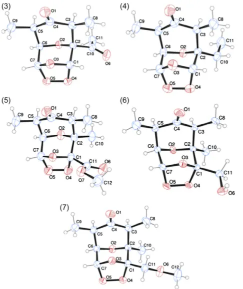
Fig. 2.ORTEP-3 views of the ozonides3–7showing the atom labeling of the tricyclic structures and 30% probability ellipsoids. H atoms are shown as small spheres of arbitrary radii.
Fig. 3.ORTEP-3 views of the ozonides8and9showing the atom labeling of the tetracyclic structures and 30% probability ellipsoids. H atoms are shown as small spheres of arbitrary radii.
with furans, afforded, upon ozonolysis, the 12-oxatricyclo[4.4.1.12,5]
dodec-3-en-11-one adducts
8
and
9
(
Scheme 1
)
[15,16]
.
As previously discussed, the yields of ozonolysis varied from
42% to 100% and no correlations between structure and yields were
observed. Even though the spectroscopic data reported
[15,16]
are
consistent with the proposed structures, a full crystal analysis
was carried out in order to shed light upon the influence of the
substituents on the tridimensional form and crystal arrangement
in the solid state.
Crystal structure analysis
The ORTEP-3 views of the tricyclic ozonides
3–7
are shown in
Fig. 2
. All compounds are chiral and crystallize in centrosymmetric
space groups (
Table 1
). Therefore, their crystal structures are a
50:50 equimolar mixture of a pair of enantiomers in a well-defined
arrangement. The enantiomers (C1(S), C2(S), C3(S), C5(R), C6(R),
C7(R)) showed for
3, 4
,
6
,
7
and (C1(R), C2(S), C3(S), C5(R), C6(R),
C7(R)) showed for
5
in
Fig. 2
were arbitrarily choose as asymmetric
units for the tricyclic ozonides.
The ORTEP-3 views of the tetracyclic ozonides
8
and
9
are
illus-trated in
Fig. 3
. As observed for compounds
3
–
7
, the ozonide
8
crystallizes in a centrosymmetric space group and therefore is a
racemic crystal. The ozonide
9
was solved in the space group
P2
12
12
1and since it is a chiral molecule crystallized in a chiral
space group containing just one molecule in the asymmetric unit,
its crystal structure contains a pure enantiomer. However, it was
not possible to determine the absolute structure of
9
using X-ray
diffraction since it contains only electron-poor atoms, which does
not have anomalous scattering large enough (using Mo K
a
radia-tion) to permit a reliably refined Flack parameter value
[33]
. The
enantiomers showed in
Fig. 3
, (C1(R), C2(R), C3(S), C5(R), C6(S),
C7(S)) and (C1(S), C2(S), C3(R), C5(S), C6(R), C7(R)) for
8
and
9
,
respectively, were arbitrarily choose as asymmetric units for the
tetracyclic ozonides.
Equivalent enantiomers of tricyclic ozonides
3–7
are
superim-posed in a capped stick fashion (
Fig. 4
a). The overlay of molecular
backbones shows the conformational similarity between
homolo-gous atoms in the three fused rings and their first neighbor atoms.
In other words, except for the moieties of the functional groups
linked to C1 and C2, the remaining molecular geometry is very
similar (see also
Tables 4 and 5
). The same features are observed
when the tetracyclic ozonides
8
and
9
are compared (
Fig. 4
b).
The overlay of molecular backbones of
4
(tricyclic) and
9
(tetracy-clic) shows that the homologous three-fused rings are also
geomet-rically identical (
Fig. 4
c). An important difference is only observed
for the orientation of one of the three non-hydrogen atoms linked
at C3 and C5 atoms, which points to front in the tricyclic ozonides
(C8 and C9) and to the back in the tetracyclic ones (C8 and C11).
Taking into account the intermolecular interactions, dimmers
assembled with inversion symmetry related molecules through
intermolecular non-classical H bonds are common building blocks
for the packing of the centrosymmetric ozonides
3–8
(
Fig. 5
). The
hydrogen-bond motifs of the supramolecular dimmers are R
22
(8)
for
3
,
4
, and
6
; R
22
(10) for
5
and
7
; R
22(12) for
8
[34]
. The linkage
via methyne strongest non-classical H bond donor
[35]
occurs to
3
,
4
,
6
, and
8
. In three of the five tricyclic ozonides,
3
,
4
and
7,
the O1 atom works as H bond acceptor forming the supramolecular
synthons in which the inversion symmetry related molecules
Fig. 4.Mercury views showing the superposition of equivalent enantiomers of (a)
3–7, (b)8and9, (c)4and9. The ozonides are labeled by color scheme. The hydrogen atoms were hidden for the representation clarity. (For interpretation of the references to color in this figure legend, the reader is referred to the web version of this article.)
Table 2
Comparison between the oxygen–oxygen bond lengths (Å) obtained by theoretical calculations and X-ray analysis.
Compound Oxygen(4)a-oxygen(5) bond length (Å)
Theoretical calculations X-ray data
Semi-empirical (PM6) (error %)b DFT (B3LYP/6-31G(d)) (error %)b
3 1.448 (2.5) 1.481 (0.4) 1.487(2)
4 1.447 (3.0) 1.481 (0.7) 1.492(2)
5 1.444 (2.8) 1.477 (0.6) 1.485(2)
6 1.442 (3.8) 1.472 (1.8) 1.499(2)
7 1.445 (3.6) 1.479 (0.9) 1.492(2)
8 1.449 (3.1) 1.482 (0.9) 1.495(3)
9 1.449 (2.3) 1.482 (0.1) 1.484(3)
a The numbers 4 and 5 in the oxygen refer only to the ORTEP representations.
b Values in brackets refer to the percentage error between experimental and theoretical computations.
Table 3
X-ray and calculated bond lengths (Å) for ozonides3–9.
Fragment 3 4 5 6 7 Fragment 8 9
Bond length DFT* Bond length DFT* Bond length DFT* Bond length DFT* Bond length DFT* Bond length DFT* Bond length DFT*
C(1)–C(2) 1.532(2) 1.548 1.528(2) 1.551 1.556(3) 1.552 1.614(2) 1.564 1.563(2) 1.568 C(1)–C(2) 1.542(2) 1.553 1.537(3) 1.551
C(1)–O(3) 1.411(2) 1.415 1.415(2) 1.418 1.413(2) 1.409 1.395(2) 1.421 1.422(2) 1.424 C(1)–O(3) 1.418(2) 1.416 1.421(3) 1.415
C(1)–O(4) 1.421(2) 1.425 1.432(2) 1.426 1.444(2) 1.444 1.381(2) 1.450 1.439(2) 1.441 C(1)–O(4) 1.428(2) 1.427 1.417(3) 1.426
C(2)–C(10) 1.530(2) 1.540 1.504(2) 1.536 1.515(3) 1.514 1.490(2) 1.526 1.508(2) 1.525 C(10)–C(11) 1.535(3) 1.542 1.523(4) 1.542
C(2)–C(3) 1.550(2) 1.563 1.543(2) 1.558 1.546(3) 1.584 1.502(2) 1.566 1.550(2) 1.565 C(2)–C(12) 1.524(2) 1.535 1.508(3) 1.524
C(2)–O(2) 1.429(2) 1.431 1.443(2) 1.439 1.433(2) 1.429 1.466(2) 1.444 1.445(2) 1.446 C(2)–C(3) 1.541(2) 1.560 1.543(3) 1.557
C(3)–C(4) 1.528(2) 1.544 1.512(2) 1.539 1.515(3) 1.538 1.509(2) 1.538 1.518(2) 1.538 C(2)–O(2) 1.435(2) 1.442 1.444(2) 1.439
C(3)–C(8) 1.522(2) 1.532 1.524(2) 1.533 1.529(3) 1.531 1.517(2) 1.533 1.524(3) 1.534 C(3)–C(4) 1.523(2) 1.528 1.513(3) 1.528
C(4)–C(5) 1.521(2) 1.535 1.526(2) 1.536 1.514(3) 1.537 1.512(3) 1.534 1.516(2) 1.535 C(3)–C(8) 1.544(2) 1.565 1.542(3) 1.563
C(4)–O(1) 1.212(2) 1.213 1.210(2) 1.215 1.218(2) 1.215 1.157(2) 1.215 1.210(2) 1.216 C(4)–C(5) 1.516(2) 1.528 1.513(3) 1.528
C(5)–C(6) 1.527(2) 1.541 1.521(3) 1.543 1.528(3) 1.545 1.475(2) 1.544 1.527(2) 1.544 C(4)–O(1) 1.214(2) 1.215 1.225(2) 1.216
C(5)–C(9) 1.522(2) 1.531 1.516(3) 1.531 1.518(3) 1.531 1.512(3) 1.530 1.518(2) 1.530 C(5)–C(11) 1.558(2) 1.562 1.566(3) 1.563
C(6)–C(7) 1.516(2) 1.539 1.519(2) 1.539 1.510(3) 1.539 1.585(2) 1.535 1.522(2) 1.534 C(5)–C(6) 1.527(2) 1.546 1.540(3) 1.557
C(6)–O(2) 1.432(2) 1.430 1.431(2) 1.426 1.425(2) 1.428 1.424(2) 1.424 1.420(2) 1.423 C(6)–C(7) 1.515(2) 1.539 1.538(3) 1.551
C(7)–O(3) 1.417(2) 1.419 1.422(2) 1418 1.418(2) 1.420 1.428(2) 1.418 1.419(2) 1.416 C(6)–O(2) 1.437(2) 1.430 1.437(2) 1.439
C(7)–O(5) 1.425(2) 1.423 1.424(2) 1.424 1.417(3) 1.420 1.382(2) 1.419 1.424(2) 1.419 C(7)–O(3) 1.422(2) 1.418 1.414(3) 1.415
O(4)–O(5) 1.487(2) 1.481 1.492(2) 1.481 1.485(2) 1.477 1.499(2) 1.472 1.492(2) 1.479 C(7)–O(5) 1.420(3) 1.424 1.423(3) 1.426
C(1)–C(11) – – – – 1.524(3) 1.535 1.572(2) 1.537 1.510(2) 1.542 C(8)–C(9) 1.515(3) 1.542 1.520(3) 1.542
C(10)–C(11) 1.481(2) 1.508 1.504(3) 1.532 – – – – – – C(9)–C(10) 1.514(3) 1.569 1.526(4) 1.568
C(10)–O(6) 1.206(2) 1.217 – – – – – – – – O(4)–O(5) 1.484(2) 1.482 1.495(3) 1.482
C(11)–O(7) – – – – 1.198(2) 1.204 – – – – C(6)–C(13) – – 1.521(3) 1.524
C(11)–O(6) – – – – 1.314(2) 1.343 1.451(2) 1.436 1.390(2) 1.406 C(12)–C(13) 1.526(2) 1.534 – –
C(12)–O(6) – – – – – – – – 1.413(3) 1.423
C(12)–O(7) – – – – 1.452(2) 1.442 – – – –
*B3LYP/6-31G(d) method.
156
R.C.
Cusati
et
al.
/Journal
of
Molecular
Structure
1082
(2015)
Table 4
X-ray and calculated bond angles () for ozonides3–9.
Fragment 3 4 5 6 7 Fragment 8 9
Bond angle DFT* Bond angle DFT* Bond angle DFT* Bond angle DFT* Bond angle DFT* Bond angle DFT* Bond angle DFT*
C(1)–C(2)–C(10) 108.99(12) 109.00 110.32(14) 109.62 113.34(16) 112.35 110.50(13) 112.29 113.23(14) 112.70 C(1)–C(2)–C(12) 110.18(13) 110.47 110.86(17) 109.77 C(1)–C(2)–C(3) 111.36(11) 111.96 110.14(14) 110.99 111.06(15) 114.48 116.60(12) 113.92 113.29(13) 114.03 C(1)–C(2)–C(3) 109.45(12) 110.93 106.25(16) 110.14 C(1)–O(3)–C(7) 99.90(10) 99.54 99.68(13) 99.38 100.67(14) 99.99 104.15(11) 100.78 101.50(12) 100.98 C(1)–O(3)–C(7) 98.83(13) 99.60 99.06(16) 99.62 C(1)–O(4)–O(5) 103.71(10) 103.99 103.54(12) 103.95 103.91(12) 104.15 105.31(10) 105.29 104.86(11) 105.28 C(1)–O(4)–O(5) 104.08(13) 103.93 103.39(17) 103.81 C(2)–C(3)–C(4) 109.50(11) 110.61 112.17(15) 111.59 112.14(15) 110.80 110.49(13) 111.77 111.23(13) 111.46 C(2)–C(3)–C(4) 112.35(11) 110.18 111.39(16) 109.86 C(2)–C(3)–C(8) 115.19(13) 115.89 114.18(14) 115.64 115.21(16) 115.76 113.49(14) 118.16 117.09(15) 118.26 C(2)–C(3)–C(8) 114.26(11) 111.59 116.57(17) 111.84 C(2)–O(2)–C(6) 110.46(10) 111.64 111.35(12) 112.19 112.03(14) 111.02 113.61(11) 112.93 112.43(11) 113.16 C(2)–O(2)–C(6) 110.74(10) 111.86 111.69(14) 112.78 C(3)–C(2)–C(10) 111.81(11) 111.32 114.12(15) 114.18 112.73(17) 111.93 110.83(13) 112.53 112.93(14) 112.51 C(3)–C(2)–C(12) 114.52(11) 114.94 114.44(17) 114.23 C(3)–C(4)–C(5) 117.18(13) 117.59 116.50(15) 116.88 117.04(17) 116.61 118.22(14) 116.59 115.17(14) 116.56 C(3)–C(4)–C(5) 117.26(12) 114.22 117.60(16) 114.20 C(4)–C(3)–C(8) 111.99(14) 111.02 112.04(15) 111.05 111.77(17) 111.96 114.57(14) 110.74 112.71(15) 111.13 C(3)–C(8)–C(9) 117.10(13) 115.16 116.50(18) 115.04 C(4)–C(5)–C(6) 110.60(12) 110.24 110.13(14) 109.97 109.44(15) 110.48 104.32(13) 109.68 109.84(13) 109.85 C(4)–C(3)–C(8) 107.37(12) 107.84 107.97(17) 108.91 C(4)–C(5)–C(9) 112.96(13) 112.15 112.77(16) 112.22 113.26(18) 112.21 116.78(17) 112.33 114.18(15) 112.34 C(4)–C(5)–C(11) 110.10(12) 109.29 108.99(19) 109.16 C(5)–C(6)–C(7) 112.03(12) 113.44 112.05(14) 112.84 112.57(17) 113.12 115.04(14) 112.67 112.03(14) 113.00 C(4)–C(5)–C(6) 110.22(13) 109.12 110.61(18) 109.86 C(6)–C(5)–C(9) 113.53(13) 114.79 114.18(17) 114.85 114.28(17) 114.77 112.23(15) 114.97 113.39(15) 114.88 C(5)–C(11)–C(10) 117.90(14) 114.93 117.70(2) 115.04 C(7)–O(5)–O(4) 103.80(10) 103.74 103.84(13) 103.76 103.99(12) 103.43 104.85(10) 103.14 103.68(11) 103.42 C(5)–C(6)–C(7) 111.63(13) 111.81 109.48(17) 110.14 O(1)–C(4)–C(3) 121.16(15) 120.96 122.06(17) 121.55 121.30(18) 121.68 121.02(17) 121.70 122.36(16) 121.75 C(6)–C(5)–C(11) 111.53(12) 110.53 113.55(17) 111.84 O(1)–C(4)–C(5) 121.55(15) 121.31 121.33(17) 121.40 121.55(19) 121.57 120.69(17) 121.57 122.39(16) 121.65 C(7)–O(5)–O(4) 103.23(12) 103.77 103.55(16) 103.81 O(2)–C(2)–C(1) 108.93(11) 108.04 106.78(13) 107.09 106.06(14) 107.14 108.37(12) 106.09 105.84(12) 106.23 C(8)–C(9)–C(10) 114.84(14) 116.50 114.83(18) 116.47 O(2)–C(2)–C(10) 106.60(11) 107.72 106.82(13) 106.14 104.75(15) 105.99 107.55(12) 104.78 103.97(13) 104.18 C(9)–C(10)–C(11) 117.22(15) 116.53 115.60(2) 116.47 O(2)–C(2)–C(3) 109.00(11) 108.64 108.33(12) 108.14 108.36(15) 108.83 102.30(12) 106.37 106.62(13) 106.23 O(1)–C(4)–C(3) 120.38(13) 122.64 120.60(2) 122.63 O(2)–C(6)–C(5) 109.62(11) 108.87 110.04(14) 109.25 110.10(16) 109.40 109.21(13) 109.49 110.78(13) 109.62 O(1)–C(4)–C(5) 122.09(14) 122.65 121.45(19) 122.63 O(2)–C(6)–C(7) 108.67(11) 108.43 108.93(13) 108.58 108.93(16) 108.56 106.89(12) 108.77 108.29(13) 108.23 O(2)–C(2)–C(1) 107.28(11) 106.59 108.09(17) 107.08 O(3)–C(1)–C(2) 109.85(11) 109.19 111.32(13) 109.95 109.10(14) 108.91 106.97(11) 108.52 109.12(12) 108.53 O(2)–C(2)–C(12) 105.90(11) 105.52 106.03(16) 106.71 O(3)–C(1)–O(4) 104.07(11) 104.62 103.70(14) 104.24 104.10(14) 104.88 101.33(12) 103.96 103.65(12) 103.69 O(2)–C(2)–C(3) 109.22(11) 107.88 111.07(15) 108.60 O(3)–C(7)–C(6) 108.98(11) 109.21 109.07(15) 108.92 109.03(15) 109.24 112.84(12) 108.98 108.37(13) 108.73 O(2)–C(6)–C(5) 110.37(11) 109.83 108.84(16) 108.60 O(3)–C(7)–O(5) 104.11(11) 104.63 103.72(14) 104.57 103.31(15) 104.07 98.17(12) 104.15 103.82(13) 104.56 O(2)–C(6)–C(7) 109.07(13) 108.07 107.98(18) 107.08 O(4)–C(1)–C(2) 110.49(11) 110.50 111.25(14) 111.38 109.41(15) 108.71 109.67(11) 108.08 108.33(13) 108.62 O(3)–C(1)–C(2) 110.34(13) 110.48 109.79(17) 110.04 O(5)–C(7)–C(6) 111.27(12) 110.85 111.39(14) 111.14 112.44(18) 111.70 113.88(13) 111.99 111.36(15) 111.49 O(3)–C(1)–O(4) 104.29(13) 104.02 104.04(19) 104.11
C(2)–C(1)–C(11) – – – – 117.30(15) 117.80 119.51(13) 120.70 120.61(14) 120.60 O(3)–C(7)–C(6) 109.42(12) 108.70 110.88(18) 110.04
C(2)–C(10)–C(11) 119.08(14) 118.09 115.72(16) 115.06 – – – – – – O(3)–C(7)–O(5) 104.36(15) 104.39 104.00(2) 104.11
C(11)–O(6)–C(12) – – – – – – – – 111.99(17) 114.86 O(4)–C(1)–C(2) 110.60(13) 110.98 112.51(17) 111.28
C(11)–O(7)–C(12) 116.34(16) 115.28 – – – – O(5)–C(7)–C(6) 112.14(15) 111.41 111.54(18) 111.28
O(3)–C(1)–C(11) – – – – 111.23(15) 109.98 114.27(12) 108.96 105.48(13) 105.93 C(5)–C(6)–C(13) – – 114.70(2) 114.23
O(4)–C(1)–C(11) – – – – 104.80(14) 105.74 103.47(12) 105.33 108.37(13) 108.18 C(7)–C(6)–C(13) – – 108.63(19) 109.77
O(6)–C(11)–C(1) – – – – 112.52(16) 109,22 115.40(14) 112.30 110.80(15) 116.10 O(2)–C(6)–C(13) – – 106.96(17) 108.60
O(6)–C(10)–C(11) 121.83(15) 123.04 – – – – – – – – C(2)–C(12)–C(13) 114.55(14) 115.68 – –
O(6)–C(10)–C(2) 119.09(14) 118.86 – – – – – – – –
O(7)–C(11)–C(1) – – – – 121.82(19) 124.99 – – – –
O(7)–C(11)–O(6) – – – – 125.66(19) 125.77 – – – –
*B3LYP/6-31G(d) method.
R.C.
Cusati
et
al.
/Journal
of
Molecular
Structure
1082
(2015)
151–161
present very similar orientation, especially for
3
and
4
. O1 atom
also appears as H bond acceptor for the tetracyclic ozonide
8
.
The centrosymmetric dimmers are themselves linked by
classi-cal (ozonide
6,
Fig. 6
) and non-classical (
e.g.
, for ozonide
4
,
Fig. 7
) H
bonds, and Van de Waals forces (for all ozonides). The whole
dim-mer staking is exemplified for ozonide
4
in
Fig. 8
. The remaining
ones are showed in Supplementary Materials.
The packing of the non-centrosymmetric structure
9
, where
the molecules are related to 2
1-screw axis is also governed by
non-classical H bond. The strongest are those involving the
C
sp3-methynes C1–H1 and C5–H5, which act as donors to the
bifur-cated acceptor O1. The linkage forms infinite double chains parallel
to [100] (
Fig. 9
).
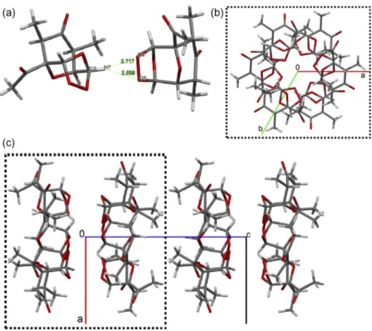
Due to the unusual supramolecular feature of compound
3
we
are going to discuss its packing system in more detail. The
centro-symmetric dimmers in
3
(
Fig. 5
a
) are themselves centrosymmetric
linked by other hydrogen-bond motifs of the type R
22
(8) forming
infinite chains along [21 1] directions (
Fig. 10
). Another interesting
Table 5
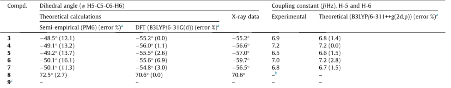
Comparison between computed and experimental values for the H5-C5-C6-H6 dihedral angle and coupling constantJ5,6.
Compd. Dihedral angle (/H5-C5-C6-H6) Coupling constant (J/Hz), H-5 and H-6
Theoretical calculations X-ray data Experimental Theoretical (B3LYP/6-311++g(2d,p)) (error %)a
Semi-empirical (PM6) (error %)a DFT (B3LYP/6-31G(d)) (error %)a
3 48.5°(12.1) 55.2°(0.0) 55.2° 6.9 6.8 (1.4)
4 49.1°(13.2) 56.0°(1.1) 56.6° 7.2 7.2 (0.0)
5 49.2°(13.7) 55.5°(2.6) 57.0° 6.5 6.6 (1.5)
6 50.1°(16.1) 55.6°(6.9) 59.7° 7.0 7.2 (2.8)
7 50.1°(11.3) 54.8°(3.0) 56.5° 6.8 6.7 (1.5)
8 72.5°(2.7) 70.6°(0.0) 70.6° –b –
9c – – – – –
aValues in brackets refer to the percentage (%) error between experimental and theoretical measures. b Do not measured due to the signal was a multiplet.
c In the ozonide9there is a methyl group on the C6.
supramolecular linkage are the H bonds having C7–H7 as
bifur-cated donor to the O4 and O5 (
Fig. 11
a), which link 6 molecules
of
3
related by the
3 axis along [0 0 1] (
Fig. 11
b). This
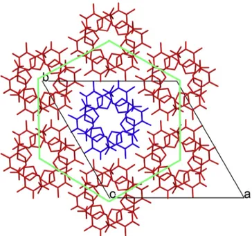
supramolec-ular synthon is stacked via Van der Waals interactions parallel to
[0 01] forming infinites channels in a hexagram fashion (
Fig. 11
b
and c). Each channel is surrounded by six other ones due to the
space group symmetry forming a very interesting honeycomb-like
structure (
Fig. 12
). Indeed, the highly symmetric hexagonal space
group observed for ozonide
3
is uncommon considering
small-molecule organic and metal–organic crystal structures. In the
Cambridge Structural Database (CSD)
[36]
only 290 of deposited
structures crystallize in R
3m, which represents
0.04% of the total
number of structures deposited (717895 entries in CSD version
5.35 and update of November 2013, February 2014, and May
2014).
Fig. 6.Partial packing of6showing the infinite chain parallel to [1 00] direction stabilized by a classical H bond, in which O4, O5 and O6 are trifurcated H bond acceptors for O–H6A donor. Some H. . .acceptor separations (Å) are shown. Hydrogen atoms not involved in the H bonds were hidden for representation clarity.
Fig. 7.Partial packing of4showing a supramolecular moiety stabilized by non-classical H bonds. Some H. . .acceptor separations (Å) are shown. Hydrogen atoms not involved in the H bonds were hidden for representation clarity.
Fig. 8.Packing of4projected ontobcplane (0 11). The centrosymmetric dimmers are dotted lines circled. The color scheme represents the symmetry operations plus translations of P21/c space group. Light gray = identity (x,y,z); green = screw axis 21( x,y+ 1/2, z+ 1/2); yellow = inversion center ( x, y, z); pink = glide plane (x,
y 1/2,z 1/2). (For interpretation of the references to color in this figure legend, the reader is referred to the web version of this article.)
Fig. 9.Partial packing of9showing the infinite double-chain parallel to [1 0 0] direction stabilized by non-classical H bonds. Some H. . .acceptor separations (Å) are shown. Hydrogen atoms not involved in the H bonds were hidden for represen-tation clarity.
After investigating in detail the structure of several ozonides by
X-ray crystallography, and having found some unusual packing
systems, we followed such work by some geometries optimization
using the semi-empirical PM6 and DFT B3LYP/6-31G(d) methods.
Of particular interest is the peroxide bond which is of fundamental
importance to the mode of action of the ozonides. In
Table 2
a list
of the oxygen–oxygen bond lengths of the peroxide bridge,
obtained by theoretical calculations and X-ray analysis, is shown.
As can be seen in
Table 2
, the oxygen–oxygen bond lengths
computed with the semi-empirical PM6 method show higher
devi-ation from the experimental values (2.3–3.8%) than that obtained
with the B3LYP/6-31G(d) approach (0.1–1.8%). In general, the O–
O bond lengths observed in the X-ray analysis (1.484–1.499 Å)
are more elongated than the standard value of 1.47 Å
[22]
. In the
cyclic structures of compounds (
3–9
) we expected a shorter bond
length due to ring tension.
In
Tables 3 and 4
additional values (including the previously
discussed) are presented for selected geometrical parameters of
the ozonides. B3LYP/6-31G(d) results are comparable with the
experimental X-ray data. These data (
Tables 3 and 4
) shows a quite
good correlation between the experimental and calculated values
for these cyclic structures
[11]
.
One important parameter is the H6–C6–C5–H5 (for
3–9
,
Fig. 13
) dihedral angle. From this angle it is possible to assign
the stereochemistry of carbons bearing the methyl groups and
this information can be obtained by H NMR data
[37]
. The
com-puted values for the dihedral angles are compared with the
experimental X-ray data in
Table 5
. As can be observed from such
data a better correlation with experimental data were obtained
from the DFT calculations. Also, the computed values for the
cou-pling constant
J
are in good agreement with the experimental
data obtained by H NMR.
Fig. 10.Partial packing of3showing the infinite chain parallel to [2 1 1] direction stabilized by non-classical H bond where O1, O6 atoms are H bond acceptors for C1–H1 and C5–H5 donors. Some H. . .acceptor separations (Å) are also shown.
Conclusion
A series of new stable ozonides were prepared in our
continu-ous efforts to find new phytotoxic molecules. In this work the
structure of seven ozonides were studied through X-ray diffraction
and also by semi-empirical (PM6) and density functional theory
methods (B3LYP). From the X-rays analyses we found that five
ozo-nides (
4
–
8
) crystallize in the monoclinic crystal system with P2
1/c
space group as racemic crystal. Compound
9
is the unique
enantio-pure crystal found in this series. The most unusual result was
obtained for compound
3
that crystallize in the centrosymmetric
space group R
3m, which represents
0.04% of the total number
of structures deposited in the Cambridge Crystallographic Data
Centre. The results from geometry optimizations using the
func-tional base B3LYP/6-31G(d) approach are in close agreement with
the experimental data obtained by X-ray and also by H NMR.
Acknowledgments
We thank Conselho Nacional de Desenvolvimento Científico e
Tecnológico (CNPq) and Fundação de Amparo à Pesquisa do Estado
de Minas Gerais (FAPEMIG) for financial support and research
fellowships (ACD, AJD, JWMC, LCAB). The authors express sincere
thanks to IFSC-USP for the X-ray facilities.
Appendix A. Supplementary material
Crystallographic data for the structural analysis of the
com-pounds discussed here have been deposited at the Cambridge
Crys-tallographic Data Centre, 12 Union Road, Cambridge CB2 1EZ, UK,
and are available on request quoting the deposition numbers
CCDC1016944, 1016932 1016945, 1016943, 1016946, 1016947,
and 1016942 for
3
,
4
,
5
,
6
,
7
,
8
, and
9
respectively. Supplementary
data associated with this article can be found, in the online version,
at
http://dx.doi.org/10.1016/j.molstruc.2014.10.082
.
References
[1]M.W. Walter, Nat. Prod. Rep. 19 (2002) 278–291. [2]O.F. Hüter, Phytochem. Rev. 10 (2011) 185–194.
[3]S.O. Duke, J.G. Romagni, F.E. Dayan, Crop Prot. 19 (2000) 583–589. [4]C.L. Cantrell, F.E. Dayan, S.O. Duke, J. Nat. Prod. 75 (2012) 1231–1242. [5]F.E. Dayan, D.K. Owens, S.O. Duke, Pest Manage. Sci. 68 (2012) 519–528. [6]P.K. Chen, G.R. Leather, J. Chem. Ecol. 16 (1990) 1867–1876.
[7]S.O. Duke, K.C. Vaughin, E.M. Croom Jr., H.N. Elsohly, Weed Sci. 35 (1987) 499– 505.
[8]F.E. Dayan, A. Hernández, S.N. Allen, R.M. Moraes, J.A. Vroman, M.A. Avery, S.O. Duke, Phytochemistry 50 (1999) 607–6014.
[9]R.C. Paramanik, B.K. Chikkaswamy, D.G. Roy, A. Paramanik, V. Kumar, J. Phytol. Res. 21 (2008) 11–18.
[10] C.W. Jefford, Drug Discov. Today 12 (2007) 487–495.
[11]A.F.C. Alcântara, D. Piló-Veloso, W.B. De Almeida, C.R.A. Maltha, L.C.A. Barbosa, J. Mol. Struct. 791 (2006) 180–185.
[12]R.R. Teixeira, L.C.A. Barbosa, J.O. Santana, D. Piló-Veloso, J. Ellena, A.C. Doriguetto, M.G.B. Drew, F.M.D. Ismail, J. Mol. Struct. 837 (2007) 197–205. [13]L.C.A. Barbosa, A.V. Costa, A.J. Demuner, A.A. Silva, J. Agric. Food Chem. 47
(2000) 4807–4814.
[14]L.C.A. Barbosa, E.S. Alvarenga, A.J. Demuner, R. Figueiredo, A.A. Silva, Pest Manage. Sci. 59 (2003) 1043–1051.
[15]L.C.A. Barbosa, U.A. Pereira, R.R. Teixeira, C.R.A. Maltha, S.A. Fernandes, G. Forlani, J. Agric. Food Chem. 56 (2008) 9434–9440.
[16]L.C.A. Barbosa, C.R.A. Maltha, R.C. Cusati, R.R. Teixeira, F.F. Rodrigues, A.A. Silva, M.G.B. Drew, F.M.D. Ismail, J. Agric. Food Chem. 57 (2009) 10107–10115. [17]L.C.A. Barbosa, D. Cutler, J. Mann, M.J. Crabbe, G.C. Kirby, D.C. Warhurst, J.
Chem. Soc. Perkin Trans. I (1992) 3251–3252.
[18]L.C.A. Barbosa, D. Cutler, J. Mann, M.J. Crabbe, G.C. Kirby, D.C. Warhurst, J. Chem. Soc. Perkin Trans. I (1996) 1101–1105.
[19]J. Kula, Chem. Health Saf. 6 (1999) 21–22.
[20] S.G.V. Ornum, R.M. Champeau, R. Pariza, Chem. Rev. 106 (2006) 2990–3001. [21]W.J. Cummins, M.G.B. Drew, J. Mann, E.B. Walsh, J. Chem. Soc. Perkin Trans. 1
(1983) 167–171.
[22]K. Griesbaum, A. Frank, K.J. McCullough, Eur. J. Org. Chem. 8 (2006) 1978– 1980.
[23] Nonius, Collect. Nonius BV, Delft, The Netherlands, 1999.
[24]Z. Otwinowski, W. Minor, Methods in enzymology, in: C.W. Carter Jr., R.M. Sweet (Eds.), Macromolecular Crystallography, Part A, vol. 276, Academic Press, New York, 1997, pp. 307–326.
[25]G.M. Sheldrick, Acta Crystallogr. A A64 (2008) 112–122. [26]L.J. Farrugia, J. Appl. Crystallogr. 45 (2012) 849–854. [27]L.J. Farrugia, J. Appl. Crystallogr. 30 (1997) 565–569.
[28]C.F. Macrae, P.R. Edgington, P. McCabe, E. Pidcock, G.P. Shields, R. Taylor, M. Towler, J. van de Streek, J. Appl. Crystallogr. 39 (2006) 453–457.
[29] Spartan ’06, Wavefunction Inc., Irvine, CA, USA.
[30] P.J. Stephens, F.J. Devlin, C.F. Chabalowski, M.J. Frisch, J. Phys. Chem. 98 (1994) 11623–11627.
[31]M.M. Francl, W.J. Pietro, W.J. Hehre, J.S. Binkley, M.S. Gordon, D.J. DeFrees, J.A. Pople, J. Chem. Phys. 77 (1982) 3654–3665.
[32] Gaussian 09, Gaussian Inc, Wallingford CT, 2009.
[33]M. Cianci, J.R. Helliwell, M. Helliwell, V. Kaucic, N.Z. Logar, G. Mali, N.N. Tusar, Crystallogr. Rev. 11 (2005) 245–335.
[34]J. Bernstein, R.E. Davis, L. Shimoni, N.L. Chang, Angew. Chem., Int. Ed. Engl. 34 (1995) 1555–1573.
[35]A. Allerhand, P. von, R. Schleyer, J. Am. Chem. Soc. 85 (1963) 1715–1723. [36]F.H. Allen, Acta Crystallogr. B 58 (2002) 380–388.
[37]H.M.R. Hoffmann, R. Henning, O.R. Lalko, Angew. Chem., Int. Ed. Engl. 12 (1973) 819–835.
Fig. 12.View of the hexagram stacking onto planeabhighlighting the honeycomb-like structure formed. Hydrogen atoms were hidden for representation clarity.
O
O
O
O
O
H
H
3C
H
R
1CH
3H
φ
O
O
O
O
O
R
3H
R
1H
3
-
7
8
-
9
R
2R
2φ
Fig. 13.Tridimensional structures of compounds3–9(R1= COCH3, CH2CH3, CH3;
R2= H, (CO)OCH3, CH2OH, CH2OCH3; R3= H, CH3) showing the dihedral angle (/) H6–C6–C5.
![Fig. 9. Partial packing of 9 showing the infinite double-chain parallel to [1 0 0]](https://thumb-eu.123doks.com/thumbv2/123dok_br/15700974.629066/9.892.192.713.816.1072/fig-partial-packing-showing-infinite-double-chain-parallel.webp)