Jamie J. Walker1,2*, Francesca Spiga1, Eleanor Waite1, Zidong Zhao1, Yvonne Kershaw1, John R. Terry1,3,
Stafford L. Lightman1
1Henry Wellcome Laboratories for Integrative Neuroscience and Endocrinology, School of Clinical Sciences, University of Bristol, Bristol, United Kingdom,2Bristol Centre for Applied Nonlinear Mathematics, Department of Engineering Mathematics, University of Bristol, Bristol, United Kingdom,3College of Engineering, Mathematics and Physical Sciences, University of Exeter, Exeter, United Kingdom
Abstract
Oscillating levels of adrenal glucocorticoid hormones are essential for optimal gene expression, and for maintaining physiological and behavioural responsiveness to stress. The biological basis for these oscillations is not known, but a neuronal ‘‘pulse generator’’ within the hypothalamus has remained a popular hypothesis. We demonstrate that pulsatile hypothalamic activity is not required for generating ultradian glucocorticoid oscillations. We show that a constant level of corticotrophin-releasing hormone (CRH) can activate a dynamic pituitary-adrenal peripheral network to produce ultradian adrenocorticotrophic hormone and glucocorticoid oscillations with a physiological frequency. This oscillatory response to CRH is dose dependent and becomes disrupted for higher levels of CRH. These data suggest that glucocorticoid oscillations result from a sub-hypothalamic pituitary-adrenal system, which functions as a deterministic peripheral hormone oscillator with a characteristic ultradian frequency. This constitutes a novel mechanism by which the level, rather than the pattern, of CRH determines the dynamics of glucocorticoid hormone secretion.
Citation:Walker JJ, Spiga F, Waite E, Zhao Z, Kershaw Y, et al. (2012) The Origin of Glucocorticoid Hormone Oscillations. PLoS Biol 10(6): e1001341. doi:10.1371/ journal.pbio.1001341
Academic Editor:Antonio J. Vidal-Puig, University of Cambridge, United Kingdom ReceivedNovember 11, 2011;AcceptedApril 20, 2012;PublishedJune 5, 2012
Copyright:ß2012 Walker et al. This is an open-access article distributed under the terms of the Creative Commons Attribution License, which permits unrestricted use, distribution, and reproduction in any medium, provided the original author and source are credited.
Funding:This work was supported by the Engineering and Physical Sciences Research Council (EPSRC) grant EP/E032249/1; the Biotechnology and Biological Sciences Research Council (BBSRC) grant BB/H015779/1; the Wellcome Trust grant 074112/Z/04/Z; and a support grant from the British Society for Neuroendocrinology (BSN). The funders had no role in study design, data collection and analysis, decision to publish, or preparation of the manuscript. Competing Interests:The authors have declared that no competing interests exist.
Abbreviations:ACTH, adrenocorticotrophic hormone; AUC, area under the curve; CORT, glucocorticoids/corticosterone; CRH, corticotrophin-releasing hormone; HPA, hypothalamic-pituitary-adrenal.
* E-mail: Jamie.Walker@bristol.ac.uk
Introduction
A fundamental requisite for survival is the ability to respond and adapt to a changing environment. This ability to respond to change or ‘‘stress’’ becomes more complex in multicellular organisms, and mammals have developed a well-integrated organization of hormonal, neural, and immunological systems that protect them from internal and external threats to their homeostatic state [1–3]. One of the most important of these systems, and one that is critical for life, is the hypothalamic-pituitary-adrenal (HPA) axis. This neuroendocrine system regu-lates the secretion of vital adrenal glucocorticoid hormones (cortisol in humans and corticosterone in rodents), which have major effects on brain and metabolic function and are essential for successful recovery and adaptation to stress [4,5].
Central regulation of glucocorticoid secretion is predominantly coordinated by the hypothalamic peptide corticotrophin-releasing hormone (CRH) [6,7], the efficacy of which can be significantly potentiated by other hypothalamic factors, most notably vasopressin [8]. CRH is synthesized by parvocellular neurons in the para-ventricular nucleus (PVN) of the hypothalamus [9], and secreted into the hypothalamic-pituitary portal circulation from axons terminating in the external zone of the median eminence [10]. Activation of CRH receptors in corticotroph cells of the anterior pituitary results in adrenocorticotrophic hormone (ACTH) secre-tion into the general circulasecre-tion, which in turn stimulates glucocorticoid-secreting cells within the adrenal cortex (Figure 1A).
Circulating glucocorticoids regulate gene expression in cells throughout the organism via activation of two widely expressed transcription factors—the glucocorticoid receptor (GR) and miner-alocorticoid receptor (MR) [11]—as well as acting directly at the cell membrane to initiate more rapid non-genomic signalling processes [12–15]. Glucocorticoids also feed back on their own regulatory system to inhibit HPA activity [16,17]. Inhibition occurs at the level of the hippocampus and hypothalamus, and with particular sensitivity at the level of the anterior pituitary (Figure 1A).
In common with other neuroendocrine systems that signal through pituitary hormone secretion, the HPA axis is character-ized by a dynamic ultradian rhythm, which is manifested by oscillating levels of ACTH [18] and glucocorticoid hormones (Figure 1B) both in the blood and in the brain [19]. At the cellular level, glucocorticoid oscillations induce ‘‘gene pulsing’’ through rapid, transient binding of glucocorticoid receptors to promoter elements of glucocorticoid-responsive genes [20,21]. This dynamic and versatile transcriptional system enables cells to rapidly detect and respond to changes in circulating glucocorticoid levels and provides a sensitive mechanism for the maintenance of homeo-stasis [22]. Indeed, when the glucocorticoid rhythm is pharmaco-logically replaced by constant levels of steroid, this results in abnormal gene expression [21], and a desensitization of physio-logical and behavioural responses to stress [23,24].
27], this has led to speculation that oscillations in the pituitary-adrenal system are a consequence of a neuronal ‘‘pulse generator’’ within the hypothalamus. However, there is a lack of concordance between pulsatile patterns of hypothalamic factors and the ultradian ACTH and glucocorticoid oscillation. In the rat, for instance, CRH pulse frequency [26] is much higher (,3 pulses/h)
than the near-hourly oscillation in ACTH [18] and glucocorticoids (Figure 1B). This suggests that episodic secretion of hypothalamic hormones is not the primary controlling factor of the ultradian rhythm and implies that oscillatory mechanisms exist at a sub-hypothalamic level. This concept of a ‘‘peripheral oscillator’’ is in keeping with in vivo lesion studies demonstrating maintenance of ultradian pulsatility following hypothalamic-pituitary disconnec-tion in the sheep [28], and a loss of circadian but not ultradian glucocorticoid oscillation following suprachiasmatic nucleus lesions in the rat (unpublished data).
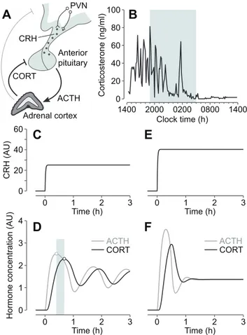
Since glucocorticoids rapidly inhibit CRH-induced ACTH secretion from the anterior pituitary [29–32], we postulated that this fast inhibitory feedback process provides a potential mecha-nism within the pituitary-adrenal system for generating oscillatory dynamics. To explore this hypothesis further, and to determine qualitatively the dynamics that result from hormonal interactions between the anterior pituitary and adrenal cortex, we previously developed a mathematical model based on differential equations that incorporates rapid glucocorticoid inhibition of ACTH secretion [33]. Numerical analysis of the model suggests that the pituitary-adrenal system can support self-sustained ACTH and glucocorticoid oscillations with a physiological ultradian frequen-cy, even under conditions of constant CRH drive to the anterior pituitary (Figure 1C–1D). In this model, the ACTH and glucocorticoid oscillations have the same frequency, but they are not synchronous—there is a small phase difference, with ACTH oscillations preceding glucocorticoid oscillations (Figure 1D). The model also predicts that the capacity for this oscillatory response depends on the degree of hypothalamic drive, with higher levels of CRH resulting in damped oscillations to steady-state (i.e., constant) levels of hormone (Figure 1E–1F).
Here we test these modelling predictions in vivo. Our data show that a constant level of CRH can activate the pituitary-adrenal system to produce ultradian hormone oscillations with a physiological frequency, and that this oscillatory activity is critically dependent on the level of hypothalamic drive, with higher levels of CRH resulting in a loss of oscillation. These results demonstrate that pulsatile secretion of hypothalamic CRH is not required for ultradian oscillatory activity in the pituitary-adrenal system, and support our theoretical hypothesis that rapid glucocorticoid inhibition at the level of the anterior pituitary is the primary factor regulating the ultradian dynamics of the system.
1400
Corticosterone (ng/ml)
0 100
20 40 60 80
1400 2000 0200 0800 Clock time (h)
0 20 40 60
CRH (AU)
Hormone concentration (AU)
0 1 2
Time (h)
3 0 1 2
Time (h) 3
ACTH
CORT
0 2 3 4
1 CORT
PVN
Adrenal cortex CRH
ACTH
A
B
C
E
Anterior pituitary
0 1 2
Time (h)
3 0 1 2
Time (h) 3
ACTH
CORT
D
F
Figure 1. Regulation of glucocorticoid hormone secretion.(A) Negative feedback in the HPA axis plays a key role in regulating glucocorticoid (CORT) secretion. Neurons of the hypothalamic para-ventricular nucleus (PVN) secrete CRH into the portal vein, which acts on the anterior pituitary to release ACTH into the general circulation. ACTH activates cells in the adrenal cortex to synthesize and secrete CORT, which in turn feeds back directly on the anterior pituitary to inhibit ACTH secretion, as well as acting at higher centres in the brain, including the hypothalamus and hippocampus. (B) Endogenous corticosterone (the main glucocorticoid in rodents) oscillations in a freely behaving male Sprague-Dawley rat. Shaded region indicates the dark phase. (C–F) Mathematical modelling predicts that ultradian ACTH and glucocorticoid (CORT) oscillations are regulated by a systems-level negative feedback mechanism in the pituitary-adrenal network, independent of pulsatile hypothalamic activity. Numerical simulations show that the pituitary-adrenal network can oscillate under conditions of constant CRH drive to the pituitary (C–D). Oscillations in ACTH and CORT are characterized by a small phase shift (shaded region indicates phase difference between oscillation peaks). For higher levels of CRH drive, the oscillations are rapidly damped to steady-state levels of hormone (E–F). AU, arbitrary units.
doi:10.1371/journal.pbio.1001341.g001 Author Summary
Results and Discussion
To test our hypothesis that the sub-hypothalamic pituitary-adrenal system functions as an ultradian hormone oscillator, we investigated the dynamics of glucocorticoid responses to different levels of constant CRH stimulation in conscious freely behaving male rats. Experiments were performed during the nadir of the circadian cycle (0700–1300 h), when there is minimal endogenous CRH [34,35], and no pulsatile secretion of corticosterone, the main glucocorticoid in rodents (Figure 1B). Animals were infused intravenously at a constant (i.e., non-pulsatile) rate for this 6-h period with concentrations of CRH in the range 0–2.5mg/h.
Circulating levels of corticosterone were measured in blood samples collected prior to and throughout the infusion using an automated blood-sampling system (see Materials and Methods).
To check that the animals were in a physiological basal state throughout the procedure, we measured corticosterone levels in a group of control animals infused with saline (Figure 2A–2B). Corticosterone levels remained low throughout the duration of the saline infusion and were not significantly different from cortico-sterone levels measured during the same time period (0700– 1300 h) in a group of untreated animals (data not shown), as assessed by analysis of the area under the curve (AUC,p.0.38).
In response to CRH infusion, corticosterone levels rose rapidly (Figure 2C–2H) and the overall effect was dose dependent (AUC, p,0.0001) (Figure 2I). There was a significant difference between the groups treated with saline and 0.5mg/h CRH (p,0.001), and between the groups treated with 0.5 and 1.0mg/h CRH
(p,0.001), but there was no significant difference between the groups treated with 1.0 and 2.5mg/h CRH (p.0.41). This suggests that both of these higher levels of CRH result in maximal pituitary-adrenal activation, which implies a systems-level ‘‘ceiling effect’’ in the pituitary-adrenal network.
Computational modelling suggests that the dynamic activity of the pituitary-adrenal system is fundamentally dependent on the level of hypothalamic stimulation [33]. In agreement with this prediction, constant infusion of CRH at different doses gave rise to different temporal patterns of corticosterone secretion. In line with the modelling hypothesis, constant infusion of CRH (0.5mg/h)
induced ultradian corticosterone oscillations that persisted throughout the infusion period (Figure 2C–2D). In contrast, and also consistent with the qualitative predictions of the model, higher doses of CRH (1.0 and 2.5mg/h) caused a rapid activation of the
adrenals, but the oscillatory component of the response was damped to constant, elevated levels of steroid (Figure 2E–2F, CRH 1.0mg/h; Figure 2G–2H, CRH 2.5mg/h).
Although CRH is the predominant ACTH secretagogue in humans and the rat [36], its ability to stimulate ACTH secretion can be potentiated by other hypothalamic neuropeptides, most notably vasopressin [8]. However, the consistency between animals in the timing of the initial corticosterone response to CRH, and the subsequent synchrony in oscillation throughout the infusion (Figure 2J) indicates that the corticosterone response is not dependent on the release of any other endogenous hypothalamic factors, including vasopressin. This is in keeping with previous observations that blocking vasopressin actions on the anterior pituitary (using a vasopressin V1b receptor antagonist) has no effect on endogenous corticosterone oscillations during the circadian peak [37], suggesting that vasopressin is not a significant factor in regulating the ultradian dynamics of the system.
If the mechanism regulating endogenous corticosterone oscilla-tions during the circadian peak is the same mechanism that is activated by the constant infusion of exogenous CRH, there should be good agreement between the characteristic frequencies
of endogenous and CRH-induced oscillations. To test this, we computed the dominant frequency component in the CRH-induced oscillations, and compared this with the dominant frequency component in endogenous corticosterone oscillations
100 200 300
Corticosterone (ng/ml)
Corticosterone (ng/ml)
Corticosterone (ng/ml)
Corticosterone (ng/ml)
Clock time (h) Clock time (h) 0600 0800 1000 1200
0
100 200 300
0
100 200 300
0
100 200 300
0
0600 0800 1000 1200
0600 0800 1000 1200 0600 0800 1000 1200
0600 0800 1000 1200 0600 0800 1000 1200
0600 0800 1000 1200 0600 0800 1000 1200
Clock time (h) 0
40 80 120 160
Corticosterone (ng/ml)
0600 0800 1000 1200 200
SAL 0.5 1.0 2.5 1200
1000
800
600
400
200
0
CRH (μg/h)
*
*
A
B
C
D
E
F
G
H
I
J
AUC
Figure 2. Glucocorticoid response to constant CRH infusion.(A– H) Individual (A, C, E, G) and mean (B, D, F, H) corticosterone responses
to constant saline (A–B) or CRH infusion (0.5mg/h, C–D; 1.0mg/h, E–F;
2.5mg/h, G–H). Grey bar indicates the period of infusion; error bars
represent mean 6 standard error of the mean (SEM) (n= 6–8 per
group). (I) Dose-dependent effect of CRH on the corticosterone response. Overall effect of the CRH infusion was significant (AUC,
p,0.0001; Kruskal-Wallis ANOVA on ranks). Error bars represent mean6
SEM (n= 6–8 per group); *p,0.001. (J) Synchronous corticosterone
oscillations (in individual rats) in response to constant CRH infusion
(0.5mg/h;n= 6). Grey bar indicates the period of infusion.
during the circadian peak (see Materials and Methods). In the CRH-induced oscillations, there was a peak frequency of ,1
pulse/h for all animals (Figure 3A–3B; mean peak frequen-cy = 0.89 pulses/h; peak frequenfrequen-cy range = 0.79–0.93 pulses/h). We then measured corticosterone levels in untreated control rats during the circadian peak, when endogenous CRH is maximal [34,35] and corticosterone is pulsatile (Figure 1B). Corticosterone oscillations were observed in all animals (Figure 3C), and frequency analysis of the data (Figure 3D) revealed no significant difference between these endogenous oscillations and the oscilla-tions induced by constant CRH infusion (p.0.57) (Figure 3E).
The consistency in oscillation frequency between different animals infused with constant CRH suggests that the oscillations are regulated by a generic mechanism at a sub-hypothalamic level.
Furthermore, the maintenance of this dominant frequency component throughout the period of CRH infusion (Figure 3F) suggests that the underlying oscillator is deterministic—as opposed to stochastic—in agreement with our modelling hypothesis [33].
Numerical simulations of the model indicate that glucocorticoid oscillations induced by constant CRH stimulation are driven by oscillations in ACTH (Figure 1C–1D). Ultradian ACTH oscilla-tions have been observed in the rat [18], and have been shown to be a critical factor in regulating pulsatile glucocorticoid secretion from the adrenal cortex [38]. Moreover, coordinated ACTH and glucocorticoid oscillations have been observed in man [39]. To confirm that the constant CRH infusion generates oscillations in both hormones, we infused CRH (0.5mg/h) from 0700–0940 h
and measured circulating levels of both ACTH and corticosterone in samples collected at 20-min intervals throughout the infusion (see Materials and Methods). In agreement with the modelling predictions, CRH induced ACTH and corticosterone oscillations that persisted throughout the infusion period (Figure 4A–4B).
A key feature of the oscillation predicted by numerical simulations is a small phase shift between the two hormones— ACTH oscillations preceding glucocorticoid oscillations (Figure 1C–1D). This small phase shift could not be detected in the CRH-induced ACTH and corticosterone oscillations (Figure 4A–4B) because of the sampling frequency (20-min inter-sample interval). Therefore, we measured both ACTH and corticosterone at a higher sampling frequency (5-min inter-sample interval) over the first 25 min of the CRH infusion (0.5mg/h),
covering the initial activation phase of the first pulse. Both hormones rose rapidly in response to CRH (ACTH,p,0.0001; corticosterone, p,0.005), with the ACTH increase preceding a delayed rise in corticosterone (Figure 4C–4D). Specifically, ACTH was significantly different from basal (time zero) by 10 min (p,0.005), whereas corticosterone was not significantly different from basal (time zero) until 20 min (p,0.05). This phase shift between ACTH and corticosterone presumably reflects the time taken for de novo biosynthesis and release of corticosterone from the adrenal cortex [40].
We then checked that this phase relationship is maintained over the full pituitary-adrenal oscillation. Since collection of large volumes of plasma (required for sensitive ACTH assay) can activate a stress response [41], this precludes high-frequency blood sampling (for ACTH measurement) for prolonged periods in the rat. We therefore used an alternative experimental approach in which animals were killed by decapitation at 10-min intervals throughout the first 70 min of the CRH infusion (0.5mg/h); ACTH and corticosterone levels were measured in plasma obtained from trunk blood (see Materials and Methods). As observed in the case of high-frequency sampling (Figure 4C–4D), the CRH-induced increase in corticosterone was delayed relative to the increase in ACTH, and this phase shift was maintained over the duration of the pulse (Figure 4E).
These results challenge the long-standing view that glucocorti-coid oscillations are a consequence of pulsatile CRH secretion from a neuronal ‘‘pulse generator’’ within the hypothalamus [26,27]. Our approach was based on the premise that feedback is a key regulatory feature of biological oscillators [42], and on our numerical results [33], suggesting that a systems-level dynamic balance between positive feedforward and negative feedback— CRH stimulation against rapid glucocorticoid inhibition of ACTH secretion—provides a mechanism for generating ultradian oscil-lations in ACTH and glucocorticoid hormone secretion. Testing this modelling prediction in vivo, our results show that constant CRH stimulation of the anterior pituitary is sufficient to generate ACTH and glucocorticoid oscillations at a physiological ultradian
A
B
F
C
D
E
120
0 20 100 80 60 40
0
Corticosterone (ng/ml)
1500 1700 1900 2100
0 0.2 0.4 0.6 0.8 1.0 1.2
Clock time (h)
2 3 4
0600 0800 1000 1200 Clock time (h)
0 1 2 3 0
Power (AU)
0 1 2 3 1
1
CRH UC Freq. (h )-1 Freq. (h )-1
0
Power (AU)
1
0600 0800 1000 1200 Clock time (h)
0 1 2 3 0
Power (AU)
1
Freq. (h )-1 0 1 2 3
0
Power (AU)
1
Freq. (h )-1
1500 1700 1900 2100 Clock time (h) 120
0 20 100 80 60 40
Corticosterone (ng/ml)
Freq. (h )
-1
0600 0800 1000 1200 Clock time (h)
0600 0800 1000 1200 Clock time (h)
Peak freq. (h )
-1
Figure 3. Frequency comparison of CRH-induced and endog-enous glucocorticoid oscillations. (A) Individual corticosterone
oscillations in response to constant CRH infusion (0.5mg/h). Grey bar
indicates the period of infusion. (B) Normalized power spectra of the corticosterone oscillations in (A). (C) Corticosterone oscillations during the circadian peak in untreated control (UC) rats. Shaded region indicates the dark phase. (D) Normalized power spectra of the corticosterone oscillations in (C). (E) Mean peak frequency (i.e., frequency corresponding to the maximum power in the spectrum) of
corticosterone oscillations in response to constant CRH infusion (0.5mg/
h; CRH;n= 6), and of corticosterone oscillations during the circadian
peak in untreated control rats (UC;n= 13). Error bars represent mean6
standard error of the mean (SEM). (F) Frequency evolution of the corticosterone oscillations in (A). AU, arbitrary units.
frequency, providing good evidence for an oscillating mechanism outside the central nervous system.
The hormone oscillations generated by this system are not simply a dynamic epiphenomenon of the pituitary-adrenal interaction, but have significant biological impact. Glucocorticoid oscillations are essential for optimal transcriptional regulation
[20,21], and are also likely to be important for more rapid non-transcriptional mechanisms of steroid action in the brain [43] that can alter behavioural function within minutes [44,45]. Exposure of tissues to abnormal glucocorticoid levels due to prolonged stress [46] and raised CRH [47,48], or due to the loss of ultradian pulsatility that has been detected in ageing animals [49], will modify glucocorticoid signalling, and could be an important factor for both stress- and age-related disease.
Materials and Methods
Subjects
All experiments were conducted on adult male Sprague-Dawley rats (Harlan Laboratories, Inc.) weighing,250 g at the time of
surgery. Animals were housed in groups of four per cage and were given at least 1 wk to acclimatize to the housing facility prior to surgery. Rats were maintained under standard environmental conditions (2161uC) under a 14-h light, 10-h dark schedule (lights on at 0500 h). Food and water were freely available throughout the experiments. All animal procedures were conducted in accordance with Home Office guidelines and the UK Animals (Scientific Procedures) Act, 1986.
Surgical Procedures
Animals were anaesthetized with a combination of Hypnorm (0.32 mg/kg fentanyl citrate and 10 mg/kg fluanisone, IM; Janssen Pharmaceuticals) and diazepam (2.6 mg/kg, IP; Phoenix Pharmaceuticals). Two silastic-tipped (Merck) polythene cannulae (Portex) were pre-filled with pyrogen-free heparinized (10 IU/ml) isotonic saline. The right jugular vein was exposed and both cannulae inserted into the vessel until they lay close to the entrance to the right atrium. This permits simultaneous intravenous blood sampling (via the sampling cannula) and substance infusion (via the infusion cannula). The free ends of the cannulae were exteriorized through a scalp incision and tunnelled through a protective spring that was anchored to the parietal bones using two stainless steel screws and self-curing dental acrylic. Following recovery, animals were individually housed in a room containing an automated blood-sampling system. The end of the protective spring was attached to a two-channel swivel (Instech Laboratories, Inc.), which rotates through 360uin the horizontal plane and 180u
in the vertical plane, providing the animals with maximal freedom of movement. Animals were given a 5-d recovery period prior to experiments. Throughout this time, both cannulae were flushed daily with heparinized saline to maintain patency.
Drug Infusions
Rats received a constant intravenous infusion of either 0.9% saline solution (vehicle control animals) or synthetic human/rat CRH (University of Bristol Peptide Synthesis Service) dissolved in 0.9% saline solution. In all experiments, drugs were infused through the infusion cannula at a volume infusion rate of 0.5 ml/h using an automated infusion pump (PHD ULTRA syringe pump; Harvard Apparatus, Ltd.).
Experimental Design
In the experiments measuring basal corticosterone levels over 24 h, corticosterone levels in response to saline or CRH infusion, or basal corticosterone oscillations during the circadian peak, blood samples were collected from the sampling cannula using an automated blood-sampling system [50]. In the experiment measuring basal corticosterone levels over 24 h (Figure 1B), blood samples were collected every 10 min from 1400–1350 h. In the experiments measuring corticosterone levels in response to saline
ACTH (pg/ml)
20 40 60 80
Corticosterone (ng/ml)
40 80
0 20 60 100 140
120
ACTH (pg/ml)
Corticosterone (ng/ml)
40 80
0 20 60 100 140
120 160
20 40 60 80 100
ACTH (pg/ml)
20 40 60 80 100 120 140
Corticosterone (ng/ml)
0 50 100 150 200 250 300 350
0700 0800 0900 0700 0800 0900 Clock time (h) Clock time (h)
Time (min)
Time (min)
0 10 20 30 40 50 60 70
ACTH
CORT
5 10 15 20 25 0 5 10 15 20 25 0
Time (min)
ACTH
CORT
ACTH
CORT
ACTH
CORT
ACTH
CORT
A
B
C
D
E
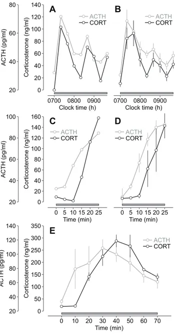
Figure 4. ACTH and glucocorticoid response to constant CRH infusion.(A–B) Individual (A) and mean (B) ACTH and corticosterone
(CORT) oscillations in response to constant CRH infusion (0.5mg/h;
n= 6). (C–D) Individual (C) and mean (D) time course of the ACTH and
corticosterone (CORT) response to constant CRH infusion (0.5mg/h)
during the initial activation phase (0–25 min) of the oscillation (n= 4).
There was a significant overall effect of the CRH infusion on both ACTH
and corticosterone (ACTH,p,0.0001; corticosterone,p,0.005; one-way
ANOVA). ACTH was significantly different from basal (time zero) by
10 min (p,0.005), whereas corticosterone was not significantly
different from basal (time zero) until 20 min (p,0.05). (E) Phase-shifted
ACTH and corticosterone (CORT) response to constant CRH infusion
(0.5mg/h) over the duration of the first pulse (n= 3–7 per time point).
Grey bar indicates the period of infusion (starting at 0700 h); error bars
represent mean6standard error of the mean (SEM).
or CRH infusion (Figures 2A–2H, 2J, and 3A), rats were constantly infused with saline or CRH (0.5–2.5mg/h) from 0700–1300 h; blood samples were collected every 5 min from 0600–1300 h. In the experiments measuring basal corticosterone oscillations during the circadian peak (Figure 3C), blood samples were collected every 5 min from 1530–2100 h. At the end of each experiment, the plasma fraction was separated by centrifugation (15 min, 3,120g, 4uC) and stored at 220uC until processed for corticosterone measurement.
In the experiments measuring ACTH and corticosterone oscillations in response to CRH infusion (Figure 4A–4B), rats received a constant CRH infusion (0.5mg/h) from 0700–0940 h
and blood samples were collected manually from the sampling cannula every 20 min throughout the infusion. In the experiments measuring ACTH and corticosterone levels in response to CRH during the initial activation phase (Figure 4C–4D), rats received a constant CRH infusion (0.5mg/h) from 0700–0725 h and blood
samples were collected manually from the sampling cannula every 5 min throughout the infusion. Blood samples from both experiments were immediately mixed with EDTA (10ml, 0.5 M,
pH 7.4) and placed on crushed ice. The plasma fraction was separated by centrifugation (15 min, 3,120g, 4uC) within 20 min of sample collection and stored at 220uC until processed for ACTH and corticosterone measurement.
In the experiment measuring ACTH and corticosterone levels in response to CRH infusion over the first pulse (Figure 4E), rats received a constant CRH infusion (0.5mg/h) from 0700–0810 h.
At 10-min intervals throughout the CRH infusion, rats were killed by decapitation following an overdose of 0.3 ml sodium pento-barbital (Euthatal, 200 mg/ml; Merial). Trunk blood was collected and immediately mixed with EDTA (50ml, 0.5 M, pH 7.4) and placed on crushed ice. The plasma fraction was separated by centrifugation (15 min, 3,120g, 4uC) within 20 min of sample collection and stored at 220uC until processed for ACTH and corticosterone measurement.
Corticosterone RIA
Corticosterone was measured in plasma by radioimmunoassay (RIA) [51]. Samples were diluted in a citrate buffer (pH 3.0) to denature the binding globulin. Antisera was supplied by G Makara (Institute of Experimental Medicine, Budapest, Hungary), and [125I] corticosterone was purchased from Izotop (Institute of Isotopes Co. Ltd., Budapest, Hungary). The intra- and inter-assay coefficients of variation of the corticosterone RIA were 14.1% and 15.3%, respectively.
ACTH IRMA
ACTH was measured in plasma by immunoradiometric assay (IRMA; DiaSorin Ltd.), in accordance with the manufacturer’s protocol. The intra- and inter-assay coefficients of variation of the ACTH IRMA were 2.8% and 6.4%, respectively.
Data Analysis
Overall corticosterone responses to saline or CRH infusion were assessed by AUC. Characterization of oscillatory corticosterone responses induced by CRH infusion, and of endogenous corticosterone oscillations recorded during the circadian peak, was performed using spectral methods. Missing data points were linearly interpolated and data were detrended using the Smooth-ness Priors Approach (SPA) [52] with the smoothing parameter set atl= 30. This parameter value was chosen so as to remove long term changes in the mean (i.e., low-frequency fluctuations), while keeping the higher-frequency ultradian fluctuations that were the focus in this study. To define the frequency of the oscillatory corticosterone data, we computed the power spectrum of the detrended data using the Discrete Fourier Transform (DFT) applied to a time window corresponding to the period of CRH infusion (0700–1300 h), or to the period of sampling (1530– 2100 h) for the basal corticosterone oscillations recorded during the circadian peak. The peak frequency was then taken as the frequency value corresponding to the maximum spectral power of the DFT, which was calculated using a quadratic interpolation. Spectrograms were computed using the Short-Time Fourier Transform (STFT).
Statistical Analysis
Statistical significance level was set at p,0.05. Saline-infused corticosterone responses and untreated corticosterone profiles were compared using the non-parametric Mann-Whitney U test. The overall effect of CRH treatment on AUC was assessed using the non-parametric Kruskal-Wallis ANOVA on ranks test, and post hoc multiple comparisons were performed using the Mann-Whitney U Test with the statistical significance level adjusted using the Bonferroni correction. Peak frequencies obtained using the DFT, for the groups with CRH-induced corticosterone oscillations and endogenous corticosterone oscillations during the circadian peak, were compared using the Mann-Whitney U test. The ACTH and corticosterone response to constant CRH infusion was analyzed using one-way ANOVA, followed by Fisher Least Significant Difference (LSD) post hoc test.
Acknowledgments
We thank David Emery, Will Mawby, and Graham Bloomberg for providing the CRH peptide.
Author Contributions
The author(s) have made the following declarations about their contributions: Conceived and designed the experiments: JJW FS EW JRT SLL. Performed the experiments: JJW FS EW ZZ YK. Analyzed the data: JJW FS. Wrote the paper: JJW FS JRT SLL.
References
1. Chrousos GP (1995) The hypothalamic-pituitary-adrenal axis and immune-mediated inflammation. N Engl J Med 332: 1351–1362.
2. Joels M, Baram TZ (2009) The neuro-symphony of stress. Nat Rev Neurosci 10: 459–466.
3. McEwen BS (2007) Physiology and neurobiology of stress and adaptation: central role of the brain. Physiol Rev 87: 873–904.
4. de Kloet ER, Joels M, Holsboer F (2005) Stress and the brain: from adaptation to disease. Nat Rev Neurosci 6: 463–475.
5. Sapolsky RM, Romero LM, Munck AU (2000) How do glucocorticoids influence stress responses? Integrating permissive, suppressive, stimulatory, and preparative actions. Endocr Rev 21: 55–89.
6. Rivier J, Spiess J, Vale W (1983) Characterization of rat hypothalamic corticotropin-releasing factor. Proc Natl Acad Sci U S A 80: 4851–4855.
7. Vale W, Spiess J, Rivier C, Rivier J (1981) Characterization of a 41-residue ovine hypothalamic peptide that stimulates secretion of corticotropin and beta-endorphin. Science 213: 1394–1397.
8. Gillies GE, Linton EA, Lowry PJ (1982) Corticotropin releasing activity of the new CRF is potentiated several times by vasopressin. Nature 299: 355–357.
9. Sawchenko PE, Swanson LW, Vale WW (1984) Co-expression of corticotropin-releasing factor and vasopressin immunoreactivity in parvocellular neurosecre-tory neurons of the adrenalectomized rat. Proc Natl Acad Sci U S A 81: 1883–1887.
11. Reul JM, de Kloet ER (1985) Two receptor systems for corticosterone in rat brain: microdistribution and differential occupation. Endocrinology 117: 2505–2511.
12. Di S, Malcher-Lopes R, Halmos KC, Tasker JG (2003) Nongenomic glucocorticoid inhibition via endocannabinoid release in the hypothalamus: a fast feedback mechanism. J Neurosci 23: 4850–4857.
13. Di S, Maxson MM, Franco A, Tasker JG (2009) Glucocorticoids regulate glutamate and GABA synapse-specific retrograde transmission via divergent nongenomic signaling pathways. J Neurosci 29: 393–401.
14. Karst H, Berger S, Turiault M, Tronche F, Schutz G, Joels M (2005) Mineralocorticoid receptors are indispensable for nongenomic modulation of hippocampal glutamate transmission by corticosterone. Proc Natl Acad Sci U S A 102: 19204–19207.
15. Orchinik M, Murray TF, Moore FL (1991) A corticosteroid receptor in neuronal membranes. Science 252: 1848–1851.
16. Dallman MF (2005) Fast glucocorticoid actions on brain: back to the future. Front Neuroendocrinol 26: 103–108.
17. Tasker JG, Herman JP (2011) Mechanisms of rapid glucocorticoid feedback inhibition of the hypothalamic-pituitary-adrenal axis. Stress 14: 398–406. 18. Carnes M, Lent S, Feyzi J, Hazel D (1989) Plasma adrenocorticotropic hormone
in the rat demonstrates three different rhythms within 24 h. Neuroendocrinol-ogy 50: 17–25.
19. Droste SK, de Groote L, Atkinson HC, Lightman SL, Reul JM, et al. (2008) Corticosterone levels in the brain show a distinct ultradian rhythm but a delayed response to forced swim stress. Endocrinology 149: 3244–3253.
20. Conway-Campbell BL, Sarabdjitsingh RA, McKenna MA, Pooley JR, Kershaw YM, et al. (2010) Glucocorticoid ultradian rhythmicity directs cyclical gene pulsing of the clock gene period 1 in rat hippocampus. J Neuroendocrinol 22: 1093–1100.
21. Stavreva DA, Wiench M, John S, Conway-Campbell BL, McKenna MA, et al. (2009) Ultradian hormone stimulation induces glucocorticoid receptor-mediated pulses of gene transcription. Nat Cell Biol 11: 1093–1102.
22. Lightman SL, Conway-Campbell BL (2010) The crucial role of pulsatile activity of the HPA axis for continuous dynamic equilibration. Nat Rev Neurosci 11: 710–718.
23. Sarabdjitsingh RA, Spiga F, Oitzl MS, Kershaw Y, Meijer OC, et al. (2010) Recovery from disrupted ultradian glucocorticoid rhythmicity reveals a dissociation between hormonal and behavioural stress responsiveness. J Neuroendocrinol 22: 862–871.
24. Sarabdjitsingh RA, Conway-Campbell BL, Leggett JD, Waite EJ, Meijer OC, et al. (2010) Stress responsiveness varies over the ultradian glucocorticoid cycle in a brain-region-specific manner. Endocrinology 151: 5369–5379.
25. Engler D, Pham T, Fullerton MJ, Ooi G, Funder JW, et al. (1989) Studies of the secretion of corticotropin-releasing factor and arginine vasopressin into the hypophysial-portal circulation of the conscious sheep. I. Effect of an audiovisual stimulus and insulin-induced hypoglycemia. Neuroendocrinology 49: 367–381. 26. Ixart G, Barbanel G, Nouguier-Soule J, Assenmacher I (1991) A quantitative study of the pulsatile parameters of CRH-41 secretion in unanesthetized free-moving rats. Exp Brain Res 87: 153–158.
27. Mershon JL, Sehlhorst CS, Rebar RW, Liu JH (1992) Evidence of a corticotropin-releasing hormone pulse generator in the macaque hypothalamus. Endocrinology 130: 2991–2996.
28. Engler D, Pham T, Liu JP, Fullerton MJ, Clarke IJ, et al. (1990) Studies of the regulation of the hypothalamic-pituitary-adrenal axis in sheep with hypotha-lamic-pituitary disconnection. II. Evidence for in vivo ultradian hypersecretion of proopiomelanocortin peptides by the isolated anterior and intermediate pituitary. Endocrinology 127: 1956–1966.
29. Hinz B, Hirschelmann R (2000) Rapid non-genomic feedback effects of glucocorticoids on CRF-induced ACTH secretion in rats. Pharm Res 17: 1273–1277.
30. Jones MT, Hillhouse EW, Burden JL (1977) Dynamics and mechanics of corticosteroid feedback at the hypothalamus and anterior pituitary gland. J Endocrinol 73: 405–417.
31. Russell GM, Henley DE, Leendertz J, Douthwaite JA, Wood SA, et al. (2010) Rapid glucocorticoid receptor-mediated inhibition of hypothalamic-pituitary-adrenal ultradian activity in healthy males. J Neurosci 30: 6106–6115.
32. Widmaier EP, Dallman MF (1984) The effects of corticotropin-releasing factor on adrenocorticotropin secretion from perifused pituitaries in vitro: rapid inhibition by glucocorticoids. Endocrinology 115: 2368–2374.
33. Walker JJ, Terry JR, Lightman SL (2010) Origin of ultradian pulsatility in the hypothalamic-pituitary-adrenal axis. Proc R Soc B 277: 1627–1633. 34. Ixart G, Siaud P, Barbanel G, Mekaouche M, Givalois L, et al. (1993) Circadian
variations in the amplitude of corticotropin-releasing hormone 41 (CRH41) episodic release measured in vivo in male rats: correlations with diurnal fluctuations in hypothalamic and median eminence CRH41 contents. J Biol Rhythms 8: 297–309.
35. Watts AG, Tanimura S, Sanchez-Watts G (2004) Corticotropin-releasing hormone and arginine vasopressin gene transcription in the hypothalamic paraventricular nucleus of unstressed rats: daily rhythms and their interactions with corticosterone. Endocrinology 145: 529–540.
36. Engler D, Redei E, Kola I (1999) The corticotropin-release inhibitory factor hypothesis: a review of the evidence for the existence of inhibitory as well as stimulatory hypophysiotropic regulation of adrenocorticotropin secretion and biosynthesis. Endocr Rev 20: 460–500.
37. Spiga F, Harrison LR, Wood S, Knight DM, MacSweeney CP, et al. (2009) Blockade of the V(1b) receptor reduces ACTH, but not corticosterone secretion induced by stress without affecting basal hypothalamic-pituitary-adrenal axis activity. J Endocrinol 200: 273–283.
38. Spiga F, Waite EJ, Liu Y, Kershaw YM, Aguilera G, et al. (2011) ACTH-dependent ultradian rhythm of corticosterone secretion. Endocrinology 152: 1448–1457.
39. Henley DE, Leendertz JA, Russell GM, Wood SA, Taheri S, et al. (2009) Development of an automated blood sampling system for use in humans. J Med Eng Technol 33: 199–208.
40. Stocco DM, Clark BJ (1996) Regulation of the acute production of steroids in steroidogenic cells. Endocr Rev 17: 221–244.
41. Carnes M, Lent SJ, Goodman B, Mueller C, Saydoff J, et al. (1990) Effects of immunoneutralization of corticotropin-releasing hormone on ultradian rhythms of plasma adrenocorticotropin. Endocrinology 126: 1904–1913.
42. Goldbeter A (2002) Computational approaches to cellular rhythms. Nature 420: 238–245.
43. Groeneweg FL, Karst H, de Kloet ER, Joels M (2011) Rapid non-genomic effects of corticosteroids and their role in the central stress response. J Endocrinol 209: 153–167.
44. Haller J, Halasz J, Mikics E, Kruk MR, Makara GB (2000) Ultradian corticosterone rhythm and the propensity to behave aggressively in male rats. J Neuroendocrinol 12: 937–940.
45. Mikics E, Kruk MR, Haller J (2004) Genomic and non-genomic effects of glucocorticoids on aggressive behavior in male rats. Psychoneuroendocrinology 29: 618–635.
46. Windle RJ, Wood SA, Kershaw YM, Lightman SL, Ingram CD, et al. (2001) Increased corticosterone pulse frequency during adjuvant-induced arthritis and its relationship to alterations in stress responsiveness. J Neuroendocrinol 13: 905–911.
47. Chrousos GP, Gold PW (1992) The concepts of stress and stress system disorders. Overview of physical and behavioral homeostasis. JAMA 267: 1244–1252.
48. Reul JM, Holsboer F (2002) Corticotropin-releasing factor receptors 1 and 2 in anxiety and depression. Curr Opin Pharmacol 2: 23–33.
49. Lightman SL, Windle RJ, Julian MD, Harbuz MS, Shanks N, et al. (2000) Significance of pulsatility in the HPA axis. Novartis Found Symp 227: 244–257. 50. Windle RJ, Wood SA, Lightman SL, Ingram CD (1998) The pulsatile characteristics of hypothalamo-pituitary-adrenal activity in female Lewis and Fischer 344 rats and its relationship to differential stress responses. Endocrinol-ogy 139: 4044–4052.
51. Spiga F, Harrison LR, Wood SA, Atkinson HC, MacSweeney CP, et al. (2007) Effect of the glucocorticoid receptor antagonist Org 34850 on basal and stress-induced corticosterone secretion. J Neuroendocrinol 19: 891–900.