294
Original Article
Combined Transplantation of Skeletal Myoblasts
and Mesenchymal Cells (Cocultivation) in
Ventricular Dysfunction After Myocardial Infarction
Luiz Cesar Guarita Souza, Katherine Athayde Teixeira de Carvalho, Carmen Rebelatto,
Alessandra Senegaglia, Marcus Furuta, Nelson Miyague, Paula Hansen, Julio C Francisco,
Paulo Roberto Slud Brofman
Curitiba, PR - Brazil
Pontifícia Universidade Católica do Paraná - Curitiba
Mailing address: Luiz César Guarita Souza - Rua Silveira Peixoto, 1062/ 162 – 80240-120 – Curitiba, PR, Brazil - E-mail: [email protected] Received for publication: 04/03/2004
Accepted for publication: 06/22/2004 English version by Stela Maris Costalonga
Despite the evidence of mitotic division in the heart 1 after
myocardial infarction, most of the time, cardiomyocytes do not regenerate, and heart failure may be one of the complications. The current treatments proposed, both clinical and surgical, funda-mentally manage the consequences of myocardial infarction.
Two new research lines have been proposed to treat the major consequence of myocardial infarction, which is loss of the con-tractile cell, aiming at myocardial regeneration: the administration of angiogenic factors and cell transplantation.
Skeletal myoblasts 2-5 and mesenchymal cells 6-8 (originating
from bone marrow cells) have been used in several studies, both experimental and clinical, with satisfactory results.
The transplantation of skeletal myoblasts has proved to be effective in the infarcted myocardium, because those cells can differentiate into viable muscle amidst myocardial fibrosis. However, the following facts may lead to questions: skeletal myoblasts do not morphologically differentiate into cardiomyocytes; the absence of intercalated disks between the transplanted cells and the native adult cardiomyocytes hinders the connection between them; and the presence of posttransplantation ventricular arrhythmia 9
(pro-bably of ischemic origin) in some patients.
On the other hand, the characteristic that bone marrow cells differentiate according to the medium they are in contact with 10
opened a new perspective for the regeneration of infarcted muscle. Bone marrow cells may undergo the following 2 processes of differentiation: the hematopoietic cell line, which generates blood cells (lymphocytes, eosinophils, basophils, neutrophils, red blood cells, and platelets); and the mesenchymal line, which may gene-rate muscle cells, hepatocytes, osteocytes, and chondrocytes 11.
Regarding acute myocardial infarction, some studies 6 have
suggested the occurrence of functional improvement after bone marrow cell transplantation; others have suggested the occurrence of differentiation into cardiomyocytes7; and, yet, others have
sug-gested only the existence of an angiogenic potential 12.
Thus, this study aimed at performing a functional and anato-micopathological assessment of combined transplantation of cocul-tivated skeletal myoblasts and mesenchymal cells for the treatment of infarcted myocardium.
Methods
All experiments were performed according to the “Guiding
Objective
Cell therapy in the myocardium has been mainly performed with satisfactory results using 2 cell types: skeletal myoblasts (myogenic) and mesenchymal cells (angiogenic). This study assessed the combined transplantation of those 2 cell types (SMM) into infarcted rats.
Methods
Myocardial infarction was induced by ligature of the left coro-nary artery in 26 Wistar rats. After one week, the animals under-went echocardiography for assessing ejection fraction (EF%) and left ventricular end-diastolic and systolic volumes (EDV, ESV, mL). After 2 days, the animals were reoperated on and divided into 2 groups: 1) control (n=10), which received 0.15 mL of culture medium; and 2) SMM (n=16), which received 7.5x10⁶
hetero-logous skeletal myoblasts and mesenchymal cells in the infarcted region. The cells were obtained from puncture of the iliac crest and biopsy of skeletal muscle, and were cultured in vitro. After one month, the animals underwent a new echocardiography.
Results
No significant difference in EF, EDV, and ESV was observed between the 2 groups on baseline echocardiographic values. One month after transplantation, the following was observed: a reduction in EF in the control group (29.31 ± 5.6% to 23.54 ± 6.51%; P=0.048); and an increase in EF in the SMM group (24.03 ± 8.68% to 31.77 ± 9.06%; P=0.011). The presence of neovascularization and muscle fibers was identified in the regions of myocardial fibrosis in the SMM group.
Conclusion
Cocultivation of skeletal myoblasts and mesenchymal cells is functionally effective.
Keywords
295
Principles in the Care and Use of Animals” of The American Physio-logical Society.
Fifty Wistar rats with a mean weight of 250g were operated upon. Anesthetic induction was performed with the intramuscular administration of ketamine, 50 mg/kg, and xylazine, 10 mg/kg. The animals underwent endotracheal intubation, without surgical exposure of the trachea, and mechanical ventilation with a volume of 2.5 mL at a frequency of 55 cycles/minute (model 683 respira-tor, Harvard, Inc., Massachusetts, USA).
All animals underwent left lateral thoracotomy, through which pericardiotomy was performed. Then, the left coronary artery was ligated with a 7/0 polypropylene thread (Ethicon, Inc., Somerville, NJ), inducing infarct in the left ventricular anterolateral wall. Effec-tiveness of the procedure was confirmed when an alteration in left ventricular wall coloration occurred. A 30% mortality was observed in the animals undergoing the procedure.
Seven days after myocardial infarction, the animals were anes-thetized with intramuscular administration of ketamine, 50 mg/ kg, and xylazine, 10mg/kg, and underwent 2-dimensional trans-thoracic echocardiography with a Sonos 5500 model device (Hew-lett Packard) with sectorial transducers S12 (5 -12 MHz) and threshold of 15L6 (7-15mHz), which allowed an analysis of up to 160Hz and was specifically produced for the ultrasound study of small animals. The transducer was positioned in the left anterola-teral portion of the thorax, and the hearts were visualized by using 2-dimensional mode with the axial view of the left ventricle, including the mitral and aortic valves and the apex in the same image. Digital conversion of the image was obtained by delimiting the interventricular septum and the left ventricular posterior wall. Then, for the calculation of the left ventricular end-systolic and end-diastolic volumes and ejection fraction, the following measu-rements were taken: left ventricular end-systolic and end-diastolic surfaces, end-systolic and end-diastolic length, and heart rate. All measures were taken 3 times by the same echocardiographer. Then, the mean for each parameter was blindly calculated, ie, the examiner ignored the phase of the study and the group the animal belonged to. The study included only the animals with left ve-ntricular ejection fraction < 40%, which characterizes veve-ntricular dysfunction. The animals with ejection fraction > 40% were exclu-ded from the study, which corresponexclu-ded to 18% of the animals.
The formula for calculating the ventricular volume was: V=8x(S)2/(3x3.1415926xC), where V=volume; S=area; and C=weight.
The formula for calculating the left ventricular ejection fraction was: EF=EDV-ESV/EDV, where EF = left ventricular ejection frac-tion; EDV = left ventricular end-diastolic volume; ESV = left ventricular end-systolic volume.
From this moment on, the animals were divided into 2 groups: control, n=10, and SMM (skeletal myoblasts and mesenchymal cells), n=16.
Skeletal myoblasts were isolated after biopsy of the skeletal muscle of the lower limb, according to the technique of Delaporte 13.
Mesenchymal cells were isolated through bone marrow aspiration of the posterosuperior iliac crest of Wistar rats 14. After cell
cen-trifugation and separation according to density using the Ficoll-Paque PLUS solution (Amersham Biosciences), the mesenchymal cells adhered to the surface of the plate, while those of hemato-poietic origin did not. After centrifugation of the bone marrow
cells, the mononuclear cells were distributed in flasks, and, after 48 hours, they were washed with PBS, and only the stromal cells remained adhered, containing the mesenchymal cells.
The assays were performed in 25-cm2 flasks, and the cells
were distributed in the proportion of 2 skeletal myoblasts to 1 mesenchymal cell (2:1), approximately 5 x10⁵/mL per 12 days.
The culture medium used was DMEM (Dulbecco´s Modified Eagle Medium) containing 15% Fetal Calf Serum (FCS), 1% of antibiotics, and 10 ng/mL of insulin growth factor (IGF-I) (Gibcco BRL, Life Technologies, Inc, Rockville, MD). The cultures were maintained at 37°C in the incubator with 5% of CO2. As this was a cocultivation,
the mesenchymal cells and skeletal myoblasts were distributed for 12 days, and the culture medium was changed every 48 hours.
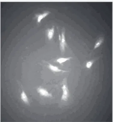
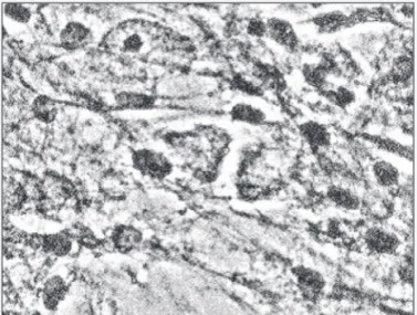
The cell culture was histologically analyzed. Identification of the cells of muscular origin and of the mesenchymal cells was performed through labeling with the anti-desmin antibody (Dako-Anti-Human Desmin-Clone D33) (fig. 1) and the anti-vimentin protein (Immunoperoxidase-Sigma, IMMH-10-Vimentin S-20, Santa Cruz Biotechnology, FITC) (fig. 2), respectively.
The viability of transplanted cells, assessed through the trypan blue stain, ranged from 85 to 95%.
The histological studies of the anatomical specimens were performed with hematoxylin-eosin and Gomori’s trichrome for iden-tifying areas of myocardial infarct and presence of new structures in myocardial fibrosis.
The BrDu antibody (Bromodeoxyuridine-monoclonal anti-body detecting cells proliferation and activation, BD) through direct immunofluorescence was used to label the cells before transplan-tation, so that they could be identified after the procedure.
Nine days after myocardial infarction, the animals were reo-perated on through a median sternotomy. After individualization of the infarcted myocardial area, 0.15 mL of culture medium (57% MEM and 0.5% BSA, Fraction V, Sigma) was subepicardially injected in the control group. The SMM group received 7.5x10⁶
296
skeletal myoblasts and heterologous mesenchymal cells diluted in 0.15 mL of culture medium. All animals received cyclosporine (15mg/kg/day) before sternotomy, and the same dose was main-tained until the day of sacrifice.
One month after cell transplantation, all animals underwent a new echocardiography, in which the same parameters used in the pretransplant examination were assessed. Then, the animals were euthanized for anatomicopathological analysis.
All data are presented as means ± SD, and the statistical significance level adopted was P < 0.05. The groups were com-pared at baseline and 1 month after transplantation with the paired t test.
After assessing homogeneity of variance and normal distribution of data, analysis of variance was performed for determining the difference between the groups in each period (baseline and one month later). The Fisher exact test was used to analyze the propor-tion of cases that improved or were stable one month after trans-plantation.
Results
No statistical difference was observed between groups, control and SMM, in regard to left ventricular ejection fraction and end-systolic and end-diastolic volumes after myocardial infarction and before transplantation. All animals were considered homogeneous (29.31 ± 5.60% and 24.03 ± 8.68%, P=0.10; 0.36 ± 0.09 and 0.37 ± 0.06, P=0.593; and 0.50 ± 0.11 mL and 0.49 ± 0.08 mL, P=0.757; respectively).
One month after cell transplantation, however, a significant difference in left ventricular ejection fraction was observed between the control and SMM groups (31.77±9.06% vs 23.54±6.51%, P=0.020) (fig. 3).
Analyzing the groups individually, left ventricular ejection fraction decreased in the control group (from 29.31±5.60% to 23.54± 6.51%, P=0.048) and increased in the SMM group (from 24.03± 8.68% to 31.77±9.06%, P=0.011) (tab. I).
Analyzing the left ventricular end-diastolic volume, no statistical difference was identified between the control and SMM groups one month after transplantation (0.69 ± 0.19mL vs 0.67 ± 0.12mL, P=0.791, respectively) (fig. 4). In isolation, however, the control and SMM groups showed an increase in the left ven-tricular end-diastolic volume (from 0.50 ± 0.11mL to 0.69 ± 0.19mL, P=0.008, and from 0.49 ± 0.08mL to 0.67 ± 0.12mL, P<0.001, respectively) (tab. II).
Analyzing the left ventricular end-systolic volume, no statistical difference was identified between the control and SMM groups one month after transplantation (0.54 ± 0.17mL vs 0.47 ± 0.12mL, P=0.233, respectively) (fig. 5). In isolation, the control and SMM groups had an increase in the left ventricular end-systolic volume (from 0.36 ± 0.09mL to 0.36 ± 0.09mL, P=0.002, and from 0.37 ± 0.06 to 0.47 ± 0.12mL, P=0.011, respectively) (tab. III).
Analyzing the animals per each group, in the SMM group, of the 16 animals, 14 (87.5%) improved their ejection fraction. In the control group, of the 10 animals, 8 worsened their ejection function, and only 2 (20%) improved their ejection fraction, although no cell was transplanted (tab. IV).
The animals were euthanized one month after cell transplan-tation, and their hearts were removed and divided into 2 parts, the apex containing the infarcted area and the base. Each part was frozen in liquid nitrogen at -80°C, divided into fractioned sec-tions every 5 microns, involving the zone of transition between the intact myocardium and the region of fibrosis.
In the SMM group, one month after intervention, transplanted cells were identified in the infarcted region by use of the
hemato-Fig. 2 - Cell cocultivation showing the presence of cells of mesenchymal origin. (Anti-vimentin, immunoperoxidase, 40x).
EF
EF (%)
p=0.101 p=0.020
Control Mean ± SD
45
40
35
30
25
20
15
10
Control
Cocultivation Cocultivation
Baseline 1 month
Fig. 3 - Ejection fraction of left ventricle between the two groups and in the two periods of evaluation.
Table I - Ejection fraction
Variable Control (n=10) SMM (n=16) p* Mean ± SD Mean ± SD
Baseline EF (%) 29.31±5.60 24.03±8.68 0.101 EF – 1 month (%) 23.54±6.51 31.77±9.06 0.020
p² 0.048 0.011
FDV
FDV (ml)
p=0.757 p=0.791
Control Mean ± SD
1.0 0.9 0.8 0.7 0.6 0.5 0.4 0.3 0.2 0.1 0.0
Control
Cocultivation Cocultivation
Baseline 1 month
297
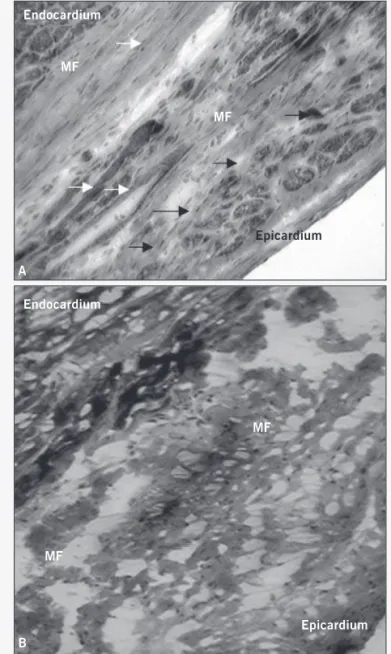
xylin-eosin and Gomori’s trichrome stains. Morphologically, the identified cells had characteristics of skeletal muscular fibers that colonize the region of fibrosis. The formation of new blood vessels was also identified in this region in the SMM group. In the control group, however, the presence of neither muscle nor blood vessels was visualized in the region of myocardial fibrosis (fig. 6a and b). In the SMM group, the transplanted cells were identified by use of the anti-BrDu antibody 24 hours after transplantation (fig. 7).
Discussion
The great majority of the studies about transplantation of skeletal myoblasts into the myocardium have aimed at recolonizing the regions of myocardial fibrosis with contractile cells. These studies, both experimental 2-4 and clinical 5, have suggested the occurrence
of functional improvement in the heart; however, the lack of diffe-rentiation of skeletal myoblasts into cardiomyocytes, the absence of GAP junctions between the transplanted cells and the intact myocardium, and the ventricular arrhythmias identified in some patients operated on 9, could not pass unquestioned.
On the other hand, the studies with bone marrow cells disa-gree in regard to their differentiation into cardiomyocytes and the
real functional benefit that the transplantation of these cells may provide. Orlic et al 8 transplanted the bone marrow cells into the
transition region between the intact and the infarcted myocardium 5 hours after occlusion of the anterior descending coronary. These authors reported cardiac functional improvement with the presence of endothelial cells and smooth muscle in the animals studied, but no bone marrow cells differentiated into cardiomyocytes were identified in the infarcted region, only in the transition zone. The smooth muscle may be justified by its presence in the tunica media of the neoformed blood vessels. Ferem et al 15 believe that
the mononuclear cells of the bone marrow, when transplanted into patients with ischemic cardiomyopathy, provide significant functional and clinical benefits.
The fundamental characteristic of the bone marrow cells, mainly the mesenchymal cells, is their medium-dependent diffe-rentiation, ie, those cells acquire the characteristics of the cells they are in contact with. For example, the bone marrow cells in contact with bone tend towards differentiating into bone; similarly, those in contact with liver cells tend towards differentiating into
Table II - Left ventricular end-diastolic volume
Variable Control (n=10) SMM (n=16) p* Mean ± SD Mean ± SD
Baseline EDV (mL) 0.50±0.11 0.49±0.08 0.757 EDV 1 month (mL) 0.69±0.19 0.67±0.12 0.791 P² 0.008 <0.001
ESV
ESV (mL)
p=0.593 p=0.233
Control Mean ± SD
0.8
0.7
0.6
0.5
0.4
0.3
0.2
0.1
0.0
Control
Cocultivation Cocultivation
Baseline 1 month
Fig. 5 – End sistolic volume of left ventricle between the groups and in the two periods evaluation.
Table III - Left ventricular end-systolic volume
Variable Control (n=10) SMM (n=16) p* Mean ± SD Mean ± SD
Baseline ESV (mL) 0.36±0.09 0.37±0.06 0.593 ESV 1 month (mL) 0.54±0.17 0.47±0.12 0.233
p² 0.002 0.011
Table IV - Percentage of improvement in ejection fraction
EF (%) Group Valor de p* Control SMM
Improvement 2 (20.0%) 14 (87.5%) 0,001 Worsening 8 (80.0%) 2 (12.5%)
Total 10 16
Fig. 6 - a) Presence of muscular fibers (black arrows) and blood vessels (white arrows) in the region of myocardial fibrosis (MF) between the epicardium and the endocardium in the SMM group (Gomori’s trichrome, 20x); b) Absence of muscular fibers and blood vessels in the region of myocardial fibrosis (MF) in the control group (Gomori’s trichrome, 20x).
Epicardium
Endocardium
MF
MF
A
Epicardium Endocardium
MF
MF
298
MF
Fig. 7 - Identification of transplanted cells (arrows) in the region of myocardial fibrosis in the SMM group (direct immunofluorescence BrDu, 20X).
hepatocytes; those in contact with cardiac cells tend towards differentiating into cardiomyocytes, and so forth 16. Following this
same rationale, when those cells are transplanted into the region of chronic myocardial infarct, ie, into the fibrotic area, they would consequently tend towards differentiating into fibrosis. Based on that principle, it is hard to believe that bone marrow cells could recolonize the chronically infarcted myocardium with new cardiac muscle fibers.
Some studies 12,17 have shown by use of anatomicopathological
analysis that, when bone marrow cells (mononuclear and/or mesenchymal cells) are transplanted into models of chronic myo-cardial infarct, neoformation of blood vessels is observed in the region of myocardial fibrosis.
The idea of using a combination of skeletal myoblasts, which do really recolonize the infarcted myocardium with new muscle fibers of skeletal origin, and cells derived from the bone marrow, which stimulate the formation of new vessels in the region of fibrosis, was based on the hypothesis of providing an angiomuscular regeneration and not only an isolated muscular or angiogenic regeneration.
Analyzing the SMM group separately, the functional improve-ment identified between the period after myocardial infarction and one month after cell transplantation was significant (form 24.03 ± 8.68% to 31.77 ± 9.06, P=0.011). The same was observed when the SMM group was compared with the control group one month after transplantation (23.54 ± 6.51% vs 31.77 ± 9.06%, P=0.020). These data suggest the effectiveness of the transplantation of cocultivated cells. However, it is difficult to identify which transplanted cell, if not both, is responsible for the increase in left ventricular ejection fraction.
Still in regard to functional analysis and considering only the animals that maintained their left ventricular ejection fraction as
stable or improved it, the placebo effect was present in 2 of the control group animals, because their ejection fraction improved even without any type of treatment. In the SMM group, 14 (87.5%) animals of the 16 improved their ejection fraction. The fact that 2 animals had no improvement in their cardiac function may be attributed to technical problems, such as a failure in cell injection or even embolization.
Despite the effectiveness of cell transplantation in the SMM group in regard to left ventricular ejection fraction, a significant increase in left ventricular end-diastolic volume (P<0.001) was observed one month after transplantation, suggesting the occur-rence of ventricular remodeling independent of improvement in cardiac function. The outcome of the control group was similar, and no significant difference was observed between both groups in regard to that parameter in the same period. Other studies 2,4
on transplantation of skeletal myoblasts into the infarcted myo-cardium, including some clinical studies 9, reported the occurrence
of ventricular remodeling despite the functional improvement. The capacity of regeneration of the infarcted myocardium with skeletal myoblasts is believed to be limited to the site of cell injection with no overall anti-ventricular remodeling effect.
In regard to left ventricular end-systolic volume, an increase was observed in the control and SMM groups one month after cell transplantation, suggesting a decrease in the contractile activity (P=0.002 and P=0.011, respectively).
Pouzet et al 18, in a study of animals undergoing transplantation
of skeletal myoblasts in association with clinical treatment with an angiotensin-converting enzyme inhibitor, reported a limitation in left ventricular dilation, probably due to the effects of clinical treatment.
The mechanisms responsible for the improvement in cardiac function after transplantation of skeletal myoblasts have not been well established. That improvement is believed to result from an active process due to the improvement in the regional contractility and a decrease in the infarcted region. However, it is worth em-phasizing that a direct relation exists between the number of cells transplanted and functional improvement 19, and that the
benefit of the intervention also depends on the quality of the cells transplanted and their survival after the procedure 20, because a
large percentage of cells die after transplantation.
Another study 17, which compared the effects of the
trans-plantation of skeletal myoblasts alone and mesenchymal cells alone into myocardial infarct, reported a significant improvement in left ventricular ejection fraction in the group receiving the skeletal myoblasts as compared with that in the control group; on the other hand, the group receiving the mesenchymal cells showed stabilization of left ventricular ejection fraction. The anatomico-pathological examination of the group receiving the mesenchymal cells revealed no new muscular fibers in the infarcted region, where the cells had been injected, but an intense neoangiogenesis. This fact may explain the stabilization of left ventricular ejection fraction, because the formation of new blood vessels can reduce apoptosis and the expansion of the infarct, thus preserving the myocardium. A similar functional result has also been reported in another study 21, which compared cells of the hematopoietic line
(CD 133+) and skeletal myoblasts.
299
1. Kajstura J, Leri A, Finato N, et al. Myocite proliferation in end-stage cardiac failure in humans. Proc Natl Acad Sci USA. 1998;95:8801-5.
2. Ghostine S, Carrion C, Guarita Souza LC, et al. Long term efficacy of myoblast transplantation on regional structure and function afer myocardial infartction. Circulation.2002;106(Suppl I):131-136.
3. Murry C, Wiseman R, Schwartz S, et al. Skeletal myoblast transplantation for repair of myocardial necrosis. J Clin Invest. 1996; 98:2512-23.
4. Scorsin M, Hagege A, Vilquin JT, et al. Comparison of the effects of fetal cardiomyo-cyte and skeletal myoblast transplatation on post-infarct left ventricular function. J Thorac and Cardiovas Surgery. 2000; 119:1169-75.
5. Menasché P,Hagége AA, Scorsin M, et al. Myoblast transplantation for heart failure. Lancet. 2001;357:279-80.
6. Strauer BE, Brehm M, Zeus T, et al. Repair of infracted myocardium by autologous intracoronary mononuclear bone marrow cell transplantation in humans. Circu-lation.2002;106:1913-8.
7. Arjun D, Shaohua W, Kimberly A, et al. Bone marow-derived cardiomyocites are present in adult human heart. Circulation. 2003;107:1247-9.
8. Orlic D, Kajstura J, Chimenti S, et al. Transplanted adult bone marrow cells repair myocardial infarcts in mice. Ann. N Y Acad Sci. 2001;938:221-9.
9. Menasché P, Hagege A, Vilquin J T, et al. Autologous skeletal myoblast transplan-tation for severe postinfarction left ventricular dysfunction. J Am. Coll Cardiol. 2003;41:1078-83.
10. Verfaillie CM. Adult stem cells: assessing the case for pluripotency. Trends Cells Bio. 2002;12:502-8.
11. Friedenstein A, Petrakova K, Kurolesova A. Heterotopic of boné marrow. Analysis of percursor cells for osteogenic and hematopoietic tissues. Transplantation. 2002;6: 230-247.
References
12. Bel A, Messas E, Agbulut O, et al. Transplantation of autologous fresh boné marrow into infarcted myocardium: A word of caution. Circulation. 2003;108 [suppl II]:247-252. 13. Delaporte C, Dehaupas M, Fardeau M. Comparison between the growth pattern of cell cultures from normal and Duchenne Dystrophy Muscle. J Neurol Sci. 1984; 64:149-60.
14. Boyüm A. Isolation of mononuclear cells and granulocytes from human blood. Scan J Clin Lab Invest. 1968; 21(suppl): 77-89.
15. Perin E, Dohman H, Radovan B, et al. Transendocardial autologous bone marrow cell transplantation for severe chronic ischemic heart failure. Circulation. 2003;107:2294-302.
16. Malouf NN, Coleman WB, Girsham JW, et al. Adult derived stem cells from the liver become myocytes in the heart in vivo. Am J Pathol, 2001:158:1929-1935. 17. Guarita Souza LC, Carvalho KAT, Rebelato C. The effectivennes of myoblast and
bone marrow cells transplantation, on post-infarct ventricular dysfunction. Circu-lation, 2003;(suppl IV) 108.
18. Pouzet B, Ghostine S, Vilquin JT et al. Is skeletal myoblast transplantation clini-cally relevant in the era of angiotensin-converting enzyme inhibitors? Circulation. 2001 Sep 18;104(12 Suppl 1):I223-8.
19. Pouzet B, Vilquin JT, Hagege AA, et al. Factors affecting functional outcome after autologous skeletal myoblast transplantation. Ann Thorac Surg. 2001;71:844-50. 20. Pouzet B, Hagege AA, Vilquin JT. Transplantation of autologous skeletal myoblasts
in ischemic cardiac insufficiency. J Soc Biol. 2001; 195: 47-9.
21. Agbulut O, Velde S, Attar N, et al. Which cell type for regeneration of chronically infarcted myocardium? A face-to-face comparison of skeletal myoblasts and marrow-derived hematopoietic stem cells. Circulation. 2003;(suppl IV): 108. 22. Zhang Y, Hartzell C, Narlow M, et al. Stem cell-derived cardiomycytes demonstrate
arrhythmic potencial. Circulation. 2002;106:1294-9. their studies with skeletal myoblasts was not an isolated finding.
Zhang et al 22 have also suggested that bone marrow embryonic
cells, when transplanted into the infarcted myocardium, may also show arrhythmogenic characteristics.
Myocardial ischemia is one of the causes of ventricular arrhy-thmias. Therefore, in the study by Menasché et al 9, the cause of
arrhythmias is believed to be ischemic, because after the trans-plantation of skeletal cells into a region of fibrosis, that region became viable, visualized on positron emission tomography (PET-SCAN). However, in that viable region, which was previously infarcted, myocardial ischemia may be suspected, because blood supply to the patients studied was absent due to one coronary artery occlusion without collateral circulation.
On anatomicopathological examination in this study, the iden-tification of new muscular fibers in the region of fibrosis and the presence of new blood vessels in the region confirm the hypothesis of angiomuscular regeneration.
Pouzet et al 18 reported the existence of a significant cell
mortality right after cell transplantation, possibly due to the local inflammatory reaction of the infarct or even of cell injection. One cause of late mortality of transplanted cells could be the chronic ischemia in the region. The presence of new blood vessels in the region into which the muscular cells were transplanted may help to preserve the new muscular fibers and reduce ischemia, atrophy, and cardiomyocyte death.
In conclusion, the combination of skeletal myoblasts and mesenchymal cells proved to be effective in the regeneration of myocardial fibrosis, with identification of new skeletal muscular fibers and new blood vessels in the region of myocardial fibrosis.