www.biogeosciences.net/8/1853/2011/ doi:10.5194/bg-8-1853-2011
© Author(s) 2011. CC Attribution 3.0 License.
Biogeosciences
Vertical and longitudinal gradients in HNA-LNA cell abundances
and cytometric characteristics in the Mediterranean Sea
F. Van Wambeke1, P. Catala2,3, M. Pujo-Pay2,3, and P. Lebaron2,3
1CNRS, Universit´e de la M´editerran´ee, Laboratoire de Microbiologie, G´eochimie et Ecologie Marines, UMR6117,
Case 901, Campus de Luminy, 13 288 Marseille cedex 9, France
2UPMC Univ Paris 06, Laboratoire ARAGO – Observatoire Oc´eanologique, 66651 Banyuls/mer, France 3CNRS, UMR7621, LOMIC, Observatoire Oc´eanologique, 66651 Banyuls/mer, France
Received: 29 October 2010 – Published in Biogeosciences Discuss.: 10 November 2010 Revised: 18 June 2011 – Accepted: 20 June 2011 – Published: 13 July 2011
Abstract. Heterotrophic bacterioplankton abundance and production were investigated with depth (down to bathy-pelagic layers) and with longitude (from 4.9◦E to 32.7◦E) along a cruise track across the Mediterranean Sea in early summer 2008. Abundances and flow cytometric character-istics (green fluorescence and side scatter signals) of high nucleic acid (HNA) and low nucleic acid (LNA) bacterial cells were determined using flow cytometry. Contrary to what is generally observed, the relative importance of HNA cells, as a percent of total cells, (%HNA, range 30–69 %) was inversely related to bacterial production (range 0.15– 44 ng C l−1h−1) although the negative relation was weak (log–log regressionr2=0.19). The %HNA as well as the mean side scatter of HNA group increased significantly with depth in the meso and bathypelagic layers. Vertical stratifica-tion played an important role in influencing the distribustratifica-tion and characteristics of bacterial cells especially with regard to layers located above, within or below the deep chlorophyll maximum. Within a given layer, the relationships between the flow cytometric characteristics and environmental vari-ables such as chlorophyll-a, nutrients or bacterial production changed. Overall, the relationships between HNA and LNA cells and environmental parameters differed vertically more than longitudinally.
1 Introduction
Flow cytometry is a powerful tool to study microbial commu-nities. In samples from aquatic environments, bacterioplank-ton cells tend to cluster into at least 2 or 3 distinct fractions
Correspondence to:F. Van Wambeke
(france.van-wambeke@univmed.fr)
based on differences in the side scatter signal (SSC, related to the size, density and morphology of the cells) and in the relative green fluorescence (related to the nucleic acid con-tent of the cells). These fractions are more generally named HNA (high nucleic acid) and LNA (low nucleic acid) cells (Lebaron et al., 2001). Many papers have reported abun-dances of HNA and LNA cells over a wide range of envi-ronmental samples, from oligotrophic to eutrophic environ-ments, since the pioneering work of Li et al. (1995). How-ever, factors influencing the relative importance of HNA to LNA cells both within and among aquatic systems are un-clear. Comparisons among studies are hindered as different protocols are used (differences in instrument, sample fixation and storage, nucleic acid stain, etc.). Thus, comparisons have generally been made on population abundance, and few have considered cytometric characteristics.
hypothesized by early cell sorting experiments (Servais et al., 1999; Lebaron et al., 2001). The general conclusion from the literature is that HNA bacteria are larger and more active on a cell basis than LNA bacteria. Overall, despite considerable amount of work, the contribution of LNA cells to total bacte-rial production is still a subject of much debate (Longnecker et al., 2006; Sherr et al., 2006; Scharek and Latasa, 2007; Moran et al., 2010).
The Mediterranean Sea is largely oligotrophic over much of the year, and is characterized by a gradient of oligotro-phy from the oligotrophic West to the ultra-oligotrophic East. The BOUM (Biogeochemistry from the Oligotrophic to the Ultra-oligotrophic Mediterranean) cruise carried out in early summer 2008, offered the opportunity to examine bacterial abundance and production with depth over a large longitu-dinal gradient. Indeed, to our knowledge, information avail-able in Mediterranean Sea about HNA and LNA cytomet-ric group distributions and properties is restcytomet-ricted to West-ern, coastal and shelf areas (Lebaron et al., 2001; Joux et al., 2005; Scharek and Latasa, 2007). Very limited data on LNA and HNA distributions within open sea areas are available: the Dyfamed site in the N.W. Mediterranean has been stud-ied on different time scales but with no reports on variability of cytometric properties like SSC or fluorescence (M´evel et al., 2008; Winter et al., 2009).
In this study we analyzed a large number of samples to in-vestigate flow cytometric characteristics in relation to bacte-rial production and environmental parameters (temperature, total chlorophyll-a, phosphate, nitrate + nitrite). Samples were gathered from surface to mesopelagic waters from 30 stations across the Mediterranean Sea. The aim of this study was to explore the factors that determine the variability in abundances and cytometric properties of HNA and LNA cells in Mediterranean Sea waters, including the deep sea and the Eastern Basin. Flow cytometric characteristics of the bacteri-oplankton were examined within layers located above, within and below the deep chlorophyll maximum, as well as longi-tudinally among different regions.
2 Material and methods
2.1 Study area and sample collection
This work was carried out during the “BOUM” cruise on the R/V Atalanteduring June–July 2008. The cruise was planned as a transect of stations encompassing a large lon-gitudinal gradient in the Mediterranean Sea (Fig. 1). Sam-ples were collected using a rosette of 24×12 l Niskin bot-tles mounted on a CTD rosette system. Sensors on the CTD provided profiles for temperature, salinity, and fluo-rescence at each station. The depth of the deep chlorophyll maximum (dcm) was determined from the fluorescence pro-file. Total chlorophyll-a(TChl-a) was analyzed using HPLC (Ras et al., 2008) and the data for vertical and horizontal
B
25 24
C
21 15
16
1 3 5 7 9 11
A
17 19
2 6 8 10 26
27
23 22
20 18
1413 12
4
Rh
We
Si
Ea
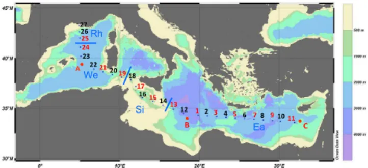
Fig. 1. Map of the BOUM transect. All stations were sampled for
bacterial abundances and TChl-a whereas only stations indicated
with a code in red were sampled for bacterial production. Blues characters and lines separates the different regions compared (see Material and Method section). Figures 1 and 3 were drawn with Ocean Data View program (Schlitzer, 2009).
profiles are presented in Moutin et al. (2011) and Crom-bet et al. (2011). Phosphate (PO34−) and nitrate + nitrite (NO−3 + NO−2) were analysed using automated colorimet-ric technique as described in Pujo-Pay et al. (2011). Detec-tion limits for the procedures were 0.01 µM for PO34− and 0.02 µM for NO−3 + NO−2. The numbered stations were oc-cupied briefly and sampled at different times of the day. All these numbered stations were investigated for bacterial abun-dance and only one half of them for bacterial production (1, 3, 5, 7, 9, 11, 13, 15, 17, 19, 21, 24, 25). Sites designated by letters (A, B and C) were gyre stations, located inside anticy-clonic gyres and each was sampled over a 4 day period. At each gyre station, up to 4 depth profiles for bacterial abun-dance and production were obtained, three at 09:00 a.m., and one at 02:00 a.m. (local time). The whole data set was used to describe bacterial abundance over 45 vertical profiles (493 samples) while relationships with BP were examined using 25 profiles (198 samples).
2.2 Flow cytometric analysis of bacteria
Aliquots of 1.8 ml were taken for heterotrophic bacterio-plankton (sensus stricto referring to heterotrophic prokary-otes), fixed with 2 % (w/v) formaldehyde (PFA) solution, stored for at least 30 min at room temperature, frozen in liq-uid nitrogen and then stored at−80◦C until samples could be processed on shore.
LNA HNA
LNA HNA
LNA HNA
LNA HNA 5 m, surface 124 m, dcm
3000 m, deep 200 m, below dcm
b b
b b
green fluorescence
side scatter
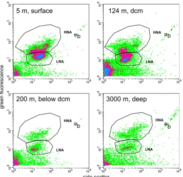
Fig. 2. Examples of four cytograms obtained after SyBR green I
staining. The data comes from Station B (Ionian Sea): surface (5 m), at the dcm (125 m), below dcm (200 m) and in deep layers (3000 m). Time of analysis was set to 1 min for the 3 first layers, and 3 min for the 3000 m layer. Windows for HNA and LNA cells are drawn and were adjusted from one sample to another. b: 1 µm beads.
of the analysis was dependant on the bacterial abundance of the sample, typically the volume analyzed was around 20 µl (low speed). The subsequent cell concentration esti-mation was determined from the flow rate, which was cal-culated by weighing one tube of milliQ water before and af-ter a 5 min run of the cytomeaf-ter (this flow rate calibration was done after each five sample tubes). Fluorescent beads (1.002 µm, Polysciences Europe) were systematically added to each sample as an internal standard and used to normalize values of single cell variables. Thus, the fluorescence and side scatter values of cells were standardized to those of the reference beads to account for potential differences in mea-surement conditions (all samples were analysed during a 6 month-period by the same analyst). In a plot of green fluo-rescence versus red fluofluo-rescence we were able to distinguish photosynthetic prokaryotes which were removed from non-photosynthetic prokaryotes. Bacteria with a high nucleic acid content (HNA cells) were discriminated from bacteria with a low nucleic acid content (LNA cells). Each subgroup was de-limited on the SSC versus green fluorescence plot by drawing a gate, and cell abundance was determined in each subgroup (Fig. 2). Windows were adjusted for each individual sample. Total abundance is determined as LNA + HNA and the per-centage of HNA cell abundance (%HNA) was calculated as HNA/(HNA + LNA).
2.3 Bacterial production
“Bacterial” production (BP – sensus stricto referring to het-erotrophic prokaryotic production –) was determined by [3H] leucine incorporation applying the centrifugation method (Smith and Azam, 1992). Duplicate 1.5 mL samples and one trichloracetic acid (TCA) killed control for blank correction were incubated with a mixture of [4,5–3H]leucine (Perkin
Elmer, specific activity 115 Ci mmol−1)and nonradioactive
leucine at final concentrations of 16 and 7 nM, respectively. Samples were incubated in the dark at the respective in situ temperatures for 2–5 h depending on the expected activity. Preliminary checks showed that the incorporation of leucine was linear with time. Incubations were stopped by adding of TCA to a final concentration of 5 %. To facilitate the pre-cipitation of proteins, bovine serum albumin (BSA, Sigma, 100 mg L−1final concentration) was added prior to centrifu-gation which was carried out at 16 000 g for 10 min. After discarding the supernatant, 1.5 ml of 5 % TCA was added and the samples were vigorously shaken using a vortex and then centrifuged again. After discarding the supernatant, 1.5 ml of 80 % ethanol was added and then the samples were shaken and centrifuged again. The supernatant was discarded, and 1.5 ml Ultimagold MW added in the centrifuge tube. The ra-dioactivity incorporated into the pellet was counted using a Packard LS 1600 Liquid Scintillation Counter. A factor of 1.5 kg C mol leucine−1was used to convert the incorporation
of leucine to carbon equivalents, assuming no isotopic dilu-tion. This was checked on 3 occasions with concentration kinetics (range of concentrations 3 to 50 nM, isotopic dilu-tion between 1.01 and 1.07). Activities within meso- and bathy-pelagic layers were also investigated at stations A, B and C, between 250 and 910 m at St C, 250–3000 m at St B and 250–2700 m at St A. These deeper samples, where a lower activity was expected, were treated by the filtration technique (Kirchman, 1993). We added 10 nM final concen-tration of [4,5–3H]leucine in 50 ml samples. Two duplicates and one formalin-killed blank were incubated in the dark at in situ temperature for 15–20 h. Live samples were termi-nated by formalin addition (1 % final concentration) and all samples were filtered onto 0.2 µm Millipore GS/WP filters before TCA extraction (5 % final) and a 80 % ethanol rinse directly on the filter tower. Filters were dissolved in 1 ml of ethyl acetate before addition of Ultima Gold MV scin-tillation cocktail. Errors associated with the variability be-tween replicate measurements (half the difference bebe-tween the two replicates) averaged 6 % and 8 % for BP values de-termined using the centrifuge (surface samples) and filtration (deep samples) methods, respectively.
2.4 Statistical analysis and groups of data
and cytometric characteristics as well as BP and environ-mental parameters (temperature, chlorophyll, nutrients) were log10 transformed in order to achieve normality and
homo-geneity of variances before making correlations or ANOVA. Respecting recommendations of Sokal and Rohlf (1981), data sets including values between zero and one were mul-tiplied by 100 (green fluorescence) or 1000 (SSC) before log transformation to avoid negative characteristics in the loga-rithms. PO34−and NO−3 + NO−2 data sets also included many zeros and their transformation was done applying the formula LOG(data*100 + 1). Model II regression was used to ex-amine relationships between 2 parameters when both (used in X and Y axis) were subjected to measurement variability. Model I was used to examine the relationship between any Y environmental dependant variable and an X independent variable as temperature or depth. Note that the coefficient of determination is the same whatever the model used.
Relationships were examined vertically following 4 differ-ent groups of samples:
– “surface” samples located between the surface and the deep chlorophyll maximum (dcm) layer,
– “dcm” samples at the dcm depth,
– “below dcm” corresponds to samples taken between the dcm and 250 m,
– “deep” corresponds to samples taken below 250 m. Indeed, the “dcm” layer represents an ideal ecological boundary in our study which enabled data to be compared from stations across the Mediterranean Sea, where the trans-parency of water, depths of nutriclines and dcm varied greatly (Pujo Pay et al., 2011; Crombet et al., 2011).
Longitudinally, we compared 4 groups defined as follows: (i) “Rh” including North Western Mediterranean Sea stations (25 to 27) which were closer to the Rhˆone river with a higher chlorophyll biomass and bacterial abundances and a chloro-phyll maximum between 3 and 50 m; (ii) “We” including sta-tions 19 to 24 and A located in open Western Mediterranean with deeper dcm depths (between 69 and 96 m); (iii) “St” with stations 14 to 17 (within the Sicily strait) and station 18 which is in the Western basin but its intermediary layers are influenced by the flow of subsurface Eastern waters (Pujo-Pay et al., 2011); and (iv) “Ea” including stations 1 to 13 and stations B and C located in open, Eastern Mediterranean (Fig. 1).
3 Results
3.1 Distribution and variability of biological and physico-chemical parameters
The stations were located between 43.2◦N, 4.93◦E (close to Rhˆone river mouth) and 33.7◦N, 32.7◦E (South of Cyprus,
A 21 19 17 3 5 7 11C
25 24 26
27 23 22 20 18 161514 1312B 1 2 4 6 8 910
bacterial production (ng C l‐1h‐1)
dcm depth
depth
(m)
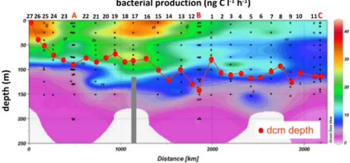
Fig. 3.Contour plot of bacterial production along the BOUM
tran-sect, from the most Northwestern station (left) to the most Eastern station (right) and for the 0–250 m layer. Depth of the deep chloro-phyll maximum (dcm) is also indicated (red dots). The bar at St 7 in the Sicily strait shows the bottom depth (116 m).
on the sea mount Erathost`ene, Fig. 1). All stations were sit-uated within the continental slope or open Sea (except at the Sicily Strait: St 14, 382 m and St 17, 116 m and close to the Rhone river mouth: St 27, 105 m), with bottom depths down to 3321 m (st 12). The diel variability of sea surface tem-perature per station was around 1◦C. Surface temperature (5 m) reached a maximum of 27.5◦C (st 12 in the Levan-tine Sea) with lowest temperatures at St 27 (17.2◦C, close to the Rhˆone river mouth) and decreasing with depth down to 13.3◦C in the Eastern Basin and 12.8◦C in the Western Basin (Table 1).
There was a classical West – East gradient of the deep-ening of the depth of the dcm (Fig. 3), which varied from 5 m at the western station 27 to 145 m at eastern station B. Maximum TChl-aconcentration was found at station 25, with 1.7 µg TChl-al−1 at 50 m depth. Based on integrated
TChl-a(concentrations measured down to 250 m), the most oligotrophic station was station 8, with 16.1 mg TChl-am−2
whereas the richest was station 25 (55.3 mg TChl-am−2).
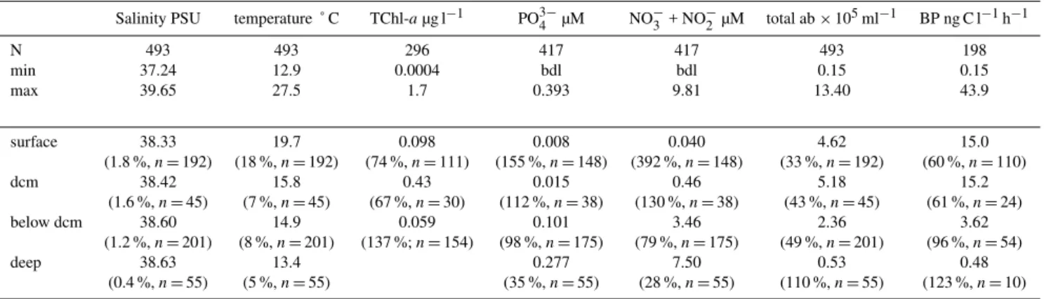
Table 1. Ranges of salinity, temperature, total chlorophyll-a(TChl-a), phosphate (PO34−), nitrate + nitrite (NO−3 + NO−2), total bacterial abundance (total ab), and bacterial production (BP) for the data set used for comparison of abundance and cytometric characteristics of HNA and LNA cells. Means, coefficient of variation and number of data for each group of water column partition: layers above the deep chlorophyll maximum (surface), layers at the deep chlorophyll maximum (“dcm”), layers below the dcm but above or equal to 250 m (“below dcm”), and layers below 250 m “deep”. bdl: below detection limits.
Salinity PSU temperature ˚ C TChl-aµg l−1 PO3−
4 µM NO−3 + NO−2 µM total ab×105ml−1 BP ng C l−1h−1
N 493 493 296 417 417 493 198
min 37.24 12.9 0.0004 bdl bdl 0.15 0.15
max 39.65 27.5 1.7 0.393 9.81 13.40 43.9
surface 38.33 19.7 0.098 0.008 0.040 4.62 15.0
(1.8 %,n=192) (18 %,n=192) (74 %,n=111) (155 %,n=148) (392 %,n=148) (33 %,n=192) (60 %,n=110)
dcm 38.42 15.8 0.43 0.015 0.46 5.18 15.2
(1.6 %,n=45) (7 %,n=45) (67 %,n=30) (112 %,n=38) (130 %,n=38) (43 %,n=45) (61 %,n=24)
below dcm 38.60 14.9 0.059 0.101 3.46 2.36 3.62
(1.2 %,n=201) (8 %,n=201) (137 %;n=154) (98 %,n=175) (79 %,n=175) (49 %,n=201) (96 %,n=54)
deep 38.63 13.4 0.277 7.50 0.53 0.48
(0.4 %,n=55) (5 %,n=55) (35 %,n=55) (28 %,n=55) (110 %,n=55) (123 %,n=10)
due to the deepening of nutriclines in the East, and for “deep” layers were due to the different origin of deep waters masses in Eastern and Western Basins (see Pujo-Pay et al., 2011). 3.2 Total bacterial abundance and production
Total bacterial abundances ranged from 0.15 to 13.4×10⁵ml−1(Table 1). BP ranged from 0.1 ng C l−1h−1
(650 m, station C) to 43.9 ng C l−1h−1 (Sicily Strait, 5 m,
station 15), and 41 ng C l−1h−1at the station sampled for BP
closest to the Rhˆone river mouth (25 m, station 25, Fig. 3). The relationship between bacterial biomass (BB, calcu-lated assuming 12 fgC per cell) and BP was high (r2=0.7) when the data were pooled all together (data not shown). r2 was lower but remained significant for different layers (r2=0.24, 0.54 and 0.47 for “surface”, “dcm” and “below dcm” layers, respectively). The slopes obtained for the whole data set (0.57) as well as for the separate water-column layers (0.44–0.55) suggest bottom-up control of bacterial biomass following Billen et al. (1990) and Ducklow (1992). TChl-a was weakly linked to BP (29 % of the variability explained, relation not shown). Notably in different layers, there was no relationship between TChl-aand BP for “surface” layers, and weak relationships for the “dcm” and “below dcm” lay-ers (r2=0.52 and 0.53). There was not any relationship be-tween BP and concentration of PO34−(log transformed data, p >0.05) regardless of layer.
3.3 HNA and LNA bacterial abundance
All samples had two HNA and LNA cell fractions discern-able by fluorescence versus SSC cytograms. HNA cell abun-dances ranged from 7.7×10³to 6.1×10⁵ml−1, and those of LNA cells from 6.5×10³to 7.3×10⁵ml−1. Both box-plot
distributions of HNA and LNA cells showed high variability
within the “surface” and “dcm” layers (Fig. 4a, b). Neither HNA nor LNA cell abundances were statistically different in the “surface” when compared to the “dcm” layers (ANOVA, threshold chosen at p=0.01), but significantly decreased “below the dcm” and in the “deep” layers (ANOVA, p < 0.01). The decrease in HNA and LNA cell abundances with depth (log-log regressions abundance-depth) were significant for “dcm”, “below dcm” and “deep” layers (r2ranged 0.33– 0.66, for all regressionsp <0.01). Surprisingly, the percent-age of HNA cell abundance (%HNA = HNA/(LNA + HNA)) increased significantly with depth in the meso- and bathy-pelagic zones (log-log regression in the “deep” layers,r2=
0.59,n=55, Fig. 5b), whereas for the other layers the corre-lation was insignificant.
Longitudinal variability was examined in the different ver-tical layers. Within the “surface” layer a West- East gradient was visible for both HNA and LNA abundances, with signif-icantly higher values in the “Rh” region and lower values in “Ea” compared to “We” region (Fig. 6a, b). However, these differences in abundances among different regions were con-comitant for both groups as longitudinally, %HNA did not change (see for example %HNA distribution in “surface lay-ers” Fig. 7a).
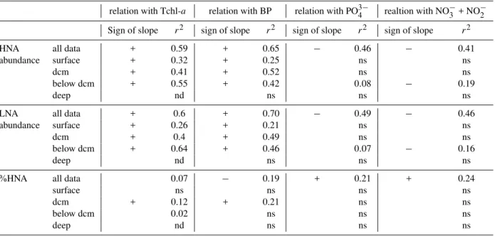
Table 2. Relationships between abundance of HNA cells, abundance of LNA cells and %HNA versus TChl-a, versus BP, versus soluble
reactive phosphorus (PO34−) and versus nitrate + nitrite (NO−3 + NO−2) concentrations. Data were transformed before fitting with linear
regressions (see methods). n: number of data,r2: determination coefficient, nd: not determined, ns: not significant (Significant threshold set
atp=0.05). For clarity, sign of slopes for weak correlations (r2<0.1) were not indicated. The number of data used in the regressions for
each type of environmental variable and for each layer are indicated in Table 1.
relation with Tchl-a relation with BP relation with PO34− realtion with NO−3 + NO−2
Sign of slope r2 sign of slope r2 sign of slope r2 sign of slope r2
HNA all data + 0.59 + 0.65 − 0.46 − 0.41
abundance surface + 0.32 + 0.25 ns ns
dcm + 0.41 + 0.52 ns ns
below dcm + 0.55 + 0.42 0.08 − 0.19
deep nd ns ns ns
LNA all data + 0.6 + 0.70 − 0.49 − 0.46
abundance surface + 0.26 + 0.21 ns ns
dcm + 0.4 + 0.49 ns ns
below dcm + 0.64 + 0.46 0.07 − 0.16
deep nd ns ns ns
%HNA all data 0.07 − 0.19 + 0.21 + 0.24
surface ns ns ns ns
dcm + 0.12 + 0.21 ns ns
below dcm 0.02 ns ns ns
deep nd ns ns ns
TChl-aappeared to explain a large variability in cell abun-dances. Both HNA and LNA cell abundances showed a significant, positive slope following TChl-a concentrations (slopes model II: 0.31, r2=0.59; 0.34, r2=0.60, respec-tively). The correlations of abundances with Tchl-a were also significant within the 3 chlorophyll layers when con-sidered independently (“surface”, “dcm” and “above dcm”). TChl-a explained slightly the variability of %HNA only at the “dcm” layer (r2=0.12, Table 2).
Abundances of HNA and LNA were also inversely re-lated to concentrations of phosphate and nitrate + nitrite (r2 ranged 0.41 to 0.49, Table 2). Some of the %HNA vari-ability was explained by varivari-ability of PO34−(r2=0.21) and
NO−3 + NO−2 (r2=0.24). Such relationships were mainly due to the co-variation with depth as this relationship weak-ened and/or disappeared when layers were considered sepa-rately (Table 2).
Finally, plotting the data all together (“surface”, “dcm”, “below dcm” and “deep”) the log-log relationship between %HNA , LNA cell abundances and HNA cell abundances with temperature was weakly significant (r2=0.19, 0.25 and 0.19, respectively). However, there was no significant rela-tionship between %HNA , LNA cell abundances or HNA cell abundances with temperature in the “surface” layers where the temperature range was the highest.
3.4 Patterns in cytometric parameters
Standardized SSC of HNA and LNA cells ranged from 0.0093 to 0.0265 and from 0.078 to 0.0185, respectively. Standardized green fluorescence of HNA and LNA cells ranged from 0.18 to 0.44 and from 0.050 to 0.097, respec-tively. The coefficient of variation of SSC was slightly higher for HNA cells (21 %) than for LNA cells (17 %). This was the same for green fluorescence (15 % versus 12 %).
Green fluorescence of LNA cells decreased slightly with depth (log–log regressions,r2=0.24,n=493, Fig. 4e and Fig. 5 e,f). In marked contrast, the green fluorescence of the HNA cells increased significantly with depth (r2=0.51,
data Fig. 4f and Fig. 5e, f) and with nutrients (r2=0.45 for
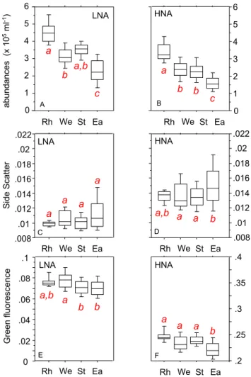
bel o w d cm dcm deep surface 0.008 0.010 0.012 0.014 0.016 0.018 0.020 0.022 0 0.02 0.04 0.06 0.08 0.10 0.008 0.010 0.012 0.014 0.016 0.018 0.020 0.022 0.20 0.25 0.30 0.35 0.40 ab un da nces (x 10 5ml -1) Side Scatter Green fluorescence LNA HNA bel o w d cm dcm deep surface LNA HNA LNA HNA 0 1 2 3 4 5 0 1 2 3 4 5 bel ow dcm dcm deep surface bel ow dcm dcm deep surface a a c a b c a b b c a
b b b
a a b c b a b a b c A B C D E F
Fig. 4. Box plots showing distribution of (A, B): abundances,
(C, D): side scatter and (E, F): green fluorescence of subgroups
LNA and HNA along different water column layers: above the deep chlorophyll maximum (“surface”), at the deep chlorophyll maxi-mum (“dcm”), below the dcm but above or equal to 250 m (“below dcm”), and below 250 m (“deep”). Groups connected by the same letter (in italics and in red) are not significantly different at the 0.01 probability level. Lower to the upper values are indicated, respec-tively, 10 %, 25 %, 50 % (median), 75 and 90 % percentiles.
While green fluorescence characteristics were very dis-tinct between the HNA and LNA cells regardless of depth, SSC differences were slight within surface layers but sur-prisingly higher in meso and bathy pelagic layers (Fig. 5d and Fig. 4d). SSC decreased with depth in “surface” layers (log–log regressions, model I,p <0.001,r2=0.37 for HNA cellsp <0.001,r2=0.46 for LNA cells, Fig. 5c). In con-trast, SSC increased in “deep” layers (p <0.001,r2=0.66 for HNA cells,p <0.001,r2=0.27 for LNA cells. This was more pronounced for HNA cells. Indeed, SSC of this group increased 6 times more rapidly with depth than the SSC of the LNA cells (Fig. 5d). The cytometric properties (SSC, flu-orescence) of HNA and LNA cells showed low or inexistent relationships with nutrient concentrations when the depth ef-fect was removed (i.e. when looking for relationships with
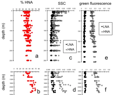
% HNA 0 50 100 150 200 250
0.005 0.01 0.015 0.02 0.025 0.03
LNA HNA 0 50 100 150 200 250
0 0.1 0.2 0.3 0.4 0.5 0.6
300 800 1300 1800 2300 2800 3300
0.0050.01 0.0150.02 0.0250.03 300 800 1300 1800 2300 2800 3300
0 0.1 0.2 0.3 0.4 0.5 0.6 0 50 100 150 200 250
01020304050 607080
300 800 1300 1800 2300 2800 3300
0 10 20 30 40 50 60 70 80
SSC green fluorescence
depth (m) depth (m)
a
b
c
d
e
f
LNA HNAFig. 5. Vertical distributions of(a, b): percentage of HNA cells;
(c, d): side scatter signal (SSC);(e, f): green fluorescence signal.
NO−3 + NO−2 or PO34−inside the different chlorophyll lay-ers). Within “surface” layer, regional effects on SSC was inexistent for LNA (Fig. 6c) or scarce for HNA (Fig. 6d). Green fluorescence of both cytometric groups slightly de-creased toward East (“St” and “Ea” regions for LNA, “Ea” region for HNA, Fig. 6e, f).
4 Discussion
4.1 Links with resources: chlorophyll and nutrient availability
0 1 2 3 4 5 6 abundances (x 10 5ml -1) .008 .01 .012 .014 .016 .018 .02 .022 Side Scatter 0 .02 .04 .06 .08 .1 Green fluorescence .008 .01 .012 .014 .016 .018 .02 .022 .2 .25 .3 .35 .4 0 1 2 3 4 5 6 LNA LNA HNA LNA HNA A B C D E F HNA a b c a,b a b b c Ea
Rh We St Rh We St Ea
Ea Rh We St
Ea
Rh We St Rh We St Ea
Ea Rh We St
a a a a a,b a b b a,b b a a a a b a
Fig. 6. Box plots showing distribution of (A, B): abundances,
(C, D): side scatter and (E, F): green fluorescence of subgroups
LNA and HNA in the “surface” layers according a regional distri-bution: “Rh” (stations 27, 26, 25), “We” (stations 24, 23, A, 22, 21, 20, 19), “St” (Stations 18, 17, 16, 15, 14) and “Ea” (stations 13, 12, B, 1, 2, 3, 4, 5, 6, 7, 8, 9, 10, C). Groups connected by the same letter (in italics and in red) are not significantly different at the 0.01 probability level.
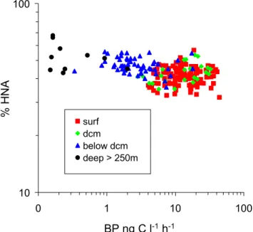
These very contrasting results suggest that the bulk activity is not only controlled by HNA cells since some active cells may be spread differentially across distinct groups of bacte-rioplankton or that other environmental variables are better predictors of the relative importance of HNA cells.
In the stratified Mediterranean Sea, the dynamics of het-erotrophic bacterial communities in surface layers is linked to the availability of inorganic resources, mostly P alone, sometimes N + P and in few occasions, N alone (Sala et al., 2002; Van Wambeke et al., 2002). Consequently, we also examined the relationships between inorganic nutrient con-centrations and the %HNA. The %HNA was slightly more strongly correlated with PO34− or NO−3 + NO−2 concentra-tions (r2=0.21 and 0.23, respectively) than with TChl-a
(r2=0.07). As the vertical variability of nutrient
concentra-b 30 40 50 60 70 Ea
Rh We St
%
HNA
a,b a,b a30 40 50 60 70 bel ow dcm dcm deep surface b c d A B a
Fig. 7. Box plots showing distributions of %HNA in the different
regions for the surface layers(A)and distribution of %HNA in the
different layers (all stations included,(B)). Within a common layer,
groups connected by the same letter (in italics and in red) are not significantly different at the 0.01 probability level.
tion is important, the longitudinal effect could only be stud-ied by comparing different layers. However, although mean nutrient concentrations showed a few statistical differences between some regions (mainly between “Ea” and “We”), we could not find any distinct patterns of %HNA longitudinally among the different regions compared. This idea is rein-forced by the fact that the %HNA was not related to PO34− or NO−3 + NO−2 concentration when considering layers in-dependently. This suggests that HNA and LNA distributions are much more affected by the vertical stratification than by the longitudinal distribution. During the BOUM cruise, the effects of inorganic nutrient additions was examined in mi-crocosm experiments in the three gyre stations using surface layer waters (Tanaka et al., 2011). In the surface samples (8 m depth), BP was stimulated by N (station B) or N + P (station C, Tanaka et al., 2011). Nishimura et al. (2005) hy-pothesized that P limitation exerts more severe constraints on the growth of bacterial groups with higher nucleic acid con-tent. However at station B, N addition resulted in a 2 fold increase of cell specific activity of all cytometric groups, in-cluding LNA, HNA with low SSC and HNA with high SSC (Talarmin et al., 2011). Thus, it appeared that all cytometric groups reacted on the same way to nutrient additions, sug-gesting that both HNA and LNA cells were nutrient limited. This could explain why the %HNA in “surface” layers did not change in the more oligotrophic conditions in the East (Fig. 7a).
% HNA
BP ng C l-1h-1 10
100
0 1 10 100
surf dcm below dcm deep > 250m
Fig. 8.Relationships between bacterial production and the
percent-age of HNA cells.
intracellular source of P) in warm, resource-limited environ-ment. But the temperature role could be discounted as the temperature range in the “surface” layers during our study was great (around 11◦C: 26◦C down to 15◦C) and across this range the %HNA was not related to the temperature.
The cytometric properties of HNA and LNA cells were affected differently below the “surface” layers. Size scat-ter of HNA cells decreases, and green fluorescence of HNA cells increases (whereas that of LNA decreased). Interest-ingly, Talarmin et al. (2011) showed, through3H leucine la-belling coupled to cell sorting, that the contribution of LNA cells to bulk leucine incorporation rates was higher in surface waters whereas that of HNA cells was higher at the vicinity of the dcm at the three sites A, B and C. This is in accor-dance with the positive and significant relationship between BP and %HNA at the dcm (Table 2). Possible explanations for these trends are a switch from nutrient to carbon limita-tion which is generally observed within the dcm in stratified conditions (Sala et al., 2002; Van Wambeke et al., 2002). This is associated with large changes in taxonomic composi-tion of heterotrophic prokaryotes (Ghiglione et al., 2008; Van Wambeke et al., 2009). Apparently the HNA group benefits more of such changes than the LNA. This suggests, possibly, HNA group as a better driver of production when C is the resource limiting BP.
4.2 Deep layers
The relationships between the cytometric parameters of HNA and LNA cells were examined throughout the water col-umn. One striking feature was the increase in the percent-age of HNA with depth in the meso and bathypelagic zones
(r2=0.59,n=55,p <0.001). Since both abundances of HNA and LNA cells decreased with depth, this increase in %HNA was due to the 2 times larger decrease in LNA cell abundances with depth than that of HNA cells. This in-crease of %HNA is accordance with other reports. In the Atlantic, the %HNA has been shown to increase from 60 to 70 % at some stations between 600–750 m and 1000 m depth (Gasol et al., 2009). In the deep North Atlantic (down to 3800 m), Reinthaler et al. (2006) also reported an increase in the %HNA with depth but the data were not shown. Varia-tions in HNA and LNA groups with depth were shown to oc-cur following de-stratification of the water column (Winter et al., 2009). Another interesting and original feature from our study was the systematic increase in SSC and green flu-orescence values of HNA cells with depth in the meso and bathypelagic zones (Fig. 5). SSC of LNA cells also increased with depth but 6 times less than that of HNA cells. SSC val-ues of both HNA and LNA cells were not correlated to BP in the deep layers, nor to nutrient (PO34−, NO−3 + NO−2) con-centrations. Since SSC variations are sometimes related to biovolume (Bouvier et al., 2001; Felip et al., 2007) the SSC increase could be interpreted as an increase in cell biovol-ume but this should be considered with caution. La Ferla et al. (2004) investigated the biovolume and lipopolysaccharide content of bacterioplankton down to 4000 m in the Ionian Sea and the mean cell volume varied in a similar range in both the euphotic and aphotic zone. It is important to consider that SSC isa complex variable not only and directly related to cell biovolume but also depends on the cell structure and the chemical composition of the outer membranes. The rea-sons underlying the increase in the %HNA and SSC of both HNA and LNA in deep layers are not clearly known. They may be related (i) to membrane properties changes due to hydrostatic pressure, (ii) to a switch from Eubacteria to Ar-chaea dominance and, (iii) to differential aspects of preda-tion and/or viral lysis on HNA and LNA cells (Wilson et al., 2001; Corzo et al., 2005; Tamburini et al., 2009; Winter et al., 2009).
5 Conclusions
across the large transect sampled. Further investigations will be needed to understand these trends among these cytomet-ric groups. There is more and more evidence suggesting that different groups are not only related to different specific activities but probably also phyologenetically, perhaps with some specific groups detected as HNA or LNA cells indepen-dent of their physiological state. The combination of tools such as cell sorting with activity measurements, subsequent cloning/sequencing (Guillebault et al., 2010), and investiga-tion of individual cell C/N/P quotas (Twining et al., 2008) will be essential to better understand the ecological meaning of the ubiquitous presence of LNA and HNA cells in aquatic environments.
Acknowledgements. This research was founded by the French INSU-CNRS and by the SESAME project (Southern European Seas: Assessing and Modelling Ecosystem Changes), EC Contract No GOCE-036949, funded by the European Commission’s Sixth Framework Programme. The authors would like to thank Thierry Moutin for leadership of the project and chief scientist of the BOUM cruise, Stella Psarra and Jos´ephine Ras for total chlorophyll sampling and analysis, Claude Courties and Christian Tamburini for discussions, John Dolan for improvement of the English and anonymous reviewers for their helpful comments in the first version of this ms.
Edited by: C. Jeanthon
The publication of this article is financed by CNRS-INSU.
References
Billen, G., Servais, P., and Becquevort, S.: Dynamics of bacteri-oplankton in oligotrophic and eutrophic aquatic environments:
bottom-up or top-down control? Hydrobiologia, 27, 37–42,
1990.
Bouvier, T., Troussellier, M., Anzil, A., Courties, C., and Servais, P.: Using light scatter signal to estimate biovolume by flow cy-tometry, Cycy-tometry, 44, 188–194, 2001.
Bouvier, T., Del Giorgio, P., and Gasol, J.: A comparative study of the cytometric characteristics of high and low nucleic acid bac-teriaoplankton cells from different aquatic ecosystems, Environ. Microbiol., 9, 2050–2066, 2007.
Corzo, A., Rodr´ıguez-G´alvez, S., Lubian, L., Sobrino, C., Sangr´a, P., and Mart´ınez, A.: Antarctic marine bacterioplankton subpop-ulations discriminated by their apparent content of nucleic acids differ in their response to ecological factors, Polar Biol., 29, 27– 39, 2005.
Crombet, Y., Leblanc, K., Qu´eguiner, B., Moutin, T., Rimmelin, P., Ras, J., Claustre, H., Leblond, N., Oriol, L., and Pujo-Pay,
M.: Deep silicon maxima in the stratified oligotrophic Mediter-ranean Sea, Biogeosciences, 8, 459–475, doi:10.5194/bg-8-459-2011, 2011.
Ducklow, H. W.: Factors regulating bottom-up control of bacterial biomass in open ocean plankton communities, Arch. Hydrobiol. Beih. Ergebn. Limnol., 37, 207–217, 1992.
Felip, M., Andreatta, S., Sommaruga, R., Straskrabova, V., and Catalan, J.: Suitability of flow cytometry for estimating bacte-rial biovolume in natural plankton samples: comparison with mi-croscopy data, Appl. Environ. Microbiol., 73, 4508–4514, 2007. Gasol, J. M., Alonso-Saez, L., Vaqu´e, D., Baltar, F., Calleja, M., Duarte, C. M., and Aristegui, J.: Mesopelagic prokaryotic bulk and single cell heterotrophic activity and community composi-tion in the NW Africa – Canary Islands coastal-transicomposi-tion zone, Prog. Oceanogr., 83, 189–196, 2009.
Ghiglione, J. F., Palacios, C., Marty, J. C., M´evel, G., Labrune, C., Conan, P., Pujo-Pay, M., Garcia, N., and Goutx, M.: Role of environmental factors for the vertical distribution (0-1000 m) of marine bacterial communities in the NW Mediterranean Sea, Biogeosciences, 5, 1751–1764, doi:10.5194/bg-5-1751-2008, 2008.
Guillebault, D., Laghdass, M., Catala, P., Obernosterer, I., and Lebaron, P.: Improved Method for Bacterial Cell Capture af-ter Flow Cytometry Cell Sorting, Appl. Environ. Microbiol, 76, 7352–7355, 2010.
Hall, E. K., Neuhauser, C., and Cotner, J.: toward a mechanistic understanding of how natural bacterial communities respond to changes in teperature in aquatic ecosystems, The ISME Journal, 2, 471–481, 2008.
Joux, F., Servais, P., Naudin, J.-J., Lebaron, P., Oriol, L., and Courties, C.: Distribution of picophytoplankton and bacterio-plankton along a river plume gradient in the Mediterranean Sea, Vie et Milieu, 55, 197–208, 2005.
Kirchman, D. L.: Leucine incorporation as a measure of biomass production by heterotrophic bacteria, in: Handbook of methods in aquatic microbial ecology, edited by: Kemp, P. F., Sherr, B. F., Sherr, E. B., and Cole, J. J., Lewis, 509–512, 1993.
La Ferla, R., Giudice, A., and Maimone, G.: Morphology and LPS content for the estimation of marine bacterioplankton biomass in the Ionian Sea, Sci. Mar., 68, 23–31, 2004.
Lebaron, P., Servais, P., Agogu´e, H., Courties, C., and Joux, F.: Does the High Nucleic Acid Content of Individual Bacterial Cells Allow Us To Discriminate between Active Cells and In-active Cells in Aquatic Systems?, Appl. Environ. Microbiol., 67, 1775–1782, 2001.
Li, W. K. W., Jellet, J. F., and Dickie, P. M.: DNA distributions in planktonic bacteria stained with TOTO or TO-PRO, Limnol. Oceanogr., 40, 1485–1495, 1995.
Longnecker, K., Sherr, B. F., and Sherr, E. B.: Activity and phylo-genetic diversity of bacterial cells with high and low nucleic acid content and electron transport system activity in an upwelling ecosystem, Appl. Environ. Microbiol., 71, 7737–7749, 2005. Longnecker, K., Sherr, B. F., and Sherr, E. B.: Variation in cell
specific rates of leucine and thymidine incorporation by marine bacteria with high and low nucleic acid content off the Oregon coast, Aquat. Microb. Ecol., 43, 113–125, 2006.
(0-1000 m), Biogeosciences, 5, 1573–1586, doi:10.5194/bg-5-1573-2008, 2008.
Moran, X. A. and Calvo-Diaz, A.: Single cell vs bulk activity prop-erties of coastal bacterioplankton over an annual cycle in a tem-perate ecosystem, FEMS Microb. Ecol., 67, 43–56, 2009. Moran, X. A., Bode, A., Suarez, L. A., and Nogueira, E.: Assessing
the relevance of nucleic acid content as an indicator of marine bacterial activity, Aquat. Microb. Ecol, 46, 141–152, 2007. Moran, X. A., Calvo-Diaz, A., and Ducklow, H. W.: Total and
phytoplankton mediated bottom-up control of bacterioplankton change with temperature in the NE Atlantic shelf waters, Aquat. Microb. Ecol., 58, 229–239, 2010.
Moutin, T., Van Wambeke, F., and Prieur, L.: Introduction to the Biogeochemistry from the Oligotrophic to the Ultraoligotrophic Mediterranean experiment: the BOUM program, Biogeosciences Discuss., in prep., 2011.
Nishimura, Y., Kim, C., and Nagata, T.: Vertical and seasonal vari-ations of bacterioplankton subgroups with different nucleic acid contents: possible regulation by phosphorus, Appl. Environ. Mi-crobiol., 71, 5828–5836, 2005.
Pujo-Pay, M., Conan, P., Oriol, L., Cornet-Barthaux, V., Falco, C., Ghiglione, J.-F., Goyet, C., Moutin, T., and Prieur, L.: In-tegrated survey of elemental stoichiometry (C, N, P) from the western to eastern Mediterranean Sea, Biogeosciences, 8, 883– 899, doi:10.5194/bg-8-883-2011, 2011.
Ras, J., Claustre, H., and Uitz, J.: Spatial variability of phytoplank-ton pigment distributions in the Subtropical South Pacific Ocean: comparison between in situ and predicted data, Biogeosciences, 5, 353–369, doi:10.5194/bg-5-353-2008, 2008.
Reinthaler, T., Van Aken, H., Veth, C., Aristegui, C., Robinson, C., Williams, P. J. le B., Lebaron, P., and Herndl, G.: Prokary-otic respiration and production in the meso and bathypelagic realm of the Eastern and Western North Atlantic Basin, Limnol. Oceanogr., 51, 1262–1273, 2006.
Sala, M. M., Peters, F., Gasol, J. M., Pedros-Alio, C., Marrasse, C., and Vaque, D.: Seasonal and spatial variations in the nu-trient limitation of bacterioplankton growth in the Northwestern Mediterranean, Aquat. Microb. Ecol., 27, 47–56, 2002. Scharek, R. and Latasa, M.: Growth, grazing and carbon flux of
high and low nucleic acid bacteria differ in surface and deep chlorophyll maximum layers in the NW Mediterranean Sea, Aquat. Microb. Ecol., 46, 153–161, 2007.
Schlitzer, R.: Ocean Data View 4, http://odv.awi.de, 2009. Servais, P., Courties, C., Lebaron, P., and Trousselier, M.: Coupling
bacterial activity measurements with cell sorting by flow cytom-etry, Microb. Ecol., 38, 180–189, 1999.
Sherr, B., Sherr, E., and Longnecker, K.: distribution of bacterial abundance and cell-specific nucleic acid content in the Northeast Pacific Ocean, Deep-Sea Res. Pt. I, 53, 713–725, 2006. Smith, D. C. and Azam, F.: A simple, economical method for
mea-suring bacterial protein synthesis rates in sea water using 3H-Leucine, Mar. Microb. Food Webs, 6, 107–114, 1992.
Sokal, R. R. and Rohlf, F. J.: Biometry, 2 Edn., edited by: Freeman W. H. and Company, New York, 1981.
Talarmin, A., Van Wambeke, F., Catala, P., Courties, C., and Lebaron, P.: Flow cytometric assessment of specific leucine in-corporation in the open Mediterranean, Biogeosciences, 8, 253– 265, doi:10.5194/bg-8-253-2011, 2011.
Tanaka, T., Thingstad, T. F., Christaki, U., Colombet, J., Cornet-Barthaux, V., Courties, C., Grattepanche, J.-D., Lagaria, A., Ne-doma, J., Oriol, L., Psarra, S., Pujo-Pay, M., and Van Wambeke, F.: Lack of P-limitation of phytoplankton and heterotrophic prokaryotes in surface waters of three anticyclonic eddies in the stratified Mediterranean Sea, Biogeosciences, 8, 525–538, doi:10.5194/bg-8-525-2011, 2011.
Tamburini, C., Garel, M., Al Ali, B., M´erigot, B., Kriwy, P., Charri`ere, B., and Budillon, G.: Distribution and activity of Bac-teria and Archaea in the different water masses of the Tyrrhenian Sea, Deep Sea Res. Pt. II, 56, 700–712, 2009.
Twining, B. S., Baines, S., Vogt, S., and de Jonge, M. D.: Explor-ing ocean biogeochemistry by sExplor-ingle cell microprobe analysis of protist elemental composition, J. Eukaryot. Microbiol., 55, 151– 162, 2008.
Van Wambeke, F., Christaki, U., Giannakourou, A., Moutin, T., and Souvemerzoglou, K.: Longitudinal and vertical trends of bacte-rial limitation by phosphorus and carbon in the Mediterranean Sea, Microb. Ecol., 43, 119–133, 2002.
Van Wambeke, F., Ghiglione, J.-F., Nedoma, J., M´evel, G.,
and Raimbault, P.: Bottom up effects on bacterioplankton
growth and composition during summer-autumn transition in the open NW Mediterranean Sea, Biogeosciences, 6, 705–720, doi:10.5194/bg-6-705-2009, 2009.
Wilson, W. W., Wade, M. M., Holman, S. C., and Champlin, F. R.: Status of methods for assessing bacterial cell surface charge properties based on zeta potential measurements, J. Microbiol. Meth., 43, 153–164, 2001.