CRISPR Content Correlates with the
Pathogenic Potential of
Escherichia coli
Enriqueta García-Gutiérrez, Cristóbal Almendros, Francisco J. M. Mojica, Noemí M. Guzmán, Jesús García-Martínez*
Departamento de Fisiología, Genética y Microbiología. Universidad de Alicante, Campus de San Vicente, 03690 Alicante, Spain
Abstract
Guide RNA molecules (crRNA) produced from clustered regularly interspaced short palin-dromic repeat (CRISPR) arrays, altogether with effector proteins (Cas) encoded by cognate cas (CRISPR associated) genes, mount an interference mechanism (CRISPR-Cas) that limits acquisition of foreign DNA inBacteriaandArchaea. The specificity of this action is pro-vided by the repeat intervening spacer carried in the crRNA, which upon hybridization with complementary sequences enables their degradation by a Cas endonuclease. Moreover, CRISPR arrays are dynamic landscapes that may gain new spacers from infecting ele-ments or lose them for example during genome replication. Thus, the spacer content of a strain determines the diversity of sequences that can be targeted by the corresponding CRISPR-Cas system reflecting its functionality. MostEscherichia colistrains possess either type I-E or I-F CRISPR-Cas systems. To evaluate their impact on the pathogenicity of the species, we inferred the pathotype and pathogenic potential of 126 strains of this and other closely related species and analyzed their repeat content. Our results revealed a negative correlation between the number of I-E CRISPR units in this system and the presence of pathogenicity traits: the median number of repeats was 2.5-fold higher for commensal iso-lates (with 29.5 units, range 0–53) than for pathogenic ones (12.0, range 0–42). Moreover, the higher the number of virulence factors within a strain, the lower the repeat content. Addi-tionally, pathogenic strains of distinct ecological niches (i.e., intestinal or extraintestinal) dif-fer in repeat counts. Altogether, these findings support an evolutionary connection between CRISPR and pathogenicity inE.coli.
Introduction
CRISPR-Cas systems are composed of at least one array of clustered regularly interspaced short palindromic repeats (CRISPR) and a set ofcas(CRISPR-associated) genes [1,2]. Several CRISPR-Cas types (denoted I, II and III) and subtypes (identified with an additional letter) are distinguished according to the identity of the associatedcasgenes [3]. Although diverse tenta-tive functions were initially postulated for particular systems [4–7], it has been demonstrated
OPEN ACCESS
Citation:García-Gutiérrez E, Almendros C, Mojica FJM, Guzmán NM, García-Martínez J (2015) CRISPR Content Correlates with the Pathogenic Potential ofEscherichia coli. PLoS ONE 10(7): e0131935. doi:10.1371/journal.pone.0131935
Editor:Tony Wang, SRI International, UNITED STATES
Received:January 21, 2015
Accepted:June 8, 2015
Published:July 2, 2015
Copyright:© 2015 García-Gutiérrez et al. This is an open access article distributed under the terms of the
Creative Commons Attribution License, which permits unrestricted use, distribution, and reproduction in any medium, provided the original author and source are credited.
Data Availability Statement:All relevant data are within the paper and its Supporting Information files.
that they constitute an RNA-based interference mechanism that prokaryotes may utilize to avert infection by foreign genetic elements [8,9]. In brief, during encounters with invading DNA, short external sequences known as protospacers are integrated into a genomic CRISPR array through the acquisition process, becoming new repeat-intervening spacers [8,10–12]. This incorporation generally takes place at the end next to the leader [2,8,13–15], defined as an AT-rich sequence that usually, with the known exception of one type II system variant [16], governs transcription of the adjacent repeat-spacer array [14,17]. Afterwards, newly incorpo-rated genetic elements with target regions matching spacer sequences will be degraded in the interference stage after annealing of the target with the complementary sequence in processed mono-spacer CRISPR RNA (crRNA) molecules [18,19]. These three main steps of CRISPR-Cas mechanism (spacer acquisition, crRNA processing and interference) require CRISPR-Cas proteins coded by thecasgenes that are part of the system [19].
As a result of the diverse encounters that a cell lineage has experienced, the spacer content (number of spacers and their particular sequence) of a given CRISPR locus may vary greatly among closely related isolates. Moreover, the number of repeat-spacer units might be influ-enced by factors such as intrinsic acquisition activity, CRISPR-Cas expression levels or func-tionality of the Cas proteins in general [20–23]. Indeed, CRISPR-carrying strains that lack associatedcasgenes and/or leader show a reduced repeat number when compared to otherwise similar complete systems [15,21,23–25]. Furthermore, the acquisition efficiency in repeat arrays of a given CRISPR system varies in line with the leader expression level and repeat sequence conservation [26]. Thus, the complexity of a CRISPR array appears to mirror its over-all activity.
CRISPR-Cas systems of I-E and I-F subtype may be found inEscherichia coli. However, someE.colimembers lack the correspondingcasgenes (casI-E and I-F respectively) and only in very rare occasions are both simultaneously found [21,24]. Based on an early classification proposed by Kunin and coworkers [27], the CRISPR units of the I-E and I-F systems are as-signed to clusters 2 and 4, respectively, of repeat types (here denoted CRISPR2 and CRISPR4). CRISPR2 are organized inE.coliin up to three arrays, accordingly named CRISPR2.1 (in CRISPR I locus, adjacent to thecasI-E genes), CRISPR2.2 and CRISPR2.3. The two latter arrays are located in the CRISPR II region, at a distance of 24 kb from CRISPR I. Occasionally a single array is found in CRISPR II, therefore called CRISPR2.2–3 [24]. Whereas CRISPR2.2 is constituted by 3 repeats and two invariable spacers, an analysis of 100 strains of the species dis-closed up to a ten-fold difference (2–3 to 29–30) of repeat counts in CRISPR2.1 and CRISPR2.3 of systems with associatedcasI-E genes [24]. Even though this diversity of CRISPR2 spacers is remarkable and the functionality of the I-E system has been demonstrated in a fewE.coli strains [17,20,28,29], its role as a relevant genetic barrier inE.coliremains uncertain [24,28, 30,31]. Referring to the I-F system, when thecasI-F genes are present, they are flanked by two CRISPR repeat arrays named CRISPR4.1 and CRISPR4.2 [24]. In contrast to I-E, these com-plete I-F systems have larger CRISPR arrays [24] and immunity to foreign elements has been detected under laboratory growth conditions without induction [20]. However, mostE.coli strains lackcasI-F genes, then containing a single array (CRISPR4.1–2), with a reduced num-ber of spacers.
A relation between CRISPR and pathogenicity has been illustrated by some remarkable observations in particularE.colipathotypes and in other species. For example, a work demon-strated that CRISPR interference prevents acquisition of capsular virulence genes in Streptococ-cus pneumoniae[32]. Also, a link of CRISPR elements with serotypes and virulence potential of Shiga toxin-producingE.colistrains has been established [33]. However, the underlying cause of this association is unknown. In the context of the immunity role, we hypothesized that reduced CRISPR activity would pose fewer constraints to the entry of foreign genetic element Competing Interests:The authors have declared
and thus would favor lateral gene transfer (LGT). LGT events constitute one of the major driv-ing forces in the evolution of prokaryotes [34–37]. Therefore, strains with limited immunity would be more prone to change their lifestyle [38], such as turning from commensal to patho-genic. Indeed, commensalE.coli(CEC) strains can become pathogens upon acquisition of vir-ulence factors [39]. Moreover, infectivity of pathogenic strains could be enhanced after gaining more of these genes. In order to test whether the association between CRISPR and pathogenic-ity is a general trend inE.coli, and to shed light on the specific nature of such connection, the number of CRISPR repeat units in strains ofE.coliand related species was compared with the presence of particular virulence genes involved in pathogenic processes [40,41]. Our results confirmed the CRISPR-pathogenicity association inE.coliand supported the defensive role of CRISPR as a driving force contributing to the emergence of pathogenic strains.
Materials and Methods
Strains and growth conditions
The microorganisms analyzed in this work comprise 126 strains (seeS1 Table) harboring homologous CRISPR-Cas systems in equivalent locations [21]. These strains were chosen to cover a comprehensive range of commensal and pathogenic types, including intestinal (EnPEC) and extraintestinal (ExPEC) representatives. They consist of 124E.coliandShigella isolates, altogether referred here to asE.coliowing to the fact that both form a coherent phylo-genetic group [21,42,43], and two strains of closely related species (Escherichia fergusonii ATCC35469 andEscherichia albertiiTW07627). The 72 members of the ECOR collection [44], are included within the above mentioned panel of 124E.coliisolates. Hereinafter, the remain-ing 54 strains will be collectively called non-ECOR. Full or almost completed genomes of these latter strains are available.
LB medium was typically used for growth of ECOR strains and incubations were carried out at 37°C for 12h with shaking. Sheep’s blood agar (bioMèrieux, Spain) was used to check hemo-lytic activity under the same temperature and time conditions.
Pathotype ascription
operon (encoding capsule and S fimbriae respectively) had been reported [40], the strain was assumed to be UPEC.
Aside from the hemolytic activity usually linked to pathogenicity islands in UPEC, some EHEC strains can also carry a plasmid-encodedhlyoperon of similar sequence [39]. Thus, ECOR strains with the exclusive combination hemolysis-vt1were considered as EHEC. ECOR strains harboring other marker gene combinations associated with both EnPEC and ExPEC were assigned to the group with a higher representation of characteristic genes.
Among non-ECOR strains, onlyShigellasp. D9 had not yet been categorized. In this case, computational searches of EnPEC and ExPEC determinants were performed to infer its affiliation.
Strains where pathogenic markers were not detected were considered as commensal.
DNA extraction and polymerase chain reactions
DNA for sequencing and polymerase chain reactions (PCR) was extracted from 5 mL LB cul-tures grown as stated above. Culcul-tures were centrifuged and pellets resuspended in 1 mL of ultrapure (milliQ) water for a total of three times. Cell suspensions were then lysed by heating at 98°C for 10 min and cell debris was removed by centrifugation. Finally, the supernatant solu-tions containing the DNA were stored at -20°C.
PCR amplifications performed to assess the pathogenic affiliation of ECOR strains were conducted with Taq polymerase (Roche) on a TC-3000 thermal cycler (Techne). Primers and conditions used are specified inS2 Table.
Retrieval, processing and analysis of sequence data
The number of CRISPR units as well as the sequences of non-ECOR strains analyzed to assess the presence of genes involved in pathogenicity (i.e.,kps,hly,pap,sfa,einv,eaeA,vt1,lt1and eagg) were obtained from previous works [21,24,41] or public databases (http://www.xbase.ac. uk/colibase/;http://www.ncbi.nlm.nih.gov/genomes/). CRISPR spacers were retrieved with CRISPRFinder [50] available athttp://crispr.u-psud.fr/Server/, and similar sequences (over 75% identity) in non-CRISPR loci were searched with the CRISPRTarget tool [51] athttp:// bioanalysis.otago.ac.nz/CRISPRTarget/crispr_analysis.html.
For the phylogenetic analysis based on multilocus sequence typing (MLST), partial sequences from ECOR strains were downloaded from the Environmental Research Institute, University of Cork (http://MLST.ucc.ie;dinB,icdA,pabB,polB,putP,trpA,trpBanduidA genes) and from the Institut Pasteur (http://www.pasteur.fr/MLST;adk,fumC,gyrB,icdA, mdh,purA, andrecAgenes) web sites. In the case of non-ECOR strains, the same sets of sequences were retrieved from the abovementioned NCBI and XBASE sites. The concatenated sequence fragments from each strain were then aligned with CLUSTALW (http://align. genome.jp/) and a phylogenetic tree was constructed with the program MEGA version 6.06 (http://www.megasoftware.net/), using the UPGMA method with distances calculated by the Jukes-Cantor model on a pairwise-deletion comparison.
Statistical analyses
among those groups compared. For robustness, these analyses were performed for groups with at least 3 strains.
To determine if significant correlations could be found, Pearson and Spearman coefficients (r) were calculated for the comparisons of different groups of strains with their respective CRISPR counts. In all cases,p-values lower than 0.05 were accepted for significance.
Results
Distribution of pathogenicity traits across
E
.
coli
and closely related
species
As a first step for the comparison between CRISPR content and pathogenicity, strains under study were classified as either commensal or within a particular pathotype (seeS1 Table). In the case of strains with a previously defined pathogenic profile, the ascription reported was adopted. Otherwise, the pathotype ofShigellasp. D9 and those ECOR strains not previously characterized was inferred following the criteria described in Materials and Methods. The robustness of these criteria was demonstrated by the high degree of coincidence between the pathotype described for categorized strains and the one predicted after the detection of the selected pathogenicity markers in the genomes of such strains (S1 Table). Seeming exceptions in EnPEC genomes were theE.colistrains P12b and 101.1, previously assigned to EPEC and EAEC respectively, where we did not find the corresponding markers (eaeAandeagg). Never-theless, these results were in agreement with reports for other strains [49,52–55], indicating thateaeAandeaggmight not be considered as signatures invariably linked to the respective pathogenic group. In the case of the UPEC/ExPEC strains, our marker-based ascriptions were also highly coincident with pathogenicity documented. The most striking difference involved strain EC23, which showed hemolytic activity (encoded by thehlyoperon) in our tests and papGwas amplified, even though these UPEC genes had not been detected in a previous South-ern analysis [40]. This inconsistency might be due to low sequence conservation in this strain of the probes used in the Southern blot analyses. Another somehow unexpected result was the finding of some UPEC traits in several strains that had been deemed to be CEC or EnPEC (S1 Table), which could be attributed to the great genome plasticity found inE.coliand the fact that genes, while present, may not necessarily be expressed [56,57]. This prompted us to ascribe pathogenicity solely based on the nature and number of the ExPEC or EnPEC virulence traits.
Comparison of repeat content with pathogenicity
non-ECOR EnPEC members (comparison D inTable 1, N = 126). This equivalence between both sets of strains confirmed the overall validity of our PCR analyses. However, it should be noted that range values (minimum and maximum no. of CRISPR units) within each group consid-ered inTable 1were larger than those found in similar studies [33,58]. This hints to a higher strain diversity within the groups considered in this work (seediscussion).
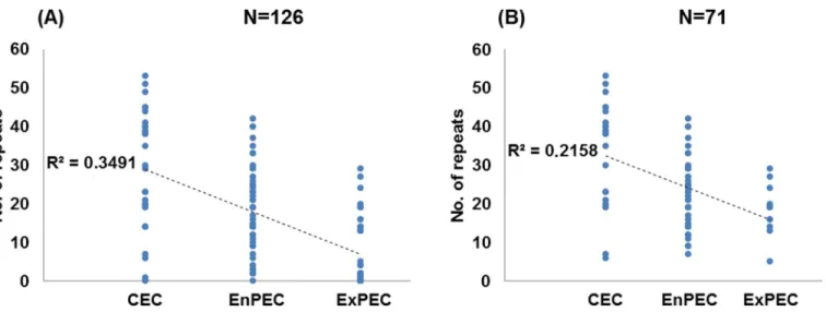
The inclusion in this study of strains lackingcasI-E genes (hence with a similarly reduced repeat number) might generate distorted results due to a possible clonal effect. However, when comparisons were performed for the subset of 71 strains carrying a complete set ofcasI-E, the results were highly coincident with those obtained for all strains (Table 1). The only exception corresponded to the lack of discrimination (p= 0.172) between EnPEC and ExPEC (Fig 1,S1 Tableand comparison B inTable 1, N = 71). However, strong negative correlation values were still found between repeat numbers and pathotype (Pearson’sr= -0.465, withp= 0.01, seeFig 2B). These results with the purged set of 71 strains suggest thatcasI-E functionality, rather than a phylogenetic (i.e. clonal) constraint, would be the main cause of the relationship found between CRISPR and pathogenicity. To provide further support to this conclusion, the distri-bution within phylogroups A and B1 of pathogenic and commensal strains with a complete set ofcasI-E genes was analyzed [21]. These two phylogenetically related MLST groups were selected for the analysis since they include the majority ofcasI-E harboring strains (N = 52). The results obtained showed that CEC and pathogenic strains were present across all the major phylogenetic subgroups within A and B1 (S1 Fig). In spite of this scattered distribution, a nega-tive correlation (seeS2 Fig) could still be observed when comparing CEC, EnPEC and ExPEC with their CRISPR repeat counts, with a Pearson coefficient ofr= -0.476 for a significance of p= 0.01. This observation in strains sharing the same phylogenetic constraints further hints that CRISPR-Cas systems may influence, at least partially, on pathogenicity.
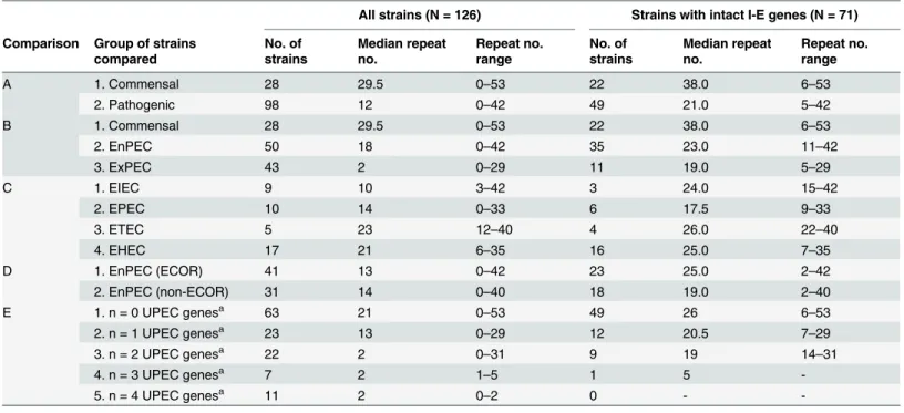
Table 1. Groups of strains studied for which statistical comparisons of repeat content and pathogenicity were performed.
All strains (N = 126) Strains with intact I-E genes (N = 71)
Comparison Group of strains compared
No. of strains
Median repeat no.
Repeat no. range
No. of strains
Median repeat no.
Repeat no. range
A 1. Commensal 28 29.5 0–53 22 38.0 6–53
2. Pathogenic 98 12 0–42 49 21.0 5–42
B 1. Commensal 28 29.5 0–53 22 38.0 6–53
2. EnPEC 50 18 0–42 35 23.0 11–42
3. ExPEC 43 2 0–29 11 19.0 5–29
C 1. EIEC 9 10 3–42 3 24.0 15–42
2. EPEC 10 14 0–33 6 17.5 9–33
3. ETEC 5 23 12–40 4 26.0 22–40
4. EHEC 17 21 6–35 16 25.0 7–35
D 1. EnPEC (ECOR) 41 13 0–42 23 25.0 2–42
2. EnPEC (non-ECOR) 31 14 0–40 18 19.0 2–40
E 1. n = 0 UPEC genesa 63 21 0–53 49 26 6–53
2. n = 1 UPEC genesa 23 13 0–29 12 20.5 7–29
3. n = 2 UPEC genesa 22 2 0
–31 9 19 14–31
4. n = 3 UPEC genesa 7 2 1
–5 1 5
-5. n = 4 UPEC genesa 11 2 0–2 0 -
-aStrains with the same total number of UPEC factors considered in the study.
Fig 1. Comparison of CRISPR counts and pathogenic categories.Median numbers of CRISPR2 units in
commensal (CEC), enteric (EnPEC) or extraintestinal (ExPEC) pathogens of theE.coliand related strains analyzed in this study, are indicated by a horizontal line. Light grey boxes represent the interquartile range values for the whole set of 126 strains (with 28, 50 and 43 isolates for each group, respectively). Dark grey boxes comprise the interquartile range values for the reduced subset of 71 strains with intactcasI-E genes
(22, 35 and 11 isolates). Vertical lines for each box denote the corresponding CRISPR2 count range. Significant differences of median values (Kruskal-Wallisp-values lower than 0.05) for the comparisons within each of these two sets of strains are indicated by an asterisk (ns, not significant).
doi:10.1371/journal.pone.0131935.g001
Fig 2. Correlation of CRISPR counts and pathogenic categories.Graphical representation of the number of CRISPR repeats in strains categorized as
commensal (CEC) or as pathogens of enteric (EnPEC) or extraintestinal (ExPEC) origins for the whole set of N = 126 strains (A) or the 71 strains with the intact set ofcasI-E genes (B). Dotted lines represent the least-square linear regressions, and their corresponding R2values are indicated.
In the case of the I-F system, the associatedcasgenes were only detected in 14 strains of those under study, the majority being pathogenic (S1 Table). This suggested a much reduced impact on pathogenicity of I-F compared to I-E.
Higher numbers of uropathogenicity genes relate to lower repeat counts
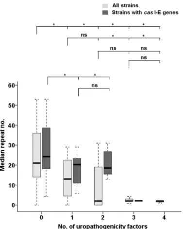
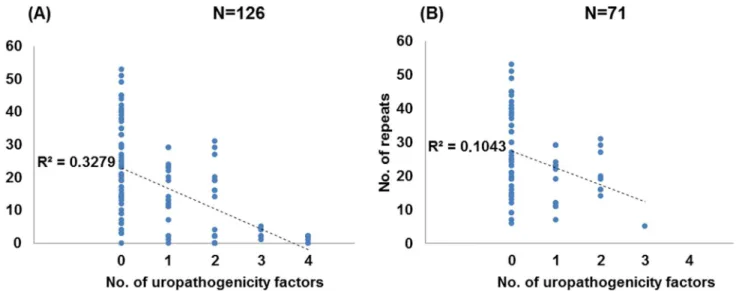
In contrast to EnPEC pathotypes where just one pathogenicity factor was considered in this study, a total of four markers were probed for UPEC. This allowed us to perform an analysis in this latter case to assess a correlation between the repeat count and the number of such patho-genic traits within each strain. This analysis showed that, regardless of their classification as pathogen or commensal, strains with the lowest number of repeats tended to bear more of such factors (Fig 3,S1 Tableand comparison E inTable 1, N = 126), showing a strong negative cor-relation (Spearman’sr= -0.622,p= 0.01, seeFig 4A). Furthermore, strains in possession of 1 uropathogenic determinant had six times more CRISPR units than those carrying 2 or more (Fig 3,S1 Tableand comparison E inTable 1, N = 126), ranging from 13 repeats (1 factor) to 2
Fig 3. Comparison of the CRISPR counts and the number of UPEC genes.Median numbers of CRISPR2 units in the strains under study, referred to the number of selected uropathogenicity genes within those strains. For each UPEC number category (x-axis), light grey boxes represent the interquartile range for the median value (horizontal line) of all strains (N = 126, with 63, 23, 22, 7 and 11 isolates for each category, respectively), while dark grey boxes indicate that value for strains with completecasI-E genes (N = 71 and 49, 12, 9, 1 and 0 isolates, respectively). Vertical lines indicate the CRISPR2 count ranges. Significant differences of median values (Kruskal-Wallisp-values lower than 0.05) for the comparisons within each set of
strains are indicated by an asterisk (ns, not significant). The categories compared are indicated in brackets, while categories with an insufficient number of isolates are not considered for comparison (seeMaterials and Methods).
(2–4 factors). This suggested a relationship between CRISPR activity and the capability to incorporate such pathogenic factors. Thus, it could be inferred that a greater virulence potential (in terms of a higher number of factors) is associated with lower repeat counts. However, while Kruskal-Wallis tests differentiated (in terms of CRISPR count) between strains with 1 or no UPEC factors from the rest, they did not discriminate between strains with 2, 3 or 4 UPEC fac-tors (p>0.05 in all cases, seeFig 3). This lack of differentiation might suggest a certain degree of specialization at least in uropathogenicity, where a critical number of virulence determinants should be required to elicit pathogenicity. This conclusion is further supported when consider-ing that, of the 16 strains with a previously defined pathotype that were in possession of just 1 UPEC factor (seeS1 Table), only in 2 was the reported pathotype UPEC/ExPEC, whereas in the rest was either CEC (4 strains) or EnPEC (10 strains). In contrast, of the 20 previously ascribed strains carrying 2 to 4 UPEC factors, 19 had been deemed as uropathogens [44,59– 63].
When strains withoutcasI-E genes were purged, an almost 4-fold difference in repeat counts between strains with 1–2 versus 3 factors (19 vs 5,S1 Tableand comparison E in Table 1, N = 71) was observed, with strong negative correlation values (Spearman’sr= -0.320, withp= 0.01, seeFig 4B). Nevertheless, the fact that just one isolate contained 3 factors did not allow us to assess significance for all the groups compared, albeitp= 0.007 was obtained to dif-ferentiate between strains carrying no UPEC determinants and those with at least one of them (Fig 3). These results for the 71 strains, coupled with those from the same subset regarding CEC, EnPEC and ExPEC groupings, strongly suggest that loss of CRISPR activity allowed ExPEC specialization, and that this loss was more often accomplished by the removal of thecas I-E genes.
Correlation between CRISPR-Cas I-E repeat numbers and
pathogenicity in other
Escherichia
species
TheE.fergusoniiATCC35469 andE.albertiiTW07 strains included in this study showed the general pattern of correlation between pathogenicity and CRISPR counts observed inE.coli (S1 Table). Thus, the commensalE.fergusoniistrain ATCC35469 [64] has a number of repeat
Fig 4. Correlation of CRISPR counts and the number of UPEC genes.Graphical representation of the number of CRISPR repeats for strains harboring 0, 1, 2, 3 or 4 UPEC factors for the whole set of N = 126 strains (A) or the 71 strains with the intact set ofcasI-E genes (B). Dotted lines represent the
least-square linear regressions, and their corresponding R2values are indicated.
units (n = 38) within the range of the median values found for CEC (n = 29.5 or n = 38, depending on the set comprising all strains or the one purged ofcas-less strains, respectively), and the CRISPR unit count in the enteropathogenicE.albertiiTW07627 [65] is on par with the median values encountered in the EnPEC isolates. Taken together, these results further support a link between the I-E CRISPR-Cas system and the pathogenicity ofE.coli-related microorganisms.
Discussion
Impact of the I-E CRISPR-Cas system on the pathogenicity of
Escherichia
A negative correlation has been established in this work between the repeat content in the I-E system and the pathogenicity ofE.coliand related strains. However, several explanations could account for this relationship. In principle, it could be interpreted as the consequence of the immunity role of CRISPR: those systems with higher numbers of spacers, as a result of a higher mean activity [26], will act as more efficient barriers against invaders, such as those carrying virulence factors that promote pathogenicity [39,46,64,66]. Although the immune function has been proven in other species, the apparently low dynamics of the CRISPR arrays ofE.coli sug-gests that they do not act as would be expected for an efficient barrier [30]. Nevertheless, the low turnover of spacers should be seen as a consequence of the stringent regulation that gov-erns expression of CRISPR-Cas I-E [17,67–70], being silenced under normal growth conditions [17,67]. Moreover, laboratory strains are able to elicit CRISPR-mediated interference against plasmids and phages [69,71] and the widespread presence inE.colistrains of spacers with iden-tities to viral and plasmid sequences [24] strongly supports the defense role of CRISPR-Cas. Indeed, a search for spacer homologs revealed that 98 out of the 114 strains studied harboring spacers have at least one that matches sequences in transmissible elements (S1 Table).
A previous work onE.colireported no meaningful association between the presence in the cell ofcasI-E genes and that of plasmids [31], arguing against a role of the I-E system as a bar-rier to the import of a genetic element. However, I-E spacers target mainly phages, with a rela-tively low proportion of plasmids [20], with a ca. five to one ratio for these elements,
respectively (seeS1 Table). These results suggest that I-E would preferentially limit viruses and, in the context of pathogenicity, CRISPR would be mainly hindering acquisition of virulence factors carried by these infectious elements. By contrast, the phage-plasmid ratio of spacer homologs in those strains carrying the less prevalent I-F is 24 to 43, albeit 15 of the plasmid homologs are found within a single CEC isolate (strain ED1a, seeS1 Table). Remarkably, ED1a andShigellasp. D9 are the only CEC strains carrying I-F whereas the rest are pathogenic. In this sense, it should be noted that, whereas some of the EnPEC markers considered in this work (namelyeinvandeagg) may be carried by plasmids, they are also present as part of chro-mosomal pathogenicity islands which, due to their size, are usually located within prophages or in association with transposons [39]. Thus, the potential association of I-F on pathogenicity, despite being more active than I-E [20] seems, due to its affinity to genetic elements and low prevalence, more negligible than I-E.
units must reflect foreign attacks (immunization), and consequent targeting activity rather than regulation of virulence factors.
These findings suggest that CRISPR activity may have hindered the emergence of pathogenic lifestyles inE.coli[73]. Alternatively, our results could be interpreted the other way around: that the pathogenic behavior promoted a reduced activity of CRISPR-Cas elements. However, the ancestral presence inEscherichiaof the CRISPR systems, altogether with the absence ofcasgenes in pathogenic groups, notably of I-E subtype in the B2 group of MLEE strains [21], disputes the latter possibility. RegardingE.coliphylogeny, the subset of strains with functional I-E systems, which mainly belong to closely related MLEE groups A and B1 [21], follows the same correlation of repeat counts and pathogenicity (as mentioned above). This fact should be considered as another indication of the role of CRISPR activity on pathogenicity, as opposed to the repeat dis-tribution being merely the result of a phylogenetic constraint.
Relationship between habitat and CRISPR-Cas activity
In the context of CRISPR acting as an immune system, differences in its activity among strains would be expected, for instance due to genetic diversity or the varied inducing factors they encounter in their respective habitat. These factors include the frequency they face invaders, the diversity of such invaders or the occurrence of mutations in the target that will prompt effi-cient acquisition [26,71,74,75]. Certainly, a link between the habitat to which the strains adapt and CRISPR activity is supported by the differences we found in the repeat content between intestinal and extraintestinal strains. However, CEC strains carry a significantly higher number of repeats than EnPEC, even though the members of both groups share habitat, being confined almost exclusively within the gut. This difference in repeat counts could be explained by a dif-ferent frequency of successful events of lateral gene transfer (LGT) in commensal and patho-genic strains. Indeed, the gut is a bacteriophage-rich environment [76,77], where strong selective pressure must exist favoring the occurrence of efficient mechanisms preventing phage infection. Nevertheless, taking into account that phages are also an important source of viru-lence factors, it is expected that EnPEC strains will have more permissive (i.e., less active) defense systems against these infective agents than CEC.
In the case of ExPEC strains, which also colonize secondary habitats where viral predators are scarcely present [78,79], less selective pressure together with the above stated advantage for a pathogen to allow LGT, would justify a further reduction in CRISPR activity.
CRISPR count diversity reveals a notable heterogeneity of pathogenic
populations of
E
.
coli
available, we inferred it by the presence of traits characteristic of a specific pathotype. Never-theless, the presence of a particular trait does not determine pathogenicity, since it might not be functional [56]. Moreover, as observed here in the case of UPEC strains, true pathogenicity might require a certain critical number of virulence traits. This biased marker-based ascription might certainly account for at least some of the apparent intra-pathogroup diversity
encountered.
Conclusions
A correlation has been established linking a reduced repeat content in the I-E system of Escher-ichia coliand related strains with a higher probability for a specific strain to exert pathogenicity (i.e. the potential ability of a microorganism to cause disease). Moreover, significant differences in the CRISPR count also correlate with the environment in which this pathogenicity is per-formed, despite all strains normally reside in the gut. However, the great variability in the num-ber of CRISPR units for strains within a pathogenic group would make its potential application for predictive studies of pathogenicity best suited as supplementary to other techniques. The increase in genomic data and a more accurate characterization of the strains (E.coliand other species) in terms of their pathogenic profile and their particular CRISPR-Cas activity will pro-vide new clues to better understand this correlation. Nevertheless, the influence of CRISPR-Cas as a barrier regulating the influx of LGT, and the subsequent impact on the diversity ofE.coli and related species, should be a factor to be considered to better understand gene exchange phenomena from an evolutionary standpoint.
Supporting Information
S1 Fig. Phylogenetic distribution of commensal and pathogenic strains.Tree showing the MLST relationships corresponding to the strains analyzed in this study belonging to phy-logroups A and B1 (see Almendroset al., 2014). Only isolates that carry a complete set ofcas I-E genes are considered. CEC, EnPEC and ExPEC strains are indicated in green, blue and red, respectively. EC58, in black, is a potentially pathogenic strain not assigned to EnPEC or ExPEC (seeS1 Table). StrainEscherichia fergusoniiATCC35469 was used as outgroup (branch length, truncated, not to scale).
(TIF)
S2 Fig. Correlation of CRISPR counts and pathogenic categories of strains in MLST groups A and B1.Graphical representation of the number of CRISPR repeats in strains categorized as commensal (CEC) or as pathogens of enteric (EnPEC) or extraintestinal (ExPEC) origin. The strains analyzed (N = 52) belong to phylogroups A and B1 and carry a complete set ofcasI-E genes. A dotted line represents the least-square linear regression. The R2value is indicated. (TIF)
S1 Table. Strain data of CRISPR counts, spacer homologs, presence ofcasI-E genes, patho-genic traits and pathopatho-genicity categories.
(XLS)
S2 Table. Primers and conditions used for amplification of pathogenicity markers. (DOC)
Acknowledgments
Author Contributions
Conceived and designed the experiments: JGM. Performed the experiments: EGG JGM. Ana-lyzed the data: JGM EGG CA NMG. Contributed reagents/materials/analysis tools: JGM CA FJMM. Wrote the paper: JGM FJMM.
References
1. Mojica FJM, Díez-Villaseñor C, Soria E, Juez G (2000) Biological significance of a family of regularly
spaced repeats in the genomes of Archaea, Bacteria and mitochondria. Mol Microbiol 36: 244–246. PMID:10760181
2. Jansen R, Embden JD, Gaastra W, Schouls LM (2002) Identification of genes that are associated with DNA repeats in prokaryotes. Mol Microbiol 43: 1565–1575. PMID:11952905
3. Makarova KS, Haft DH, Barrangou R, Brouns SJ, Charpentier E, et al. (2011) Evolution and classifica-tion of the CRISPR–Cas systems. Nat Rev Microbiol 9: 467–477. doi:10.1038/nrmicro2577PMID:
21552286
4. Babu M, Beloglazova N, Flick R, Graham C, Skarina T, et al. (2011) A dual function of the CRISPR– Cas system in bacterial antivirus immunity and DNA repair. Mol Microbiol 79: 484–502. doi:10.1111/j. 1365-2958.2010.07465.xPMID:21219465
5. Mojica FJM, Ferrer C, Juez G, Rodríguez-Valera F (1995) Long stretches of short tandem repeats are present in the largest replicons of the ArchaeaHaloferax mediterraneiandHaloferax volcaniiand could be involved in replicon partitioning. Mol Microbiol 17: 85–93. PMID:7476211
6. Viswanathan P, Murphy K, Julien B, Garza AG, Kroos L (2007) Regulation ofdev, an operon that
includes genes essential forMyxococcus xanthusdevelopment and CRISPR-associated genes and repeats. J Bacteriol 189: 3738–3750. PMID:17369305
7. Zegans ME, Wagner JC, Cady KC, Murphy DM, Hammond JH, et al. (2009) Interaction between bacte-riophage DMS3 and host CRISPR region inhibits group behaviors ofPseudomonas aeruginosa. J Bac-teriol 191: 210–219. doi:10.1128/JB.00797-08PMID:18952788
8. Barrangou R, Fremaux C, Deveau H, Richards M, Boyaval P, et al. (2007) CRISPR provides acquired
resistance against viruses in prokaryotes. Science 315: 1709–1712. PMID:17379808
9. Marraffini LA, Sontheimer EJ (2008) CRISPR interference limits horizontal gene transfer in Staphylo-cocciby targeting RNA. Science 322: 1843–1845. doi:10.1126/science.1165771PMID:19095942
10. Mojica FJM, Díez-Villaseñor C, García-Martínez J, Soria E (2005) Intervening sequences of regularly
spaced prokaryotic repeats derive from foreign genetic elements. J Mol Evol 60: 174–182. PMID:
15791728
11. Pourcel C, Salvignol G, Vergnaud G (2005) CRISPR elements inYersinia pestisacquire new repeats by preferential uptake of bacteriophage DNA, and provide additional tools for evolutionary studies. Microbiology 151: 653–663. PMID:15758212
12. Goren MG, Yosef I, Auster O, Qimron U (2012) Experimental definition of a clustered regularly inter-spaced short palindromic duplicon inEscherichia coli. J Mol Biol 423: 14–16. doi:10.1016/j.jmb.2012.
06.037PMID:22771574
13. Erdmann S, Garrett RA (2012) Selective and hyperactive uptake of foreign DNA by adaptive immune systems of an archaeon via two distinct mechanisms. Mol Microbiol 85: 1044–1056. doi:10.1111/j. 1365-2958.2012.08171.xPMID:22834906
14. Lillestol RK, Shah SA, Brugger K, Redder P, Phan H, et al. (2009) CRISPR families of the crenarchaeal genusSulfolobus: bidirectional transcription and dynamic properties. Mol Microbiol 72: 259–272. doi:
10.1111/j.1365-2958.2009.06641.xPMID:19239620
15. Lopez-Sanchez MJ, Sauvage E, Da Cunha V, Clermont D, Ratsima Hariniaina E, et al. (2012) The highly dynamic CRISPR1 system ofStreptococcus agalactiaecontrols the diversity of its mobilome. Mol Microbiol 85: 1057–1071. doi:10.1111/j.1365-2958.2012.08172.xPMID:22834929
16. Zhang Y, Heidrich N, Ampattu BJ, Gunderson CW, Seifert HS, et al. (2013) Processing-independent CRISPR RNAs limit natural transformation inNeisseria meningitidis. Mol Cell 50: 488–503. doi:10.
1016/j.molcel.2013.05.001PMID:23706818
17. Pougach K, Semenova E, Bogdanova E, Datsenko KA, Djordjevic M, et al. (2010) Transcription, pro-cessing and function of CRISPR cassettes inEscherichia coli. Mol Microbiol 77: 1367–1379. doi:10. 1111/j.1365-2958.2010.07265.xPMID:20624226
19. Westra ER, van Erp PBG, Künne T, Wong SP, Staals RHJ, et al. (2012) CRISPR immunity relies on the consecutive binding and degradation of negatively supercoiled invader DNA by Cascade and Cas3. Mol Cell 46: 595–605. doi:10.1016/j.molcel.2012.03.018PMID:22521689
20. Almendros C, Guzmán NM, Díez-Villaseñor C, García-Martínez J, Mojica FJM (2012) Target motifs
affecting natural immunity by a constitutive CRISPR-Cas System inEscherichia coli. PLoS ONE 7: e50797. doi:10.1371/journal.pone.0050797PMID:23189210
21. Almendros C, Mojica FJM, Díez-Villaseñor C, Guzmán NM, García-Martínez J (2014) CRISPR-Cas
functional module exchange inEscherichia coli. mBio 5: e00767–00713. doi:10.1128/mBio.00767-13
PMID:24473126
22. Magadán AH, Dupuis M-È, Villion M, Moineau S (2012) Cleavage of phage DNA by theStreptococcus thermophilusCRISPR3-Cas system. PLoS ONE 7: e40913. doi:10.1371/journal.pone.0040913
PMID:22911717
23. Horvath P, Romero DA, Coûté-Monvoisin A-C, Richards M, Deveau H, et al. (2008) Diversity, activity, and evolution of CRISPR loci inStreptococcus thermophilus. J Bacteriol 190: 1401–1412. PMID:
18065539
24. Díez-Villaseñor C, Almendros C, García-Martínez J, Mojica FJM (2010) Diversity of CRISPR loci in
Escherichia coli. Microbiology 156: 1351–1361. doi:10.1099/mic.0.036046-0PMID:20133361
25. Horvath P, Coûté-Monvoisin A-C, Romero DA, Boyaval P, Fremaux C, et al. (2009) Comparative analy-sis of CRISPR loci in lactic acid bacteria genomes. Int J Food Microbiol 131: 62–70. doi:10.1016/j. ijfoodmicro.2008.05.030PMID:18635282
26. Richter C, Dy RL, McKenzie RE, Watson BN, Taylor C, et al. (2014) Priming in the Type I-F CRISPR-Cas system triggers strand-independent spacer acquisition, bi-directionally from the primed protospa-cer. Nucleic Acids Res 42: 8516–8526. doi:10.1093/nar/gku527PMID:24990370
27. Kunin V, Sorek R, Hugenholtz P (2007) Evolutionary conservation of sequence and secondary struc-tures in CRISPR repeats. Genome Biol 8: R61. PMID:17442114
28. Díez-Villaseñor C, Guzmán NM, Almendros C, García-Martínez J, Mojica FJM (2013) CRISPR-spacer
integration reporter plasmids reveal distinct genuine acquisition specificities among CRISPR-Cas I-E variants ofEscherichia coli. RNA Biol 10: 792–802. doi:10.4161/rna.24023PMID:23445770
29. Brouns SJ, Jore MM, Lundgren M, Westra ER, Slijkhuis RJ, et al. (2008) Small CRISPR RNAs guide antiviral defense in prokaryotes. Science 321: 960–964. doi:10.1126/science.1159689PMID:
18703739
30. Touchon M, Charpentier S, Clermont O, Rocha EP, Denamur E, et al. (2011) CRISPR distribution within theEscherichia colispecies is not suggestive of immunity-associated diversifying selection. J
Bacteriol 193: 2460–2467. doi:10.1128/JB.01307-10PMID:21421763
31. Touchon M, Charpentier S, Pognard D, Picard B, Arlet G, et al. (2012) Antibiotic resistance plasmids spread among natural isolates ofEscherichia coliin spite of CRISPR elements. Microbiology 158: 2997–3004. doi:10.1099/mic.0.060814-0PMID:23059972
32. Bikard D, Hatoum-Aslan A, Mucida D, Marraffini LA (2012) CRISPR interference can prevent natural transformation and virulence acquisition during in vivo bacterial infection. Cell Host Microbe 12: 177– 186. doi:10.1016/j.chom.2012.06.003PMID:22901538
33. Toro M, Cao G, Ju W, Allard M, Barrangou R, et al. (2014) Association of CRISPR elements with sero-types and virulence potential of Shiga toxin-producingEscherichia coli. Appl Environ Microbiol 80: 1411–1420. doi:10.1128/AEM.03018-13PMID:24334663
34. Gupta RS (2000) The phylogeny of proteobacteria: relationships to other eubacterial phyla and eukary-otes. FEMS Microbiol Rev 27: 367–402.
35. Philippe H, Budin K, Moreira D (1999) Horizontal transfers confuse the prokaryotic phylogeny based on the HSP70 protein family. Mol Microbiol 31: 1007–1009. PMID:10048042
36. Boucher Y, Douady CJ, Papke RT, Walsh DA, Boudreau ME, et al. (2003) Lateral gene transfer and the origins of prokaryotic groups. Annu Rev Genet 37: 283–328. PMID:14616063
37. Dagan T, Martin W (2007) Ancestral genome sizes specify the minimum rate of lateral gene transfer during prokaryote evolution. Proc Natl Acad Sci U S A 104: 870–875. PMID:17213324
38. Newton ILG, Bordenstein SR (2011) Correlations between bacterial ecology and mobile DNA. Curr Microbiol 62: 198–208. doi:10.1007/s00284-010-9693-3PMID:20577742
39. Kaper JB, Nataro JP, Mobley HL (2004) PathogenicEscherichia coli. Nat Rev Microbiol 2: 123–140. PMID:15040260
41. Ahmed W, Tucker J, Bettelheim KA, Neller R, Katouli M (2007) Detection of virulence genes in Escheri-chia coliof an existing metabolic fingerprint database to predict the sources of pathogenicE.coliin sur-face waters. Water Res 41: 3785–3791. PMID:17289107
42. Fricke WF, Wright MS, Lindell AH, Harkins DM, Baker-Austin C, et al. (2008) Insights into the environ-mental resistance gene pool from the genome sequence of the multidrug-resistant environenviron-mental iso-lateEscherichia coliSMS-3-5. J Bacteriol 190: 6779–6794. doi:10.1128/JB.00661-08PMID:
18708504
43. Pupo GM, Lan R, Reeves PR (2000) Multiple independent origins ofShigellaclones ofEscherichia coli
and convergent evolution of many of their characteristics. Proc Natl Acad Sci U S A 97: 10567–10572. PMID:10954745
44. Ochman H, Selander RK (1984) Standard reference strains ofEscherichia colifrom natural populations. J Bacteriol 157: 690–693. PMID:6363394
45. García-Martínez J, Martínez-Murcia AJ, Rodríguez-Valera F, Zorraquino A (1996) Molecular evidence supporting the existence of two major groups in uropathogenicEscherichia coli. FEMS Immunol Med Microbiol 14: 231–244. PMID:8856322
46. Dobrindt U (2005) (Patho-)Genomics ofEscherichia coli. Int J Med Microbiol 295: 357–371. PMID:
16238013
47. Ewers C, Li G, Wilking H, Kiessling S, Alt K, et al. (2007) Avian pathogenic, uropathogenic, and new-born meningitis-causingEscherichia coli: how closely related are they? Int J Med Microbiol 297: 163– 176. PMID:17374506
48. Russo TA, Johnson JR (2000) Proposal for a new inclusive designation for extraintestinal pathogenic isolates ofEscherichia coli: ExPEC. J Infect Dis 181: 1753–1754. PMID:10823778
49. Ochoa TJ, Mercado EH, Durand D, Rivera FP, Mosquito S, et al. (2011) Frequency and pathotypes of diarrheagenicEscherichia coliin peruvian children with and without diarrhea (in Spanish). Rev Peru Med Exp Salud Publica 28: 13–20. PMID:21537764
50. Grissa I, Vergnaud G, Pourcel C (2007) CRISPRFinder: a web tool to identify clustered regularly inter-spaced short palindromic repeats. Nucleic Acids Res 35: W52–W57. PMID:17537822
51. Biswas A, Gagnon JN, Brouns SJJ, Fineran PC, Brown CM (2013) CRISPRTarget: Bioinformatic pre-diction and analysis of crRNA targets. RNA Biol 10: 817–827. doi:10.4161/rna.24046PMID:
23492433
52. Bin Kingombe CI, Cerqueira-Campos M- L, Farber JM (2005) Molecular Strategies for the Detection, Identification, and Differentiation between EnteroinvasiveEscherichia coli andShigellaspp. J Food
Prot 68: 239–245. PMID:15726963
53. Beutin L, Gleier K, Kontny I, Echeverria P, Scheutz F (1997) Origin and characteristics of enteroinva-sive strains ofEscherichia coli (EIEC) isolated in Germany. Epidemiol Infect 118: 199–205. PMID:
9207729
54. Rivera FP, Ochoa TJ, Maves RC, Bernal M, Medina AM, et al. (2010) Genotypic and Phenotypic Char-acterization of EnterotoxigenicEscherichia coliStrains Isolated from Peruvian Children. J Clin Microbiol
48: 3198–3203. doi:10.1128/JCM.00644-10PMID:20631096
55. Schmidt H, Knop C, Franke S, Aleksic S, Heesemann J, et al. (1995) Development of PCR for Screen-ing of EnteroaggregativeEscherichia coli. J Clin Microbiol 33: 701–705. PMID:7751380
56. Jeong H, Barbe V, Lee CH, Vallenet D, Yu DS, et al. (2009) Genome sequences ofEscherichia coliB strains REL606 and BL21(DE3). J Mol Biol 394: 644–652. doi:10.1016/j.jmb.2009.09.052PMID:
19786035
57. Rasko DA, Rosovitz MJ, Myers GS, Mongodin EF, Fricke WF, et al. (2008) The pangenome structure ofEscherichia coli: comparative genomic analysis ofE.colicommensal and pathogenic isolates. J Bac-teriol 190: 6881–6893. doi:10.1128/JB.00619-08PMID:18676672
58. Dang TND, Zhang L, Zöllner S, Srinivasan U, Abbas K, et al. (2013) UropathogenicEscherichia coliare less likely than paired fecalE.colito have CRISPR loci. Infect Genet Evol 19: 212–218. doi:10.1016/j. meegid.2013.07.017PMID:23891665
59. Chen SL, Hung CS, Xu J, Reigstad CS, Magrini V, et al. (2006) Identification of genes subject to posi-tive selection in uropathogenic strains ofEscherichia coli: a comparative genomics approach. Proc Natl Acad Sci U S A 103: 5977–5982. PMID:16585510
60. Hochhut B, Wilde C, Balling G, Middendorf B, Dobrindt U, et al. (2006) Role of pathogenicity island-associated integrases in the genome plasticity of uropathogenicEscherichia colistrain 536. Mol Micro-biol 61: 584–595. PMID:16879640
62. Welch RA, Burland V, Plunkett G III, Redford P, Roesch P, et al. (2002) Extensive mosaic structure revealed by the complete genome sequence of uropathogenicEscherichia coli. Proc Natl Acad Sci U S A 99: 17020–17024. PMID:12471157
63. Zdziarski J, Brzuszkiewicz E, Wullt B, Liesegang H, Biran D, et al. (2010) Host imprints on bacterial genomes—rapid, divergent evolution in individual patients. PLoS PATHOG 6: e1001078. doi:10.1371/ journal.ppat.1001078PMID:20865122
64. Touchon M, Hoede C, Tenaillon O, Barbe V, Baeriswyl S, et al. (2009) Organised genome dynamics in theEscherichia colispecies results in highly diverse adaptive paths. PLoS GENET 5: e1000344. doi:
10.1371/journal.pgen.1000344PMID:19165319
65. Ooka T, Seto K, Kawano K, Kobayashi H, Etoh Y, et al. (2012) Clinical significance ofEscherichia alber-tii. Emerg Infect Dis 18: 488–492. doi:10.3201/eid1803.111401PMID:22377117
66. Tenaillon O, Skurnik D, Picard B, Denamur E (2010) The population genetics of commensal Escheri-chia coli. Nat Rev Microbiol 8: 207–217. doi:10.1038/nrmicro2298PMID:20157339
67. Pul U, Wurm R, Arslan Z, Geissen R, Hofmann N, et al. (2010) Identification and characterization ofE. coliCRISPR-cas promoters and their silencing by H-NS. Mol Microbiol 75: 1495–1512. doi:10.1111/j. 1365-2958.2010.07073.xPMID:20132443
68. Mojica FJM, Díez-Villaseñor C (2010) The on-off switch of CRISPR immunity against phages in Escher-ichia coli. Mol Microbiol 77: 1341–1345. PMID:20860086
69. Yang CD, Chen YH, Huang HY, Huang HD, Tseng CP (2014) CRP represses the CRISPR/Cas system inEscherichia coli: evidence that endogenous CRISPR spacers impede phage P1 replication. Mol
Microbiol 92: 1072–1091. doi:10.1111/mmi.12614PMID:24720807
70. Westra ER, Pul U, Heidrich N, Jore MM, Lundgren M, et al. (2010) H-NS-mediated repression of CRISPR-based immunity inEscherichia coliK12 can be relieved by the transcription activator LeuO.
Mol Microbiol 77: 1380–1393. doi:10.1111/j.1365-2958.2010.07315.xPMID:20659289
71. Swarts DC, Mosterd C, van Passel MWJ, Brouns SJJ (2012) CRISPR interference directs strand spe-cific spacer acquisition. PLoS ONE 7: e35888. doi:10.1371/journal.pone.0035888PMID:22558257
72. Sampson TR, Napier BA, Schroeder MR, Louwen R, Zhao J, et al. (2014) A CRISPR-Cas system enhances envelope integrity mediating antibiotic resistance and inflammasome evasion. Proc Natl Acad Sci U S A 111: 11163–11168. doi:10.1073/pnas.1323025111PMID:25024199
73. Louwen R, Staals RHJ, Endtz HP, van Baarlen P, Van der Oost J (2014) The role of CRISPR-Cas sys-tems in virulence of pathogenic bacteria. Microbiol Mol Biol Rev 78: 74–88. doi:10.1128/MMBR. 00039-13PMID:24600041
74. Datsenko KA, Pougach K, Tikhonov A, Wanner BL, Severinov K, et al. (2012) Molecular memory of prior infections activates the CRISPR/Cas adaptive bacterial immunity system. Nat Commun 3: 945. doi:10.1038/ncomms1937PMID:22781758
75. Savitskaya E, Semenova E, Dedkov V, Metlitskaya A, Severinov K (2013) High-throughput analysis of type I-E CRISPR/Cas spacer acquisition inE.coli. RNA Biol 10: 716–725. doi:10.4161/rna.24325
PMID:23619643
76. Chibani-Chennoufi S, Bruttin A, Dillmann M-L, Brüssow H (2004) Phage-host interaction: an ecological perspective. J Bacteriol 186: 3677–3686. PMID:15175280
77. Ventura M, Sozzi T, Turroni F, Matteuzzi D, van Sinderen D (2011) The impact of bacteriophages on probiotic bacteria and gut microflora diversity. Genes Nutr 6: 205–207. doi: 10.1007/s12263-010-0188-4PMID:21484155
78. Tanji Y, Mizoguchi K, Yoichi M, Morita M, Hori K, et al. (2002) Fate of coliphage in a wastewater treat-ment process. J Biosci Bioeng 94: 172–174. PMID:16233288
79. Tanji Y, Mizoguchi K, Yoichi M, Morita M, Kijima N, et al. (2003) Seasonal change and fate of coli-phages infected toEscherichia coliO157:H7 in a wastewater treatment plant. Water Res 37: 1136– 1142. PMID:12553989
80. Krause DO, Little AC, Dowd SE, Bernstein CN (2011) Complete genome sequence of adherent inva-siveEscherichia coliUM146 isolated from ileal Crohn's disease biopsy tissue. J Bacteriol 193: 583– 583. doi:10.1128/JB.01290-10PMID:21075930
81. Nash JH, Villegas A, Kropinski AM, Aguilar-Valenzuela R, Konczy P, et al. (2010) Genome sequence of adherent-invasiveEscherichia coliand comparative genomic analysis with otherE.colipathotypes. BMC Genomics 11: 667. doi:10.1186/1471-2164-11-667PMID:21108814