Ear lobe rejuvenation: technical description
and indications
Rejuvenescimento de lóbulo de orelha: descrição da técnica e indicações
This study was performed at the Universidade Federal do Triângulo Mineiro (Federal University of Triangulo Mineiro), Uberaba, MG, Brazil.
Submitted to SGP (Sistema de Gestão de Publicações/Manager Publications System) of RBCP (Revista Brasileira de Cirurgia Plástica/Brazilian Journal of Plastic Surgery).
Article received: March 30, 2013 Article accepted: May 19, 2013 Luciana RodRigues da cunha coLombo1
PauLo magno santos guimaRães1
ivan aRaújo motta1
maRco túLio RodRigues da cunha2
manoeL PeReiRa siLva neto3
ABSTRACT
The earlobe occupies a unique position among the structures of the face and is a very impor-tant region in aesthetic composition of the ear. The age alters the shape, width and length of the lobe due to sagging tissue and thus is at odds ratio with other aesthetic elements of the ear, requiring correction. Very little has been described to guide the surgeon in rejuvena ting the ear. The correction aged lobe can be made alone or in combination with surgery of facial rejuvenation, as well as by secondary lobe rhytidoplasties inadequately secured to the face. The surgery is fairly simple, rapid recovery, with interesting results. The basic aim of the work is the description of the technique and its indications. The strategic positioning of the inal scar in continuity with a depression anatomical ear that is escafa, and hidden by antitragus ensures a proper outcome, with little apparent scars. The technique described is a tactical option to reshape and rejuvenate the ear.
Keywords: Ear/surgery. Rejuvenation. Rhytidoplasty.
RESUMO
O lóbulo de orelha ocupa posição única entre as estruturas da face e é uma região impor-tantíssima na composição estética da orelha. A idade altera o formato, a largura e o com-primento do lóbulo, em decorrência da lacidez dos tecidos e, assim, ica em desarmonia de proporção com outros elementos estéticos da orelha, exigindo correção. Muito pouco tem sido descrito para orientar o cirurgião no rejuvenescimento da orelha. A correção do lóbulo envelhecido pode ser feita isoladamente ou em combinação com uma cirurgia de rejuvenes-cimento facial, bem como em ritidoplastias secundárias por lóbulo inadequadamente preso à face. A cirurgia é bastante simples, de recuperação rápida, com resultados interessantes. O objetivo básico do trabalho consiste na descrição da técnica de rejuvenescimento do lóbulo da orelha e suas indicações. O posicionamento estratégico da cicatriz inal em continuidade com uma depressão anatômica da orelha, que é a escafa, e ocultada pelo antitrágus garante resultado inal adequado, com cicatrizes pouco aparentes. A técnica descrita representa uma opção tática para reformular e rejuvenescer a orelha.
Descritores: Orelha/cirurgia. Rejuvenescimento. Ritidoplastia.
1. Resident Physician of Plastic Surgery at the Universidade Federal do Triângulo Mineiro (Federal University of Triangulo Mineiro - UFTM), Uberaba, MG, Brazil.
2. Plastic surgeon, full member of the Sociedade Brasileira de Cirurga Plástica (Brazilian Society of Plastic Surgery - SBCP), head of the Department of Plastic Surgery at UFTM, Uberaba, MG, Brazil.
3. Plastic surgeon, member of the SBCP, professor in the Department of Plastic Surgery at UFTM, Uberaba, MG, Brazil. INTRODUCTION
The human ear is a deining characteristic of the face. Their structures subtle transmit signals age and sex, which are unmistakable, however, not easily deined.
or pit, which is a long depression that lies below the propel ler. Escafa below, is called an elevation antihelix, which ends forked into two branches, one cavity lying between them, called the fossa navicularis or triangular. The lobe is a small portion of soft tissue which is in the lower lag. The earlobe is a small prominence, known in anatomy as wolves (diminutive “lobe”). Located at the upper lobe, are the tragus and antitrago, the irst located in the opening of the external auditory canal and the second, just above the lobe.
The earlobe occupies a unique position among the struc tures of the face and is important due to the secular tra dition of the use of ornaments and jewelry on this site. The practice of piercing the earlobe date from ancient times, and, depending on the culture, may have occurred even as a social obligation. The Latin American societies are routine ly used in earrings newborn female to differentiate the male. In Ivan and Kayan tribes in Africa, the lobes were adorned with large and heavy earrings, causing an increase of the hole and stretching of the lobe. The earlobe is a very important region in aesthetic composition of the ear. The age alters the shape, width and length of the lobe due to sagging tissue and thus is at odds ratio with other aesthetic elements of the ear, requiring correction. As we get older, the loose portions of the ear increase in size. The earlobes can also “delate”, and this results in an increase in wrinkles and creases. As in other parts of the face, the skin of the earlobe is susceptible to sun damage, contributing to wrinkling of the ear.
The anatomy of the external ear was detailed by many authors as Brent1-3 and others, who thoroughly described
the shape of the outer ear and appropriate ways to rebuild it. Very little has been reported, however, to guide the sur -geon in rejuvenating the ear. Tolleth4 in 1978 published a
paper that describes the proportion of the normal ear. He described the position of the ear, as well as its various forms. Your article gives general proportions as surgical guides, however, its derivation is based on the author’s artistic perception. Sullivan in 2010, in turn, described the aesthetic proportions of the human ear based on cross-sectional data, showing aesthetic and anatomical differences between men and women, as well as changes in the morphology of the ear with age. The ear transmits information both about the age and gender and other characteristics that deine the face. It should be treated as such by the plastic surgeon to the de -si red ideal facial rejuvenation, be-sides not masculinize or femini ze inappropriately. With the appropriate data, we hope to better deine the approach to rejuvenation of the ear.
Although the guidelines for the size and orientation of the ear have been studied, minimal attention has been directed to the earlobe. The height of the lobe covers approximately 25% of the length of the ear, and is deined by the distance of the groove subauricular antitragus, and can vary between 1.5 and 2.0 cm. The earlobe ptosis is a condition which alone contributes to facial aging. In 1972, Loeb5 acknowledged the
need to reduce lobe, as an addition to rhytidectomy. Most techniques involve facelift incisions and post-auricular inci-sion connected by a infralobular (perilobular). It is important to pay attention to appearance preoperative earlobe and not distort the lobe postoperatively. Distortion or malposition of the earlobe is a noticeable and unpleasant complication of a facelift badly executed. To avoid improper positioning of the ear lobe, care must be taken when the incision is designed and made, the skin is dry, and the skin and deep tissues re -paired. Regardless of the dissection plane and vector lifting, deep retention sutures should be placed to minimize wound tension and avoid distortions earlobe.
When planning a facelift is important to take into account length, width and shape of the earlobe. Older techniques relied heavily traction of the skin to achieve a high adequate tissue. But the skin has a tendency to stretch to accommodate the tension, over time the earlobe is pulled down toward the angle of the jaw. Due to stretching, the loose portion of the ear lobe, then becomes bound to the face. This is known as a deformity type “devil’s ear” (pixie earlobe), since it seems too long and pointy. Therefore, one should avoid placing tension on the earlobe through deep sutures to support the earlobe.
The correction aged lobe can be made alone or in com -bination with surgery of facial rejuvenation, as well as by secondary lobe rhytidoplasties inadequately secured to the face. Scars are strategically positioned and, thanks to that become less apparent. The rejuvenation surgery lobe is quite simple, extremely fast recovery, with very interesting results. The purpose of this work is to methodize the technique for rejuvenating the earlobe, with the aim of to facilitate the technique for contralateral symmetrization and methods reproducibility.
METHODS
Initially a preoperative assessment of patients candidates to surgery earlobe rejuvenation was performed, on an outpa-tient basis. Paoutpa-tients with complaints of atrophy, wrinkling and stretching the earlobe were enrolled. Often the complaint is based on the earring hole widened, with unsatisfactory positioning of adornment, lower than desired. However in some cases of patients with older age, there is not necessa-rily increase the hole but sagging earlobe, which under trac-tion and weight of the earring hole stretches and assumes a vertical contour simulating a widening hole or lobule biid incomplete (Figure 1).
In a third situation, there were patients in postoperative rhytidectomy, with sequels stigmatizing lobe arrested or pulled with deformity “ear of the devil”. These patients also beneit from the reduction and repositioning of the lobe to be described as technical. Provides a size and contour of the ear more pleasant and jovial.
Thus, the earlobe rejuvenation represented a tactical option with three nominations:
• surgery to correct isolated sagging and aging lobe, which may or may not be associated with enlarged earring hole;
• surgery combined to primary rhytidectomy as adjunct treatment in cervico-facial aesthetic impro-vement;
• surgery to correct a sequel lobes tensile post-face -lift, secondary approach.
After accurate surgical indication, patient’s consent and preoperative tests suitable part to the implementation of the procedure itself, in any of the situations listed above.
In isolated procedures, local anesthesia was performedon an outpatient basis, because it is a procedure of low complexity and fast. However, if associated with lifting cervicofa -cial anesthesia of choice is usually.
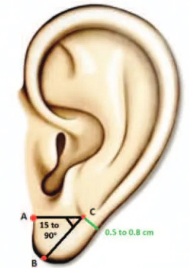
The utilezed technique to recast the ear begins with the preoperative marking. We determined the insertion point A in the face of the lobe, point B is established from a trans-verse line followed by the point A to maintain a pedicle 0.5 to 0.8 cm residual thickness of the free edge of the ear. And finally the point C is defined from an angle variable section circumference of 15º to 90º, this extension determined as the excess skin (Figures 2 and 3). The main incision is lo -cated in transverse position on the lobe, continuing to escafa below the anti-tragus, the line of deployment of the ear on the face. Delimits a triangle in varying proportions as shown excess skin in each case. In the demarcated area is resected in total plan, wedge, covering skin and subcutaneous tissue of the lobe (Figures 4 and 5). The caudal lobe projection
is excised. Frequently, the earring hole, extended or not, is encompassed in the resected area, later to be made a new hole. Do not let the amount of dry skin necessary at the ex -pense of preserving the previous hole, as it is correcting the laxity which ensures satisfactory final result. Consequently there is a reduction and repositioning of the lobe.
After hemostasis and resection is performed posterior suture the edges of bloodshed, respecting the insertion of the ear on the face. The suture is performed with simple points on the anterior and posterior lobe with nylon 6.0 suture (Figure 6).
Immediately dressing is made with microporous tape, and the stitches are removed between 7 to 10 days.
RESULTS
The strategic positioning of the inal scar in continuity with a depression anatomical ear that is escafa, and hidden by antitragus ensures a proper outcome, with little apparent
Figure 1 – Appearance ear lobe with atrophy and sagging, showing earring hole with vertical aspect.
Figure 2 – Scheme of the preoperative marking.
scars. The restoration of the shape, size and contour of the lobe of the ear translates the joviality, and consequently the face with aesthetic beneit and patient satisfaction.
The results were considered satisfactory by both the me -dical team, as the patients, subjectively.
No complications such as dehiscence points, wound in -fection, cicatricial changes like widening scar, hypertrophic, keloid or poor scar positioning.
This technique has been used in a number of cases not yet quantiied.
DISCUSSION
McKinney et al.6, in 1993, speciically addressed the
treat ment of ear in rhytidectomy and found no signiicant correlation between the height of the earlobe and aging. No difference in the size of the lobe between men and women was reported. Although the total size of the ear is greater in men, the height and width of the earlobe remain almost identical to women. This suggests that increasing the height of the ear with age is not entirely due to the effect of weight of earrings. Stretching the earlobe is most probably an ine vitable result of aging.
Many authors have discussed the importance of proper positioning of the earlobe in rhytidectomy7-12. Considera
-tions such as the morphology of the ear, tension and scar con traction should be taken into account to ensure an acceptable outcome13,14. Several authors have recommen
ded specific parameters for insertion of the earlobe during rhytidecto my. Its application in various clinical situations, however, can often be very subjective. The natural tendency Figure 4 – Intraoperative aspect of wedge resection
on overall plan of the excess of the lobe.
Figure 5 – View the resected specimen.
Figure 6 – Immediate postoperative appearance, indicating restoration of the form, contour and size of the lobe.
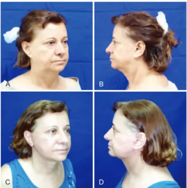
Figure 7 – In A and B, preoperative appearance of secondary rhytidectomy associated with rejuvenation of the earlobe, pulled aspect of the lobe, respectively, in right oblique position and
left proile. In C and D, 30 days postoperative appearance of secondary rhytidectomy and rejuvenation of the lobe,
showing little apparent auricular scar, respectively, in right oblique position and left proile.
A
C
B
is to migrate caudally with previous and wound contraction. With proper positioning, however, the final position of the lobe should remain in the main axis of the ear.
While recognizing that the condition of a woman’s hands and neck may reveal his true age, few people know that the appearance of our ears is another telltale sign. Like other parts of the body, because they change the natural process of aging and constant exposure to the sun. As the amount of collagen decreases and the elasticity of the skin, ears often become elongated, thin and labby with wrinkle on the surface. Women who use heavy earrings are at particular risk because the weight of ear jewelery inevitably pulling down and causes the skin to become weaker and stretched. Through a simple procedure can permanently reverse the pro blem, called “the earlobe rejuvenation.” The procedure is simple and quick when performed alone. Incision is made in a hidden area near the insertion of ear lush , and removed a small amount of tissue and skin.
In aesthetic ear reshaping the ultimate goal is to achieve a more youthful appearance. The ear lobe of a young man pos sesses on average 1.8 cm long and 2 cm wide at its midpoint. In general, the earlobe is gently curved in distal extension of the helix toward the base of the lobe. Following these ana tomical principles becomes easier to reach the end result.
It is important to note the relationship between the mor -phology of the earlobe with advancing age, and the notion that elongated ears are an undesirable result of aging. The stan dard lobe clearly changes from a short, wide frame in which youth for a long and narrow in old age. There are, however, many individuals who have large ears as one of their individual characteristics. When these ears become even greater with the aging problem arises much more visible.
CONCLUSIONS
The described technique of earlobe rejuvenation is a tactical option to reshape and rejuvenate the ear, with indi-cations for correction of sagging and aging lobe, with or without rhytidectomy, as well as for correction of the sequel of lobes under traction. It consists of a reproducible, easy and fast option, with inconspicuous scar.
REFERENCES
1. Brent B. Reconstruction of the auricle. In: McCarthy JG, eds. Plastic surgery: the face. Vol. 3. Philadelphia: W. B. Saunders; 1990. p. 2095-152. 2. Brent B. Auricular repair with autogenous rib cartilage grafts: two decades of experience with 600 cases. Plast Reconstr Surg. 1992;90(3):355-74. 3. Brent B. Technical advances in ear reconstruction with autogenous rib cartilage grafts: personal experience with 1200 cases. Plast Reconstr Surg. 1999;104(2):319-34.
4. Tolleth H. Artistic anatomy, dimensions, and proportions of the external ear. Clin Plast Surg. 1978;5(3):337-45.
5. Loeb R. Earlobe tailoring during facial rhytidoplasties. Plast Reconstr Surg. 1972;49(5):485-9.
6. McKinney P, Giese S, Placik O. Management of the ear in rhytidectomy. Plast Reconstr Surg. 1993;92(5):858-66.
7. Tolleth H. A hierarchy of values in the design and construction of the ear. Clin Plast Surg. 1990;17(2):193-207.
8. Connell BF, Martin TJ. Facial rejuvenation: facelift. In: Cohen M, ed. Mastery of plastic and reconstructive surgery. Boston: Little, Brown and Company; 1994. p. 1873-902.
9. McKinney P, Cunningham BL. Aesthetic facial surgery. New York: Churchill Livingstone; 2002. p. 222.
10. Lassus C. Another technique for the reduction of the earlobe. Aesthetic Plast Surg. 1982;6(1):43-5.
11. Tipton JB. A simple technique for reduction of the earlobe. Plast Re-constr Surg. 1980;66(4):630-2.
12. Constant E. Reduction of hypertrophic earlobe. Plast Reconstr Surg. 1979;64(2):264-7.
13. Farkas LG. Anthropometry of the normal and defective ear. Clin Plast Surg. 1990;17(2):213-21.
14. Farkas LG, Posnick JC, Hreczko TM. Anthropometric growth study of the ear. Cleft Palate Craniofac J. 1992;29(4):324-9.
Correspondence to: Luciana Rodrigues da Cunha Colombo