ABSTRACT
The three iodothyronine deiodinases catalyze the initiation (D1, D2) and termination (D3) of thyroid hormone effects in vertebrates. A recently conceived 3-dimensional model predicts that these enzymes share a similar structural organization and belong to the thioredoxin (TRX) fold superfamily. Their active center is a selenocysteine-containing pocket defined by the β1-α1-β2 motifs of the TRX fold and a domain that shares strong similarities with the active site of iduronidase, a member of the clan GH-A fold of glycoside hydrolases. While D1 and D3 are long-lived plasma membrane proteins, D2 is an endoplasmic reticulum resident pro-tein with a half-life of only 20min. D2 inactivation is mediated by selective UBC-7-mediated conjugation to ubiquitin, a process that is accelerated by T4catalysis, thus maintaining local T3homeostasis. In addition, D2 inter-acts with and is a substrate of the pVHL-interacting deubiquitinating enzymes (VDU1 and VDU2); thus deubiquitination regulates the supply of active thyroid hormone in D2-expressing cells. (Arq Bras Endocrinol Metab 2004;48/1:16-24)
Keywords:Deiodinases; Thyroid hormones; Selenocysteine; T3homeostasis
RESUMO
Trigêmeas! As Três Enzimas que Catalisam a Iniciação e Terminação dos Efeitos dos Hormônios Tiroideanos Exibem Semelhança Estrutural Ines-perada.
As três desiodases de iodotironinas catalisam a iniciação (D1, D2) e o tér-mino (D3) dos efeitos dos hormônios tiroideanos em vertebrados. Um modelo tridimensional concebido recentemente propõe que essas en-zimas apresentam organização estrutural similar e pertençam à super-família da thioredoxin (TRX)-fold. O sítio ativo é formado por um bolso contendo selenocisteína, definido pelos motivos β1-α1-β2 do TRX-fold, e um domínio similar ao sítio ativo da iduronidase, um membro do clan GH-A-fold das hidrolases de glicosídeos. Enquanto D1 e D3 são proteínas de meia-vida longa localizadas na membrana plasmática, a D2 é uma proteína residente do retículo endoplasmático com meia-vida de ape-nas 20min. A inativação da D2 é mediada por conjugação seletiva à ubiquitina, catalisada pela UBC-7, um processo que é acelerado pela catálise do T4, mantendo assim a homeostase local do T3. Além disso, a D2 interage e é substrato das VDU1 e VDU2 (pVHL-interacting deubiqui-tinating enzymes), fazendo com que sua desubiquitinação regule o suprimento de hormônio tiroideano ativo em células que expressam D2. (Arq Bras Endocrinol Metab 2004;48/1:16-24)
Descritores: Desiodases; Hormônios tireoidianos; Selenocisteína; T3 homeostase
atualização
Initiation and Termination of Thyroid
Hormone Effects
Antonio C. Bianco
Thyroid Section, Division of Endocrinology, Diabetes and Hypertension, Brigham and Women’s Hospital and Harvard Medical School, Boston,
Massachusetts 02115, USA.
T
HE THREE DEIODINASES, enzymes that activatethyroxine (T4) and inactivate both T4and T3, are
present in all vertebrates studied so far, indicating that thyroid hormone deiodination is an intrinsic compo-nent of the thyroid hormone homeostasis. In a nut-shell, their relevance resides on the fact that T4 is a
long-lived (t1/2 is ~7 days in humans) pro-hormone
molecule that must be activated by deiodination to the short-lived biologically active T3 (t1/2 is ~1 day) in
order to initiate thyroid hormone action. T3
modu-lates gene expression in virtually every vertebrate tissue through ligand-dependent transcription factors, the thyroid hormone receptors (TR). The deiodination of T4 to T3occurs in the phenolic (outer or 5’)-ring of
the T4molecule and is catalyzed by two iodothyronine
deiodinases, i.e. D1 and D2. As a counter point to the activation pathway, both T4and T3can be irreversibly
inactivated by deiodination of the tyrosyl (inner or 5)-ring, a reaction catalyzed by D3, the third member of the deiodinase group.
In both experimental animals and humans the coordinated changes in the expression and activity of these enzymes ensure thyroid hormone homeostasis and the constancy of T3 production, constituting a
major mechanism for adaptation to changes in the ingestion of iodine, starvation and changes in environ-mental temperature (reviewed in [1]). The study of animals with natural deficiency of D1 (C3H mouse) or targeted disruption of D2 (Dio2-/-) or D3 (D3-/-)
genes has not only confirmed but revealed new intri-cacies about the critical role played by these enzymes in thyroid hormone homeostasis (2-5).
Until recently, a frustrating aspect of the deiod-inase field was the lack of clinical entities associated with deiodinases dysfunction. This can be explained by the existence of multiple redundant mechanisms involved in T3 homeostasis. In order to have clinical
manifestations of deiodinase dysfunction a patient would have to have alterations in serum T3 causing
hypo- or hyperthyroidism. However, serum T3is
con-tributed not only by D1 and D2, but also by direct thyroidal secretion. One could argue that a genetic deficiency of either enzyme would be clinically silent because the other three remaining mechanisms would compensate for the missing pathway. This hypotheti-cal symptoms-free human phenotype is confirmed in C3H (2,6-8) and in the Dio2-/-mice (3,5). However,
a preliminary characterization of the Dio3-/- mouse
indicates a phenotype of central hypothyroidism, sug-gesting that the hypothalamic clearance of T3is
com-promised due to the lack of D3 activity (9). In addi-tion, the continued search for such conditions has
recently paid off and overexpression or the presence of a deiodinase gene polymorphism is indeed associated with pathological states such as consumptive hypothy-roidism in newborns (10) and adults (11), mesothe-lioma (12), obesity and insulin resistance (13), empha-sizing the key role played by the deiodinases in thyroid hormone homeostasis.
A Breakthrough: The 3D Structure of the Deiodinases is Conserved
The three deiodinase proteins (D1, D2 and D3) show considerable similarity (~50% sequence identity). All are integral membrane proteins of 29-33kDa, and have regions of high homology in the area surround-ing the active center (14-16). However, because they have such differences in substrate preference, kinetic properties, half-lives and sensitivity to PTU, the gen-eral idea was that the three dimensional (3D) structure of these proteins was substantially different, a concept that was kept alive by the inherent difficulties in deter-mining high-resolution structural data for a functional membrane-anchored enzyme.
Insights into the structures of these proteins were obtained through the fortuitous use of protein modeling through hydrophobic cluster analysis (HCA) (17). This is a sensitive method based on the fundamental princi-ples of protein fold and on a two-dimensional transposi-tion of sequences, allowing the resolutransposi-tion of a sequence into its regular secondary structures, centered on the so-defined hydrophobic clusters.
(19). Putatively, this local structural mimicry between deiodinases and iduronidase may rely partly on the overall similarity of their substrates, T4(or T3) and
sul-fated α-L-iduronic acid, respectively, both based on O-linked hexagonal rings substituted by bulky groups ortho to the linker.
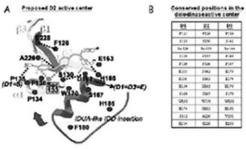
The 3D general model of the deiodinases pre-dicts that the active center is a pocket defined by the
β1-α1-β2 motifs of the TRX-fold and the IDUA-like insertion (figure 2). The most striking feature of this pocket is the presence of the rare amino acid Seleno-cysteine (Sec), critical for the deiodination reaction catalyzed by all three deiodinases. This was first iden-tified when the rat D1 cDNA became available, the analysis of which revealed the presence of the Sec encoded by UGA, which is recognized in the vast majority of mRNAs as a STOP codon (20). However, a specific RNA stem-loop immediately downstream of the UGA codon allows for the Sec incorporation in the STOP codon. This structure is termed the Sec Insertion Sequence, or SECIS element, which is pre-sent in the three deiodinases and all other selenopro-teins (21).
There is sound evidence that all three deiodi-nases can form homodimers when transiently expressed (22,23). However, it is not clear that dime-rization is necessary for function or that it even occurs when the enzymes are expressed at endogenous levels. The latter is supported by the finding of activity in higher molecular weight forms (~65kDa) than would be predicted from their respective deduced amino acid sequences (29kDa) (24,25). However, these higher
molecular weight forms could reflect associations with other cellular protein(s) not primarily involved in their catalytic function, but which could, for example, regu-late half-life, transport or subcellular localization. More studies are necessary to clarify the functional sig-nificance of deiodinase dimerization.
Topology and Subcellular Localization: Physiological Implications
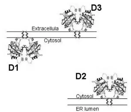
The D1 and D2 monomers are Type 1 integral mem-brane proteins oriented with a small NH2-terminal
extension in the endoplasmic reticulum (ER) lumen and a single transmembrane domain exiting the ER at about position 40 (26,27). This puts the active center of both D1 and D2 in the cytosol. D3, on the other hand, is a Type 2 integral membrane protein with the opposite orientation of D1 and D2, putting its active center inside the ER lumen (during synthesis) and finally the extracellular space (28) (figure 3).
The subcellular location of mature D1 is the plasma membrane. This has been specifically demon-strated in a number of cell types endogenously or tran-siently expressing the enzyme (22,29,30). Recently, using confocal laser microscopy of human and mouse cell lines, transiently expressed FLAG-tagged D1 was found in the plasma membrane (28). D1 does not co-localize with the ER resident protein BiP, as does D2 (27). This plasma membrane localization has been confirmed by specific biotinylation of D1 by cell-impermeable agents (28). D2, on the other hand, is an
Figure 1.3D structures and secondary structure organiza-tions of the archetype TRX enzyme and of the overall rough model of the deiodinases deduced from the HCA-based alignment. The transmembrane, hinge and intervening sequences are circled (modified from 17).
ER-resident protein. Immunofluorescent confocal microscopy of human and mouse cells transiently expressing FLAG-tagged D2 show that D2 local-izes with BiP. Endogenously expressed D2 also co-localizes with BiP in a human mesothelioma cell line (MSTO-211H cells) (12).
The subcellular localization of D3 was studied using similar techniques (28). Endogenously and tran-siently expressed FLAG-tagged D3 was identified in the plasma membrane. It co-localizes with Na, K-ATPase α, with the early endosomal marker EEA-1 and clathrin but not with two ER resident proteins. Most of the D3 molecule is extracellular and its plas-ma membrane assignment is confirmed by biotinyla-tion with a cell-impermeant probe. There is constant internalization of D3 that is blocked by sucrose or methyl-β-cyclodextrin containing medium. Exposing cells to a weak base such as primaquine increases the pool of internalized D3, suggesting that D3 is recycled between plasma membrane and early endosomes. Such recycling probably accounts for the much longer half-life of D3 (12h) than the thyroxine (T4) activating
members of the selenodeiodinase family, Type 1 (D1; 8h) or Type 2 (D2; 2h) deiodinase.
Thus, a plasma membrane location for D1 could be viewed teleologically as offering ready access of circulating reverse T3and T4to the enzyme as well
facilitating the re-entry of the T3 produced from T4
back into the plasma. The localization of D1 in the plasma membrane is in striking contrast to the ER localization of D2 (27). This differential subcellular localization of D1 and D2 may explain why there is such a minimal contribution of the T3 generated by
D1 to the intranuclear T3in contrast to large fraction
of D2-generated T3to this compartment (31-34). The
extracellular location of D3 gives ready access to cir-culating thyroid hormones, explaining its capacity for rapid inactivation of circulating T4 and T3in patients
with hemangiomas and its blockade of the access of maternal thyroid hormones to the human fetus (10,35,36).
The Ubiquitination Pathway Inactivates D2
D2 is considered the critical homeostatic T3
-generat-ing deiodinase due to its substantial physiological plas-ticity. For example, D2 responsiveness to cAMP con-stitutes the basis for its rapid adrenergic stimulation in BAT, human skeletal muscle and thyroid, linking D2 expression with the sympathetic nervous system and widening the spectrum of environmental and endoge-nous stimuli that can potentially influence adaptive T3
production (1 for review).
A number of transcriptional and post-transla-tional mechanisms have evolved to ensure limited expression and tight control of D2 levels, which is inherent to its homeostatic function. The D2 mRNA in higher vertebrates is more than 6kb in length, con-taining long 5’ and 3’ untranslated regions (UTRs). The D2 5’UTRs are greater than 600 nucleotides and contain 3–5 short open reading frames (sORFs), which reduce D2 expression by as much as 5-fold (37). Alternative splicing is another mechanism that regulates D2 level, as mRNA transcripts similar in size to the major 6- to 7-kb D2 mRNAs, but not encoding an active enzyme, are present in both human and chicken tissues. D2 levels can also be regulated by AUUUA instability motifs located in the 3’UTR of D2 mRNA as deletion of 3.7-kb from this region increases D2 activity approximately 3.8-fold due to an increase in D2 mRNA half-life (37).
D2 activity/mRNA ratios are variable, indica-ting that there is significant post-translational regula-tion of D2 expression (38). In fact, the decisive D2 property that characterizes its homeostatic behavior is a short half-life (~20min) (39) that can be further reduced by exposure to physiological concentrations of its substrate, T4, and in experimental situations,
reverse T3 or T3 (39-45). This constitutes a rapid,
potent generalized regulatory feedback loop that effi-ciently controls T3 production and intracellular T3
concentration based on how much T4is available. The
potency of the iodothyronines to induce loss of D2 activity mirrors the enzyme’s affinity for the substrate, indicating that enzyme-substrate interaction must occur in order to induce loss of D2 activity.
How is loss of D2 activity mediated? Important metabolic pathways often contain key rate-limiting enzymes whose half-lives can be modified by selective proteolysis. This process is frequently mediated by the ubiquitin (Ub)-proteasome system by which target pro-teins are marked for destruction by conjugation to Ub, a ~8kDa protein. The ubiquitinated proteins are subse-quently recognized and degraded by the proteasomes (46,47). Indeed, ubiquitination and proteasomal degra-dation are deeply implicated in the post-translational regulation of D2 activity. The first evidence was obtained in GH4C1 cells in which the half-life of the endogenous D2 was noted to be stabilized by MG132, a proteasome inhibitor (48). Substrate-induced loss of D2 activity was also inhibited by MG132 in such cells, indicating that both pathways affecting loss of D2 activ-ity were mediated by the proteasomes. This implies that the loss of D2 activity is, at least partially, due to prote-olysis, a premise that was confirmed after the levels of immunoprecipitable labeled D2 were shown to parallel D2 activity, both under basal conditions and after expo-sure to substrate (49).
Selection of specific proteins for proteolysis is usually achieved at the level of Ub conjugation, a pro-cess that involves recognition of amino acid-sequences in the target protein by the ubiquitinating enzymatic machinery. No such sequence(s) was identified for D2 yet. The first step is activation of Ub by ATP, a process catalyzed by the E1 enzyme. The next step, target recognition, is coordinated by the combined actions of a series of Ub-conjugating enzymes (E2s) and Ub-li-gases (E3s). There are approximately a dozen E2s or E2-related proteins, which share a conserved catalytic domain of approximately 150 amino acids (50). Indi-vidual E2s are involved in different cellular processes and, therefore, in the ubiquitination of different class-es of substrate proteins. E3s, on the other hand, are more abundant and with no overt sequence homolo-gy, are thought to be largely responsible for the high degree of specificity of protein ubiquitination (47).
Evidence of D2 ubiquitination and E1 involve-ment was obtained in cells expressing a temperature sensitive E1. At the restrictive high temperature, which inactivates the E1, D2 activity and protein levels are stabilized, even when protein synthesis is inhibited or D2 substrates are present (51). Ub-D2 conjugates are high molecular weight proteins (100-300kDa) easily identified in lysates of cells transiently expressing an epitope tagged D2. In such system, Ub-D2 conjugates behave as expected, i.e. they are increased in cells exposed to D2 substrates such as T4 and decreased if
E1 activity is blocked (51). Exposure to the
protea-some uptake inhibitor MG132 also increases Ub-D2 conjugates because proteasome blockade does not interfere with ubiquitination. An important observa-tion is that, under a number of condiobserva-tions, D2 activity in a cell lysate correlates with the levels of D2 protein and not Ub-D2, indicating that D2 is inactivated by ubiquitination. Interestingly, under the same condi-tions, D1 and D3 are not ubiquitinated that is in agree-ment with their relatively long (>12h) half-lives (51).
To identify additional proteins involved in D2 ubiquitination, a Cysteine mutant of D2 was expressed in the yeast S. cerevisiae, a cell model in which the Ub-proteasome system is well characterized and D2 dis-plays its typical cellular and molecular properties. Especially important, D2 was highly ubiquitinated and maintained its short half-life and sensitivity to subs-trate exposure, and its degradation was blocked by inhibitors of the proteasome uptake. Interestingly, D2 was stabilized in yeast strains that lack specific E2s, Ubc6p or Ubc7p. Both of these E2s are involved in the ER-associated degradation (ERAD) process, in agreement with the fact that D2 is an ER-resident tein. Importantly, in the yeast that lacked the E2 teins the substrate-induced loss of D2 activity and pro-teolysis were also impaired. On the other hand, no dif-ference in D2 levels was detected in a yeast strain defi-cient in Ubc1p; an E2 involved in the degradation of unfolded protein, thus confirming that D2 ubiquitina-tion in the yeast system is specific (52).
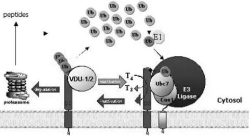
The recent identification of murine homo-logues of UBC6 and UBC7, MmUBC6 and MmUBC7 (53), has made it feasible to investigate the relevance of UBC6 and UBC7 for D2 ERAD in mam-malian cells. The major novel finding is that there is a high affinity, specific physical association between MmUBC7 and a carboxy region (aa 169-234) of the D2 enzyme (54), a region that is exposed to the cytosol according to prior topological analysis (27). While direct binding of E2s has been proposed for cer-tain substrates (47,55-57), current models of ubiquiti-nation suggest that E2s play only a secondary role in substrate recognition, with E3 enzymes being the major specificity determinant (47). It is therefore like-ly that the D2-MmUBC7 association is mediated by an as yet unknown E3 or other adaptor protein(s) pre-sent in the reticulocyte lysate.
by changes in D2 protein half-life (54). That both UBC6 and UBC7 must be neutralized in order to achieve D2 stabilization suggests that redundancy exists at the level of the E2, i.e. either UBC6 or UBC7 can catalyze D2 ubiquitination. Thus, co-expression of the individual E2 mutants had no effect on D2 ERAD. However, the fact that MmUBC6 does not bind with the same affinity to the D2 ERAD complex, as does MmUBC7 suggests that there may be a preference for UBC7-mediated D2 ERAD (54) (figure 4). In fact, it has been suggested that UBC7 rather than UBC6 is dominant in ERAD (53,58,59), and controversy exists as to the role of UBC6 in the ubiquitination of some ERAD substrates including the T-cell receptor sub-units αand CD3-δ(60).
Reversible Ubiquitination Rescues D2 From Irreversible Proteolysis
Fusion of the 8-amino acid FLAG sequence to the COOH but not the NH2terminus of D2 prolongs its
activity half-life and increases the size of the Ub-D2 pool 20- to 30 fold (51), suggesting that D2 ubiqui-tination is reversible, as not all Ub-D2 undergoes proteolysis. Enzymatic de-ubiquitination of Ub-D2 occurs in vitro (61) and could explain recycling in vivo. Using the COOH-terminal portion of D2 in a yeast-two-hybrid screening we recently identified D2 as the only known specific substrate of the Von Hip-pel Lindau protein (pVHL)-interacting deubiquiti-nating enzyme (VDU1) and VDU2 (62), which in turn are the first ubiquitin-specific processing pro-teases (UBP) known to specifically deubiquitinate an
ERAD substrate. These results show that protein recognition is not only involved in the E3-mediated ubiquitination process but also in the deubiquitina-tion pathway catalyzed by UBPs. Both VDUs are downstream targets for ubiquitination by pVHL E3 ligase, and VHL mutations that disrupt the interac-tion between the VDUs and pVHL abrogate their ubiquitination (63,64). Although hundreds of UBP enzymes have been cloned, only a few examples of substrate recognition by UBP enzymes have been reported and, to our knowledge, none are ER-resi-dent proteins (65-69). Confocal studies indicate that both VDUs co-localize with D2, itself an integral membrane ER-resident protein. Although present in the particulate fraction and not in cytosol it is not clear based on their hydrophobic profile whether VDU1/2 are integral membrane proteins (62). Their physical colocalization with D2, however, provides the opportunity for catalysis and D2 deubiquitina-tion.
VDU1 catalyzed D2 deubiquitination is an important part of the adaptive mechanism that regu-lates thyroid hormone action (figure 4). In stimulated brown adipocytes normal D2 induction increases intracellular T3production, resulting in isolated tissue
thyrotoxicosis (5,34,70). This is an important mecha-nism for cold acclimatization in that mice with targe-ted inactivation of the D2 gene develop hypothermia and marked weight loss during cold exposure due to impaired BAT thermogenesis (5). Our results demon-strate that increased VDU1-catalyzed deubiquitina-tion of a pool of Ub-D2 and its rescued from protea-somal degradation is an integral part of this mecha-nism. VDU1 mRNA levels are markedly up-regulated by cold exposure or NE, amplifying the transcription-al increase in D2 activity and hence T3production by
approximately 2.5 fold. Even though UBP induction is known to play a physiological role in a number of cellular processes (71-75), this is the first example of enzyme reactivation due to deubiquitination.
Thus, due to the intrinsic inefficiency of the selenoprotein synthesis, the availability of a reversible ubiquitination-dependent mechanism to control the activity of D2 constitutes a biochemical and physio-logical advantage that allows for rapid control of thy-roid hormone activation. The finding that VDU1 and VDU2 are co-expressed with D2 in many human tis-sues, including brain, heart and skeletal muscle (1,63), indicates that the importance of this mechanism may extend well beyond thermal homeostasis to include brain development, cardiac performance, glucose uti-lization and energy expenditure.
ACKNOWLEDGEMENT
Supported by NIH grant DK58538.
REFERENCES
1. Bianco AC, Salvatore D, Gereben B, Berry MJ, Larsen PR. Biochemistry, cellular and molecular biology and physi-ological roles of the iodothyronine selenodeiodinases. Endocr Rev 2002;23:38-89.
2. Berry MJ, Grieco D, Taylor BA, et al. Physiological and genetic analyses of inbred mouse strains with a type I iodothyronine 5’ deiodinase deficiency. J Clin Invest 1993;92:1517-28.
3. Schneider MJ, Fiering SN, Pallud SE, Parlow AF, St. Ger-main DL, Galton VA. Targeted disruption of the type 2 selenodeiodinase gene (Dio2) results in a phenotype of pituitary resistance to T4. Mol Endocrinol 2001;15:2137-48.
4. Hernandez A, Schneider M, Galton V, St. Germain D. Physiological Consequences of Deficiencies of the Type 2 (D2) and Type 3 (D3) Deiodinases. In: 85th Annual Meeting of the Endocrine Society. Philadelphia, PA; 2003.
5. de Jesus LA, Carvalho SD, Ribeiro MO, et al. The type 2 iodothyronine deiodinase is essential for adaptive ther-mogenesis in brown adipose tissue. J Clin Invest 2001;108:1379-85.
6. Schoenmakers CHH, Pigmans IGAJ, Poland A, Visser TJ. Impairment of the selenoenzyme type I iodothyronine deiodinase in C3H/He mice. Endocrinology 1993; 132:357-61.
7. Maia AL, Kieffer JD, Harney JW, Larsen PR. Effect of 3,5,3’-Triiodothyronine (T3) administration on dio1 gene expression and T3 metabolism in normal and type 1 deiodinase-deficient mice. Endocrinology 1995; 136:4842-9.
8. Maia AL, Berry MJ, Sabbag R, Harney JW, Larsen PR. Structural and functional differences in the dio1 gene in mice with inherited type 1 deiodinase deficiency. Mol Endocrinol 1995;9:969-80.
9. Hernandez A, Schneider MJ, Galton VA, St Germain DL. Physiological consequences of deficiencies of the type 2 (D2) and type 3 (D3) deiodinases. In: Endo2003. Philadelphia; 2003.
10. Huang SA, Tu HM, Harney JW, et al. Severe hypothy-roidism caused by type 3 iodothyronine deiodinase in infantile hemangiomas. N Eng J Med 2000;343:185-9.
11. Huang SA, Fish SA, Dorfman DM, et al. A 21-year-old woman with consumptive hypothyroidism due to a vas-cular tumor expressing type 3 iodothyronine deiodinase. J Clin Endocrinol Metab 2002;87:4457-61.
12. Curcio C, Baqui MMA, Salvatore D, et al. The human type 2 iodothyronine deiodinase is a selenoprotein high-ly expressed in a mesothelioma cell line. J Biol Chem 2001;276:30183-7.
13. Mentuccia D, Proietti-Pannunzi L, Tanner K, et al. Associ-ation between a novel variant of the human type 2 deiodinase gene Thr92Ala and insulin resistance:
evi-dence of interaction with the Trp64Arg variant of the beta-3-adrenergic receptor. Diabetes 2002;51:880-3.
14. Berry MJ, Kieffer JD, Harney JW, Larsen PR. Selenocysteine confers the biochemical properties of the type I iodothy-ronine deiodinase. J Biol Chem 1991;266:14155-8.
15. Croteau W, Whittemore SL, Schneider MJ, St Germain DL. Cloning and expression of a cDNA for a mammalian type III iodothyronine deiodinase. J Biol Chem 1995;270:16569-75.
16. Buettner C, Harney JW, Larsen PR. The role of selenocys-teine 133 in catalysis by the human type 2 iodothyronine deiodinase. Endocrinology 2000;141:4606-12.
17. Callebaut I, Curcio-Morelli C, Mornon JP, et al. The iodothyronine selenodeiodinases are thioredoxin-fold family proteins containing a glycoside hydrolase-clan GH-A-like structure. J Biol Chem 2003;278:36887-96.
18. Martin JL. Thioredoxin - a fold for all reasons. Structure 1995;3:245-50.
19. Carbohydrate-Active Enzymes. Server at http://afmb.cnrs-mrs.fr/~cazy/CAZY/index.html. 1999. (Accessed at http://afmb.cnrs-mrs.fr/~cazy/ CAZY/ index.html.)
20. Berry MJ, Banu L, Larsen PR. Type I iodothyronine deiod-inase is a selenocysteine-containing enzyme. Nature 1991;349:438-40.
21. Berry MJ, Banu L, Chen YY, et al. Recognition of UGA as a selenocysteine codon in type I deiodinase requires sequences in the 3’ untranslated region. Nature 1991;353:273-6.
22. Leonard JL, Visser TJ, Leonard DM. Characterization of the subunit structure of the catalytically active type I iodothyronine deiodinase. J Biol Chem 2000;276:2600-7.
23. Curcio-Morelli C, Gereben B, Zavacki AM, et al. In vivo dimerization of types 1, 2, and 3 iodothyronine selen-odeiodinases. Endocrinology 2003;144:3438-43.
24. Leonard JL, Rosenberg IN. Solubilization of a phospho-lipid-requiring enzyme, iodothyronine 5’-deiodinase, from rat kidney membranes. Biochim Biophys Acta 1981;659:205-18.
25. Safran M, Leonard JL. Comparison of the physicochem-ical properties of type I and type II iodothyronine 5’-deiodinase. J Biol Chem 1991;266:3233-8.
26. Toyoda N, Berry MJ, Harney JW, Larsen PR. Topological analysis of the integral membrane protein, type 1 iodothyronine deiodinase (D1). J Biol Chem 1995;270:12310-8.
27. Baqui MM, Gereben B, Harney JW, Larsen PR, Bianco AC. Distinct subcellular localization of transiently expressed types 1 and 2 iodothyronine deiodinases as determined by immunofluorescence confocal microscopy. Endocrinology 2000;141:4309-12.
28. Baqui MM, Botero D, Gereben B, et al. Human type 3 iodothyronine selenodeiodinase is located in the plas-ma membrane and undergoes rapid internalization to endosomes. J Biol Chem 2003;278:1206-11.
30. Prabakaran D, Ahima RS, Harney JW, Berry MJ, Larsen PR, Arvan P. Polarized targeting of epithelial cell proteins in thyrocytes and MDCK cells. J Cell Sci 1999;112(Pt):1247-56.
31. Larsen PR, Silva JE, Kaplan MM. Relationships between cir-culating and intracellular thyroid hormones: physiological and clinical implications. Endocr Rev 1981;2:87-102.
32. Silva JE, Larsen PR. Contributions of plasma triiodothyro-nine and local thyroxine monodeiodination to tri-iodothyronine to nuclear tritri-iodothyronine receptor satu-ration in pituitary, liver, and kidney of hypothyroid rats. Further evidence relating saturation of pituitary nuclear triiodothyronine receptors and the acute inhibition of thyroid- stimulating hormone release. J Clin Invest 1978;61:1247-59.
33. Bianco AC, Silva JE. Nuclear 3,5,3’-triiiodothyronine (T3) in brown adipose tissue: receptor occupancy and sources of T3 as determined by in vivo techniques. Endocrinology 1987;120:55-62.
34. Bianco AC, Silva JE. Cold exposure rapidly induces virtu-al saturation of brown adipose tissue nuclear T3 recep-tors. Am J Physiol 1988;255:E496-E503.
35. Wasco EC, Martinez E, Grant KS, St Germain EA, St Ger-main DL, Galton VA. Determinants of iodothyronine deiodinase activities in rodent uterus. Endocrinology 2003;144:4253-61.
36. Huang SA, Dorfman DM, Genest DR, Salvatore D, Larsen PR. Type 3 Iodothyronine deiodinase is highly expressed in the human uteroplacental unit and in fetal epitheli-um. J Clin Endocrinol Metab 2003 (in press).
37. Gereben B, Kollar A, Harney JW, Larsen PR. The mRNA structure has potent regulatory effects on type 2 iodothyronine deiodinase expression. Mol Endocrinol 2002;16:1667-79.
38. Burmeister LA, Pachucki J, St. Germain DL. Thyroid hor-mones inhibit type 2 iodothyronine deiodinase in the rat cerebral cortex by both pre- and posttranslational mechanisms. Endocrinology 1997;138:5231-7.
39. St. Germain DL. The effects and interactions of sub-strates, inhibitors, and the cellular thiol-disulfide balance on the regulation of type II iodothyronine 5’-deiodinase. Endocrinology 1988;122:1860-8.
40. Leonard JL, Kaplan MM, Visser TJ, Silva JE, Larsen PR. Cerebral cortex responds rapidly to thyroid hormones. Science 1981;214:571-3.
41. Koenig RJ, Leonard JL, Senator D, Rappaport N, Watson AY, Larsen PR. Regulation of thyroxine 5’-deiodinase activity by 3,5,3’- triiodothyronine in cultured rat anterior pituitary cells. Endocrinology 1984;115:324-9.
42. Silva JE, Leonard JL. Regulation of rat cerebrocortical and adenohypophyseal type II 5’- deiodinase by thy-roxine, triiodothyronine, and reverse triiodothyronine. Endocrinology 1985;116:1627-35.
43. Halperin Y, Shapiro LE, Surks MI. Down-regulation of type II L-thyroxine, 5’-monodeiodinase in cultured GC cells: dif-ferent pathways of regulation by L-triiodothyronine and 3,3’,5’-triiodo-L-thyronine. Endocrinology 1994;135:1464-9.
44. Leonard JL, Silva JE, Kaplan MM, Mellen SA, Visser TJ, Larsen PR. Acute posttranscriptional regulation of cere-brocortical and pituitary iodothyronine 5’-deiodinases by thyroid hormone. Endocrinology 1984;114:998-1004.
45. Obregon MJ, Larsen PR, Silva JE. The role of 3,3’,5’-tri-iodothyronine in the regulation of type II 3,3’,5’-tri-iodothyronine 5’-deiodinase in the rat cerebral cortex. Endocrinology 1986;119:2186-92.
46. Coux O, Tanaka K, Goldberg AL. Structure and functions of the 20S and 26S proteasomes. Ann Rev Biochem 1996;65:801-47.
47. Hershko A, Ciechanover A. The ubiquitin system. Ann Rev Biochem 1998;67:425-79.
48. Steinsapir J, Harney J, Larsen PR. Type 2 iodothyronine deiodinase in rat pituitary tumor cells is inactivated in proteasomes. J Clin Invest 1998;102:1895-9.
49. Steinsapir J, Bianco AC, Buettner C, Harney J, Larsen PR. Substrate-induced down-regulation of human type 2 deiodinase (hD2) is mediated through proteasomal degradation and requires interaction with the enzyme’s active center. Endocrinology 2000;141:1127-35.
50. Scheffner M, Smith S, Jentsch S. The ubiquitin-conjuga-tion system. In: Peters J-M, Harris JR, Finley D, eds. Ubiq-uitin and the biology of the cell. New York: Plenun Press; 1998.p.65-91.
51. Gereben B, Gonçalves C, Harney JW, Larsen PR, Bianco AC. Selective proteolysis of human type 2 deiodinase: a novel ubiquitin-proteasomal mediated mechanism for regulation of hormone activation. Mol Endocrinol 2000;14:1697-708.
52. Botero D, Gereben B, Gonçalves C, de Jesus LA, Harney JW, Bianco AC. Ubc6p and Ubc7p are required for nor-mal and substrate-induced endoplasmic reticulum-associated degradation of the human selenoprotein type 2 iodothyronine monodeiodinase. Mol Endocrinol 2002;16:1999-2007.
53. Tiwari S, Weissman AM. Endoplasmic reticulum (ER)-asso-ciated degradation of T cell receptor subunits. Involve-ment of ER-associated ubiquitin-conjugating enzymes (E2s). J Biol Chem 2001;276:16193-200.
54. Kim BW, Zavacki AM, Curcio-Morelli C, et al. ER-associ-ated degradation of the human type 2 iodothyronine deiodinase (D2) is mediated via an association between mammalian UBC7 and the carboxyl region of D2. Mol Endocrinol 2003(in press).
55. Weissman AM. Themes and variations on ubiquitylation. Nat Rev Mol Cell Biol 2001;2:169-78.
56. Gosink MM, Vierstra RD. Redirecting the specificity of ubiquitination by modifying ubiquitin-conjugating enzymes. Proc Natl Acad Sci USA 1995;92:9117-21.
57. Kalchman MA, Graham RK, Xia G, et al. Huntingtin is ubiquitinated and interacts with a specific ubiquitin-conjugating enzyme. J Biol Chem 1996;271:19385-94.
58. Wilhovsky S, Gardner R, Hampton R. HRD gene depen-dence of endoplasmic reticulum-associated degrada-tion. Mol Biol Cell 2000;11:1697-708.
59. Webster JM, Tiwari S, Weissman AM, Wojcikiewicz RJ. Inos-itol 1,4,5-trisphosphate receptor ubiquitination is mediated by mammalian Ubc7, a component of the Endoplasmic Reticulum-associated degradation pathway, and is inhib-ited by chelation of intracellular Zn2+. J Biol Chem 2003.Oct 3;278(40):38238-46
61. Bianco AC, Harney J, Larsen PR. Identification of ubiqui-tinated forms of human type 2 deidinase (hD2). In: 72nd
Annual Meeting of The American Thyroid Association. Palm Beach, FL; 1999.p.5.
62. Curcio-Morelli C, Zavacki AM, Christofollete M, et al. Deubiquitination of type 2 iodothyronine deiodinase by pVHL-interacting deubiquitinating enzymes regulates thyroid hormone activation. J Clin Invest 2003 (in press).
63. Li Z, Na X, Wang D, Schoen SR, Messing EM, Wu G. Ubiq-uitination of a novel deubiquitinating enzyme requires direct binding to von Hippel-Lindau tumor suppressor protein. J Biol Chem 2002;277:4656-62.
64. Li Z, Wang D, Na X, Schoen SR, Messing EM, Wu G. Iden-tification of a deubiquitinating enzyme subfamily as sub-strates of the von Hippel-Lindau tumor suppressor. Biochem Biophys Res Commun 2002;294:700-9.
65. Taya S, Yamamoto T, Kanai-Azuma M, Wood SA, Kaibuchi K. The deubiquitinating enzyme Fam interacts with and stabilizes beta-catenin. Genes Cells 1999;4:757-67.
66. Gnesutta N, Ceriani M, Innocenti M, et al. Cloning and characterization of mouse UBPy, a deubiquitinating enzyme that interacts with the ras guanine nucleotide exchange factor CDC25(Mm)/Ras-GRF1. J Biol Chem 2001;276:39448-54.
67. Taya S, Yamamoto T, Kano K, et al. The Ras target AF-6 is a substrate of the fam deubiquitinating enzyme. J Cell Biol 1998;142:1053-62.
68. Chen X, Zhang B, Fischer JA. A specific protein substrate for a deubiquitinating enzyme: Liquid facets are the sub-strate of Fat facets. Genes Dev 2002;16:289-94.
69. Ideguchi H, Ueda A, Tanaka M, et al. Structural and functional characterization of the USP11 deubiquitinat-ing enzyme, which interacts with the RanGTP-associat-ed protein RanBPM. Biochem J 2002;367(Pt 1):87-95.
70. Bianco AC, Silva JE. Intracellular conversion of thyroxine to triiodothyronine is required for the optimal thermo-genic function of brown adipose tissue. J Clin Invest 1987;79:295-300.
71. Huang Y, Baker RT, Fischer-Vize JA. Control of cell fate by a deubiquitinating enzyme encoded by the fat facets gene. Science 1995;270:1828-31.
72. Moazed D, Johnson D. A deubiquitinating enzyme inter-acts with SIR4 and regulates silencing in S. cerevisiae. Cell 1996;86:667-77.
73. Zhu Y, Carroll M, Papa FR, Hochstrasser M, D’Andrea AD. DUB-1, a deubiquitinating enzyme with growth-suppress-ing activity. Proc Natl Acad Sci USA 1996;93:3275-9.
74. Naviglio S, Mattecucci C, Matoskova B, et al. UBPY: a growth-regulated human ubiquitin isopeptidase. EMBO J 1998;17:3241-50.
75. Park KC, Kim JH, Choi EJ, et al. Antagonistic regulation of myogenesis by two deubiquitinating enzymes, UBP45 and UBP69. Proc Natl Acad Sci USA 2002;99:9733-8.
76. Bianco AC, Larsen PR. Thyroxine regulates its own acti-vation by feed back control of type 2 iodothyronine deiodinase (D2) proteolysis. Hot Thyroidology 2002;6.
Endereço para correspondência:
Antonio C. Bianco Thyroid Section,
Division of Endocrinology, Diabetes and Hypertension, Brigham and Women’s Hospital and Harvard Medical School. 77 Avenue Louis Pasteur; HIM Bldg. #643
Boston MA 02115