Article
J. Braz. Chem. Soc., Vol. 21, No. 7, 1293-1302, 2010. Printed in Brazil - ©2010 Sociedade Brasileira de Química 0103 - 5053 $6.00+0.00*e-mail: [email protected]
Theoretical Studies of the Tautomerism in 3-(2-R-Phenylhydrazono)-naphthalene-
1,2,4-triones: Synthesis of Copper(II) Complexes and Studies of Antibacterial
and Antitumor Activities
Acácio I. Francisco,a Maria D. Vargas,*,a Thaís P. Fragoso,a J. Walkimar de M. Carneiro,a Annelise Casellato,b Fernando de C. da Silva,a Vitor F. Ferreira,a Jussara P. Barbosa,c Claudia Pessoa,d
Letícia V. Costa-Lotufo,d José D. B. Marinho Filho,d Manoel O. de Moraesd and Antonio S. Mangriche
aInstituto de Química, Universidade Federal Fluminense, Campus do Valonguinho, Centro, 24020-150 Niterói-RJ, Brazil
bInstituto de Química, Universidade Federal do Rio de Janeiro, Ilha do Fundão, 21945-970 Rio de Janeiro-RJ, Brazil
cInstituto Oswaldo Cruz, FIOCRUZ, CP 926, 21045-900 Rio de Janeiro-RJ, Brazil
dUniversidade Federal do Ceará, Depto de Fisiologia e Farmacologia, Campus do Porangabussu, 60430-270 Fortaleza-CE, Brazil
eDepartamento de Química da Universidade Federal do Paraná, 81531-990 Curitiba-PR, Brazil
Cálculos teóricos utilizando os funcionais B3LYP e PBE1PBE e as bases 6-31G(d) e 6-311+G(2d,p) para o sistema 3-(2-fenil-hidrazona)-naftaleno-1,2,4-triona, em solução (dmso) e em fase gasosa, evidenciaram, em ambos os casos, a maior estabilidade da forma ceto-hidrazona (rotâmeros Ia e Ib) comparada às formas enol-azo (rotâmeros IIa/IIb, por volta de 14 kcal mol-1)
e III (aproximadamente 6 kcal mol-1). A natureza do substituinte no grupo fenil não inluenciou a
estabilidade relativa dos tautômeros. Estes resultados foram conirmados por dados espectroscópicos dos derivados HL1-HL13, sintetizados a partir da 2-hidroxi-1,4-naftoquinona e arilaminas (R = 4-OMe, 4-N2-C6H5, 4-Cl, 4-I, 3-I, 2-I, 4-COOH, 3-COOH, 4-CN, 3-CN, 4-NO2, 3-NO2, 2-NO2).
A avaliação da atividade anticâncer in vitro (contra linhagens de células cancerosas SF-295, HCT-8, MDAMB-435 e HL-60) e bactericida (Bacillus cereus, Bacillus subtilis, Enterococcus faecalis,
Staphylococcus aureus, Escherichia coli, Klebsiella pneumonia e Pseudomonas aeruginosa) dos compostos HL1-HL13 e dos seus respectivos complexos de cobre(II), [Cu(L1-13)2], foi avaliada. Em geral a atividade bactericida foi baixa, exceto para o derivado HL5 (R = 3-I), mais ativo do que o controle; entretanto, seu complexo não foi ativo. Por outro lado, a complexação levou, em geral, ao aumento da atividade antitumoral dos pré-ligantes. O complexo [Cu(L13)2] (R = 3-NO2) apresentou moderada citotoxicidade contra leucemia humana (HL-60).
DFT calculations using the B3LYP and PBE1PBE functionals with the standard 6-31G(d) and 6-311+G(2d,p) basis sets were carried out for the 3-(2-phenylhydrazone)-naphthalene-1,2,4-trione system in solution (dmso) and in the gas phase, and showed the keto-hydrazone forms (rotamers Ia
and Ib) to be more stable than the enol-azo forms (rotamers IIa and IIb, by about 14 kcal mol-1) and
III (by approximately 6 kcal mol-1), independently of the nature of the substituent in the phenylene
ring. These results were conirmed by spectroscopic data on the derivatives HL1-HL13, obtained from 2-hydroxy-1,4-naphthoquinone and arylamines (R = 4-OMe, 4-N2-C6H5, 4-Cl, 4-I, 3-I, 2-I, 4-COOH,
3-COOH, 4-CN, 3-CN, 4-NO2, 3-NO2, 2-NO2). The in vitro antitumor (against SF-295, HCT-8,
MDAMB-435 and HL-60 cancer cell lines) and antibacterial activities (Bacillus cereus, Bacillus subtilis, Enterococcus faecalis, Staphylococcus aureus, Escherichia coli, Klebsiella pneumonia and
Pseudomonas aeruginosa) of compounds HL1-HL13 and of their respective copper(II) complexes, [Cu(L1-13)2], were tested. In general, these compounds exhibited low antibacterial activity, except for HL5 (R = 3-I), more active than the control; however, the corresponding complex was inactive. In contrast, increased cytotoxicity was observed upon complexation. Complex [Cu(L13)2] (R = 3-NO2) presented moderate cytotoxicity against human leukemia (HL-60).
Theoretical Studies of the Tautomerism in 3-(2-R-Phenylhydrazono)-naphthalene-1,2,4-triones J. Braz. Chem. Soc. 1294
Introduction
The 1,2 and 1,4-naphthoquinone nuclei are commonly encountered in natural products1,2 and their
derivatives are found to exhibit an interesting range of pharmacological properties including antibacterial,3-6
antiviral,7,8 trypanocidal,9-11 anticancer,12-14 antimalarial,15-17
antifungal18,19 and moluscicide20activities. Such properties
are due to the interference of quinones in the electron transport chain by electron reduction processes, generating semiquinone radical (Q•−) and hydroquinone anion (Q2−).21,22
Azo dyes with ortho and para hydroxy substituents to the azo linker generally exhibit azo-hydrazone tautomerism, which involves transfer of the hydroxyl hydrogen to one of the nitrogens in the azo group.23 Tautomeric forms can be
identiied from their spectral properties24 and the stability
of such forms in solution may be inluenced by the nature of the substituents present in the molecules.25
Incorporation of an azo group into 2-hydroxy-1,4-naphthoquinone has led to promising antimalarial and anticancer agents, in which metal complexation with copper(II) resulted in increased cytotoxicity.26,27 The
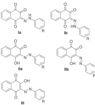
derivatives can be represented by several tautomeric structures illustrated in Figure 1: 3-(2-R-phenylhydrazono)-naphthalene-1,2,4-triones, Ia and Ib, 3-R-arylazo-4-hydroxy-1,2-naphthoquinones, IIa and IIb, and 3-R-arylazo-2-hydroxy-1,4-naphthoquinone III. A poor X-ray diffraction study of the derivative containing R=3-Me suggested that this compound exists as tautomer IIa in the solid state.26
Knowledge of the geometry and electronic structures, as well as the relative stabilities of the various tautomeric forms in solution, is of utmost importance as it provides a basis for understanding the pharmacological properties of the molecules and designing new derivatives with improved activity.26,27 In the present work we describe the results
of our theoretical calculations using density functional theory (DFT) carried out to investigate the relative stability of the tautomeric forms of 3-(2-R-phenylhydrazono)-naphthalene-1,2,4-trione derivatives (Figure 1) as a function of the substituent, both in the gas phase and in solution. Low solubility prevented experimental NMR experiments.25 Some of these compounds have been
previously described,26,27,30,31 but theirtautomerism has
not yet been investigated. In addition to reporting the synthesis of novel compounds (HL1-HL13), we also present the results of in vitro antitumor screening and antibacterial activity of these compounds and of their copper(II) complexes (Scheme 1) against several cancer cell lines (SF-295, HCT-8, MDAMB-435 and HL-60) and bacteria strains (Bacillus cereus, Bacillus subtilis,
Enterococcus faecalis, Staphylococcus aureus, Escherichia coli, Klebsiella pneumonia and Pseudomonas aeruginosa).
Experimental
Materials and methods
Reagents and solvents were used without further puriication, except for triethylamine, which was previously distilled. Microanalyses were performed using a Perkin-Elmer CHN 2400 micro analyser at the Central Analítica - Instituto de Química, USP-São Paulo, Brazil. Melting points were obtained with a Mel-Temp II, Laboratory Devices - USA apparatus and are uncorrected. IR spectra (KBr pellets) were recorded on a FT-IR Spectrum One (Perkin Elmer) spectrophotometer. 1H and 13C NMR spectra were
recorded with a Varian Unit Plus 300 MHz spectrometer in dmso-d6; coupling constants are reported in Hertz (Hz) and chemical shifts in parts per million (ppm) relative to internal standard Me4Si. The hydrogen signals were attributed through coupling constant values and 1H × 1H - COSY
experiments. Electronic spectra were taken on a Diode Array 8452A (Hewlett Packard - HP) spectrophotometer using spectroscopic grade solvents (Tedia Brazil) in 10-3
and 10-4 mol L-1 solutions. Electron paramagnetic resonance
(EPR) spectra of the frozen copper(II) samples dissolved in dmso at 77 K were obtained on a Bruker EMX equipment with modulation frequency of 100 kHz operating at about 9.5 GHz (X-band), using quartz tubes accommodated in a quartz Dewar. The EPR parameter values were obtained by O O O N H N R O O O N HN R O O OH N N R O OH O N N R Ia Ib IIa III O O OH N IIb N R
Francisco et al. 1295 Vol. 21, No. 7, 2010
treating and simulating the experimental spectra using the Windows software programs WINEPR and SIMFONIA (Bruker), and the WEAK PITCH BRUKER sample pattern. Cyclic voltammograms were obtained on an Epsilon - BAS potentiostat-galvanostat from 1×10⁻³ mol L-1
solutions in dmso containing 0.1 mol L-1 of (Bu 4N)BF4
as supporting electrolyte, at room temperature and under argon atmosphere. A standard three component system was used: a carbon-glassy working electrode, a platinum wire auxiliary electrode, and an Ag/AgCl reference electrode for organic media. Ferrocene was used as an internal standard (E1/2 0.40 V vs. NHE, normal hydrogen electrode).
Density functional calculations were carried out using the Gaussian03W molecular orbital package.32 Geometries
were fully optimized using the B3LYP functional33 with the
standard 6-31G(d) basis set.34 The electronic spectra were
calculated using the TD (Time Dependent) methodology available in Gaussian with the PBE1PBE functional and the 6-311+G(2d,p) basis set. For calculation of the electronic spectra, solvent effects (dmso) were included by mean of the continuum solvation model using the conductor-like polarisable continuum model35 (CPCM). The dielectric
constant of dmso, ε, is 46.7. Relative energies are reported at the PBE1PBE/6-311+G(2d,p) level with inclusion of solvent effects.
Synthesis of the hydrazono compounds HL1-HL13
Compounds HL1-HL13 (Figure 2) were synthesised according to the general procedure described in the
literature.26,27,30,31HL1, HL7, HL11, HL12 and HL13
were described previously.26,30 See Supplementary
Information for description on the synthesis, analytical and spectroscopic data of the novel products.
Synthesis of complexes [Cu(L1- L13)2]from HL1- HL13
To a suspension of 1 mmol of the hydrazono proligand HL in 30 mL MeOH, was added a solution of CuCl2•2H
2O
(83 mg, 0.5 mmol) in 1 mL MeOH. After addition of Et3N (0.14 mL, 1 mmol), the suspension turned into a solution, followed immediately by the formation of a brown solid. The reaction mixture was left under stirring in the dark for 24 h at room temperature. The resulting solids were iltered off, washed with cold methanol and dried under vacuum. Complexes 1-13 (Figure 3) have not been described previously. See Supplementary Information for analytical and spectroscopic data.
Antibacterial assays
The antibacterial evaluation was performed with Gram-positive (Bacillus cereus ATCC 33019, Bacillus subtilis ATCC 6633, Enterococcus faecalis ATCC 29212,
Staphylococcus aureus ATCC 25923) and Gram-negative (Escherichia coli ATCC 25922, Klebsiella pneumoniae
ATCC 700603, Pseudomonas aeruginosa ATCC 27853) bacteria as test-microorganisms, as previously described (see Supplementary Information).15,36,37 The tests were
performed in triplicate. O
O
OH
N2+Cl– O
O O N H N R R O O N OCuN O N N O O R R + NaOH EtOH
CuCl22H2O
NEt3 / MeOH
HL1-HL13 1-13
.
Scheme 1. Synthesis of hydrazono compounds HL1-HL13 and of their respective complexes 1-13; R = 4-OMe, 4-N=NC6H5, 4-Cl, 4-I, 3-I, 2-I, 4-COOH, 3-COOH, 4-CN, 3-CN, 4-NO2, 3-NO2 and 2-NO2.
R =
4-OMe, HL1 4-N=N-C6H5,HL2 4-Cl, HL3 4-I, HL4 3-I, HL5 2-I, HL6 4-COOH, HL7
3-COOH, HL8 4-CN, HL9 3-CN, HL10 4-NO2, HL11 3-NO2, HL12 2-NO2, HL13 2 3 4 5 6 7 8 O O O N H N R 1
Theoretical Studies of the Tautomerism in 3-(2-R-Phenylhydrazono)-naphthalene-1,2,4-triones J. Braz. Chem. Soc. 1296
Antitumoral assays
The compounds (1-5 µg mL-1) were tested for cytotoxic
activity against three cancer cell lines: SF-295 (central nervous system), HCT-8 (colon) and MDAMB-435 (breast). All cell lines were maintained in RPMI 1640 medium supplemented with 10% fetal bovine serum, 2 mmol L-1 glutamine, 100 U mL-1 penicillin, and
100 µg mL-1 streptomycin at 37 ºC with 5% CO 2. Each
compound was dissolved in dmso and diluted with water to obtain a concentration of 1 mg mL-1. They were incubated
with the cells for 72 h. The negative control received the same amount of dmso (0.5% in the highest concentration). Doxorubicin (0.1-0.58 µg mL-1) was used as a positive
control. The cell viability was determined by reduction of the yellow dye 3-(4,5-dimethyl-2-thiazol)-2,5-phenyl-2H-tetrazolium bromide (MTT) to a blue formazan product as described by Mosmann.38
Results and Discussion
Synthesis and characterization of the hydrazono compounds
Compounds HL1-HL13 (Figure 2) were synthesized from the diazonium salts of the respective arylamines, followed by coupling with lawsone C3 in ethanol under stirring at room temperature. After addition of the diazonium salts to the lausonate solution, the orange products precipitated immediately. Following iltration and washing with cold EtOH, the products were washed with hot acetonitrile, dried under vacuum and obtained in pure form, in yields ranging from 71-92%. Compounds HL1- HL13 are stable in the solid state and in solution. Their structures were formulated on the basis of analytical and spectroscopic data (see Supplementary Information). The 1H spectra of compounds HL1-HL13 were obtained
in hot dmso-d6, due to poor solubility in other solvents. The spectra exhibit characteristic signals attributed to the four hydrogens of the naphthoquinone unit (H5-H8), which
appear in the d7.5-8.5 region as dd or bd (H5 and H8) and td (H5 and H7) (see Figure 2 for numbering). The substituted phenylene ring hydrogens appear as broad doublets (substituents in the para position) and multiplets/ triplet (substituents in the meta/ortho positions); in most cases the naphthoquinone hydrogen signals overlap with those of the arylamine ring, resulting in multiplet signals. Attributions were made on the basis of 1H × 1H - COSY
experiments, J values and multiplicity. The 13C NMR
spectra of the previously reported 2-, 3- and 4- substituted methyl derivatives26 have not been described. In the 13C NMR spectra of compounds HL1-HL13 the number of
signals does not correspond to the number of the expected carbons, either because of the high relaxation time of some of the carbon nuclei, or due to the poor solubility of the compounds (see Supplementary Information). For this reason tautomerism studies using this technique could not be carried out as described previously for similar systems;25
instead, density functional calculations involving relative stability of the possible tautomers and full geometry optimization for the ground state were carried out and are described below.
Theoretical calculations
Calculation of several tautomers of the unsubstituted derivative, e.g., the enol-azo forms II and III and the alternative keto-hydrazone Ia and Ib forms (Figure 1) showed that Ia and Ib are much more stable than the enol-azo forms IIa (presumably the molecular structure determined by an X-ray diffraction study, see below) and IIb, by at least 14 kcal mol-1 (Table 1). Indeed geometry
optimization of the enol-azo tautomer IIb directly converges to the keto-hydrazone form Ib. The two rotamers Ia and Ib are almost isoenergetic, with relative energies in the order or below 0.5 kcal mol-1 (see Supplementary
Information for detailed theoretical calculations data). This result is in agreement with those reported on a similar system, viz. 2-arylazo-1-hydroxycyclohex-1-en-3-one.39
Figure 3. Complexes [Cu(L1-L13)2].
O O N OCuN O N N O O R R R =
4-OMe, 1
4-N=N-C6H5,2 4-Cl, 3
4-I, 4
3-I, 5
2-I, 6
4-COOH, 7
3-COOH, 8
4-CN, 9
3-CN, 10
Francisco et al. 1297 Vol. 21, No. 7, 2010
The nature of the substituent in position 4 (electron withdrawing substituents NO2 and CN versus electron donor OMe) does not affect appreciably this stability order (Ib ≅Ia >> II). This agrees with previous calculations on tautomeric equilibrium for 4-anilino-1,2-naphthoquinones40
and is in contrast with the theoretical and experimental results reported for the analogous tautomeric equilibrium of the azo derivatives of 2-naphthol for which electron withdrawing groups were found to strongly stabilize the keto-hydrazone tautomer, whereas electron donor groups shift the equilibrium toward the enol-azo tautomer.25
According to the reported X-ray diffraction study of the R = 3-Me (HL-3-Me) derivative,26 this compound exists in
the solid state as the enol-azo tautomer IIa (see Figure 1). The authors reported that the molecule occupies two semi-populated sites related by crystallographic inversion centers, and because many atoms coincided, it was necessary to apply bond length constraints to reine the structure. The cif list does not make clear which atoms presented problems (an analysis of the data would be necessary); however, it shows that C(1) and H(14) on the one hand, and C(4) and N(2) on the other (see Figure 4 for the numbering) have been reined in the same positions. It is possible, therefore, that the C(1)-O(1), C(4)-O(4) and N(1)-N(2) distances reported were influenced by the applied restrictions. Considering that the structure was proposed to be that of tautomer IIa (1,2- rather than a 1,4-naphthoquinone and enol-azo instead of keto-hydrazone) based on C-O, C-N and N-N bond length analysis, and that our theoretical calculations indicate that this is the highest energy tautomer of all forms investigated, we propose that the structure of this compound is best described as a mixture of the keto-hydrazone tautomers(rotamers Ia and Ib). Unfortunately suitable crystals of HL1-HL13 for an X-ray diffraction study could not be obtained in a variety of solvents and conditions to conirm this proposal.
FTIR spectra
The FTIR spectra of compounds HL1-HL13 support the proposed structure in the solid state. The broad
band around 3450 cm-1, previously attributed to the
intramolecular hydrogen bond between C(4) hydroxyl group and N(1) of the azo linkage,26 may be attributed to
intramolecular hydrogen bonded N-H stretching present in the two rotamers Ia and Ib,41 since elemental analyses
do not show the presence of water (see Supplementary Information). Calculation of the vibrational spectrum at the B3LYP/6-31G(d) level indicate that OH stretching in the enol-azo tautomers appears at a much lower wavenumber (around 2830 cm-1 for R=H) than the NH stretching in the
keto-hydrazone forms which appears at about 3300 cm-1
(R=H).41 The ν(C=O) stretches appear as a single band
around 1690-1655 cm-1,26 as the result of coalescence of
the carbonyl bands; in the cases of compounds HL3, HL4 and HL10, in whose spectra this band appears at the lower end of this energy range, an additional ν(C=O) band is observed around 1695 cm-1. The absorption due to ν(N=N)
vibrations, expected at about 1420-1450 cm-1 according to
the literature40 (calculated 1462 cm-1) was not observed,
although a very weak band around 970 cm-1 previously
attributed to ν(C-N=N-C) was observed in the spectra of all compounds and may arise from some tautomerization.
UV-Vis spectra
The UV-Vis spectra of HL1-HL13, obtained in dmso, show two absorption bands: one very intense in the 257-298 nm region and a broad low-energy band in the visible region of the spectrum between 459-411 nm. The high intensity of the band in the 257-298 nm region, attributed to the aromatic and quinone π-π* transitions,
is associated to the high conjugation performed by the arylazo group. The low-energy band is inluenced by the nature of the substituent on the phenylene ring, electron-releasing groups (−OMe) shifting it to higher wavelengths (bathochromic shift) and electron-withdrawing groups blue shifting it (hypsochromic effect). Furthermore, substituents in ortho and para positions were found to affect λmax more signiicantly than groups in the meta position.
The calculated electronic spectra of the most stable tautomers (rotamers Ia and Ib) were obtained using the TDDFT methodology together with the
PBE1PBE/6-Table 1. Relative energies (PBE1PBE/6-311+G(2d,p), including solvent effect dmso) of tautomers Ia, Ib, IIa and III with different substituents (kcal mol-1)
R Ia Ib IIa III
4-OMe 0.36 0.0 13.60 5.89
H 0.23 0.0 14.39 6.98
4-CN −0.01 0.0 14.13 7.49
4-NO2 −0.03 0.0 14.20 7.74
Figure 4. Molecular structure of the ‘azo compound’ HL-3-Me as previously described.26
Theoretical Studies of the Tautomerism in 3-(2-R-Phenylhydrazono)-naphthalene-1,2,4-triones J. Braz. Chem. Soc. 1298
311+G(2d,p) method. In agreement with the experimental observations, they show two main intense absorption bands due to π→π* transitions (HOMO → LUMO and
HOMO → LUMO+1). HOMO is one of the π orbitals of the phenyl ring whereas LUMO and LUMO+1 both are π* orbitals of the quinone ring (see Supplementary
Information for details of the calculated electronic spectra and illustrations of these orbitals).
Synthesis and characterization of the copper(II) complexes
The copper(II) complexes of proligands HL1-HL13 were synthesized in order to investigate their antibacterial and antitumor activities, since complexation of the methyl substituted compounds was found to result in increased cytotoxicity.26,27
Complexes 1-13 (Figure 3) were obtained by addition of triethylamine to a methanol suspension of HLn (n = 1-13) and CuCl2•2H
2O (2:2:1), under stirring at room temperature,
for 24 h, in yields varying from 63 to 88%. Elemental analyses conirmed the same formulation, [CuL2], as that of the analogous complexes of the 2-, 3- and 4- substituted methyl derivatives (HL-Me) whose structures were established by an X-ray diffraction study of the 3-Me derivative.27 Due to low solubility in methanol, acetonitrile
and water, conductivity measurements could not be carried out. All compounds were characterized by IR, UV-Vis and EPR spectroscopy (see Supplementary Information).
The FTIR spectra of the complexes are in accordance with those described for the complexes of the HL-Me derivatives.27 All spectra exhibit a weak broad absorption
band around 3450-3500 cm-1, assigned to the ν(O-H)
stretching vibrations of the hydration water molecules in the complexes, whose presence in all samples was conirmed by elemental analyses (see Supplementary Information). As the result of complexation to the oxygen phenolate and the nitrogen bonded to the phenylene ring, two sharp ν(C=O) bands (instead of one in the spectra of most proligands) are observed in the spectra of most complexes, except for compounds 3, 11-13 that exhibit a broad absorption band (Table 2). Absorptions due to ν(N=N) and ν(C-N=N-C) vibrations were not observed in most spectra, due to the apolar nature of these bonds.40
The UV-Vis spectra of complexes 1-13 were recorded in dmso and compared to those of their respective proligands. Upon complexation, the low-energy band in the visible region (416-473 nm) only shifts slightly, except in the cases of compounds 5, 11, 12 and 13, for which high bathochromic shifts are observed. The band around 257-298 nm is not altered by coordination. In general, increase in the intensities of the bands of all complexes is
observed. The expected charge transfer L→M band, around 306-370 nm,42 is not observed and is possibly masked by
the broad energy band of the solvent. Only in the spectra of compounds 2, 5, 6, 7 and 8 (10-3 mol L-1) itwas possible
to observe a very broad and low intensity band, between 500-600 nm, assigned to d-d transitions of the copper(II) ion. As most complexes are poorly soluble, even in dmso, diffuse relectance spectra of some of the compounds were obtained (complexes 1, 3, 4, 9, 10, 12, 13), which show a band around 586-637 nm.
The EPR spectra of all compounds exhibit g|| > g⊥ > 2 values, in accordance with approximately square-planar geometry, in some cases with some tetrahedral distortion. The EPR parallel parameters ratio (g||/A||) has been used as a convenient empirical index of tetrahedral distortion in the CuL4 chromophore units.43 This value ranges from ca. 105 to 135 cm for the square-planar structure, and increases upon increasing tetrahedral distortion. Complexes 3, 4 and 11 have the highest g||/A|| ratio values which indicates that these compounds present the weakest ligand ield strength in dmso solutions, as expected, due to the presence of electron withdrawing groups in the 4-position (see Supplementary Information for Spin-Hamiltonian parameters).
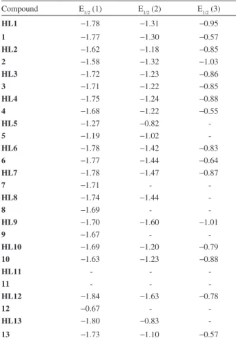
Cyclic voltammetry studies of HL1-HL13 and complexes 1-13
The redox behavior of compounds HL1-HL13 and their respective complexes 1-13 was evaluated by cyclic voltammetry (CV) at room temperature in dmso/(Bu4N)BF4 (0.1 mol L-1). The CVs were obtained in the potential range
Table 2. Carbonyl absorptions (cm-1) of the proligands HL1-HL13 and their copper(II) complexes 1-13
Proligand ν(C=O) Complexes ν(C=O)
HL1 1678 1 1683, 1658
HL2 1688 2 1685, 1663
HL3 1669 3 1675 (br)
HL4 1663 4 1691, 1640
HL5 1688 5 1690, 1640
HL6 1688 6 1690, 1640
HL7 1689 7 1688, 1639
HL8 1689 8 1690, 1640
HL9 1695 9 1689, 1650
HL10 1655 10 1693, 1647
HL11 1686 11 1668 (br)
HL12 1675 12 1672 (br)
Francisco et al. 1299 Vol. 21, No. 7, 2010
from +1.5 to –1.8V vs. FcH/FcH+ (Table 3). For most cases
three quasi-reversible pairs of waves were observed for the hydrazonocompounds in the negative region of the CV, which is attributed to the electron transfer of the hydrazono forms Ia/Ib and/or process associated to deprotonation of the hydroxyl group in forms IIa/III26 (Figure 1). The
redox potentials of the naphthoquinone unit are directly influenced by the substituents in the phenylene ring: electron-donor groups present lower E1/2 when compared to electron-releasing groups. The complexity of the CV observed for HL11 and complex 11 indicated that reduction potentials for the nitro group and the naphthoquinone are similar. Upon complexation, the E1/2(1) peak is slightly shifted to more positive potentials.
Antibacterial activity
The antibacterial activity of the hydrazonocompounds HL1-HL13 and their copper(II) complexes 1-13 was evaluated against seven strains of bacteria: Bacillus cereus (BC), Bacillus subtilis (BS), Escherichia coli (EC),
Enterococcus faecalis (EF), Klebsiella pneumoniae (KP),
Pseudomonas aeruginosa (PA) and Staphylococcus aureus
(SA). The results show that only compound HL5 exhibited signiicant activity against three strains of bacteria (BC, BS and EC). It inhibited EC growth at 20 µmol L-1, i.e., at lower
concentration than the positive control (chloramphenicol, 40-90 µmol L-1) and exhibited similar activity (90 µmol L-1)
to chloramphenicol against BC and BS strains. Furthermore, this compound was far more active against EC than the analogous 3-hydrazino-naphthoquinones derived from 3-diazo-naphthalene-1,2,4-trione.6 None of the complexes
showed signiicant antibacterial activity (> 200 µmol L-1);
furthermore, complex 5 was less active than its proligand HL5. Lawsone and CuCl2•2H
2O, tested for comparison,
only inhibited bacterial growth at high concentrations (see Supporting Information for detailed antibacterial assay data). Low activity of both proligands and complexes may be associated to their poor solubility and, therefore, low bioavailability. Same behavior was observed for the copper(II) complexes of aminonaphthoquinone Mannich bases derived from lawsone,3 and the metal complexes of
the anion of 5-amino-8-hydroxy-1,4-naphthoquinone.44
Antitumor activity
The antitumor screening of proligands HL1-HL13 and complexes 1-6, 9, 10, 12 and 13 was initially carried out against three strains of cancer cell lines: SF-295 (central nervous system), HCT-8 (colon) and MDAMB-435 (breast). Doxorubicin (dox) was used as a positive control. Lawsone was also tested for comparison (see Supplementary Information for antitumor activity data). Only the hydrazono compound HL6 exhibited higher growth inhibition of colon cancer cells HCT-8 (96.03%) than the positive control dox (91.67%); however, its complex, 6, did not show any signiicant activity. Complex 13 also exhibited higher antitumor activity (96.03 %) than dox. In four cases (HL2, HL4, HL9 and HL13) complexation resulted in increased antitumor activity against all cancer cell lines. The results indicate, in this system, that the presence of NO2 and I groups in ortho and
para positions, respectively, is relevant for the antitumor activity. Due to the high antitumor activity of compounds HL6 and 13, they were selected for IC50 determination against four cancer cell lines: SF-295 (central nervous system), HCT-8 (colon), MDAMB-435 (breast) and HL-60 (leukemia) and the results are presented in Table 4. Compound 13 (Entry 2) shows moderate cytotoxic activity against leukemia cell line.
Interestingly, the most active compound against HL-60 cell line (human leukemia), complex 13 (R = 2-NO2), also presents the lowest EPR parameter g||/A|| ratio, i.e., the weakest ield ligand (see Supporting Information). The EPR
Table 3. Voltammetric data (V) for HL1-HL13 and 1-13 vs. FcH/FcH+
Compound E1/2 (1) E1/2 (2) E1/2 (3)
HL1 −1.78 −1.31 −0.95
1 −1.77 −1.30 −0.57
HL2 −1.62 −1.18 −0.85
2 −1.58 −1.32 −1.03
HL3 −1.72 −1.23 −0.86
3 −1.71 −1.22 −0.85
HL4 −1.75 −1.24 −0.88
4 −1.68 −1.22 −0.55
HL5 −1.27 −0.82
-5 −1.19 −1.02
-HL6 −1.78 −1.42 −0.83
6 −1.77 −1.44 −0.64
HL7 −1.78 −1.47 −0.87
7 −1.71 -
-HL8 −1.74 −1.44 -
8 −1.69 -
-HL9 −1.70 −1.60 −1.01
9 −1.67 - -
HL10 −1.69 −1.20 −0.79
10 −1.63 −1.23 −0.88
HL11 - -
-11 - -
-HL12 −1.84 −1.63 −0.78
12 −0.67 -
-HL13 −1.80 −0.83
Theoretical Studies of the Tautomerism in 3-(2-R-Phenylhydrazono)-naphthalene-1,2,4-triones J. Braz. Chem. Soc. 1300
spectrum of this complex also shows a typical organic free radical line over the perpendicular copper(II) spectrum that is absent in the spectra of complexes 11 (R = 4-NO2) and 12 (R = 3-NO2) and may be responsible for the antitumor activity of complex 13.
Conclusions
The predominance of the keto-hydrazone tautomers Ia/Ib, as opposed to the previously reported enol-azo IIb, was conirmed by theoretical calculations that also established that, differently from the analogous 2-naphthol azo system, the nature of the substituent in the phenylene ring does not affect this stability order (Ib ≅Ia >> II). In spite of the higher stability of the keto-hydrazone Ia and Ib forms, deprotonation, followed by reaction with Cu2+, results in
the formation of enolate complexes of the azo form IIb. The antibacterial assays revealed that of all compounds only proligand HL5 (R = 3-I) inhibited the growth of EC at lower concentration than the positive control (chloramphenicol), thus structure/reduction potential-activity correlations could not be attempted. In the antitumor screening, in general, complexes were found to be more active than the respective proligands; however, only complex 13 (R = 2-NO2) showed moderate cytotoxic activity against HL-60 cells. Formation of an organic free radical observed in the EPR spectrum of this complex might be responsible for this cytotoxic behaviour.
Supplementary Information
Supplementary information associated with this paper contains the results of the theoretical calculations, spectroscopic data, NMR spectra (1H and 13C), EPR,
voltammograms, antibacterial and antitumor assay results of compounds HL1-HL13 and the complexes 1-13. These data are available free of charge at http://jbcs.sbq.org.br, as a PDF ile.
Acknowledgments
The authors thank the Brazilian agencies Conselho Nacional de Desenvolvimento Cientíico e Tecnológico (CNPq) and Fundação de Amparo à Pesquisa do Estado
do Rio de Janeiro (FAPERJ) for financial support. Pronex-FAPERJ (grant number E-26/171.512/2006) is acknowledged. J. W. D. C, M. D. V, V. F. F., A. S. M. and L. V. C.-L. are recipients of CNPq research fellowships. A. I. F. and A. C. were beneited with CAPES fellowships and J. P. B. is grateful to FAPERJ for a visitor research fellowship.
References
1. Nagaoka, K.; Kishi, Y.; Tetrahedron 1981, 37, 3783. 2. Furusaki, A.; Watanabi, T.; Chem. Pharm. Bull. 1973, 21, 931. 3. Neves, A. P.; Barbosa, C. C.; Greco, S. J.; Vargas, M. D.;
Visentin, L. C.; Pinheiro, C. B.; Mangrich, A. S.; Barbosa, J. P.; da Costa, G. L.; J. Braz. Chem. Soc. 2009, 20, 712. 4. Tandon, V. K.; Maurya, H. K.; Mishra, N.; Shukla, P. K.; Eur.
J. Med. Chem. 2009, 44, 3130.
5. Kim, H.-W.; Lee, C.-H.; Lee, H.-S.; Food. Sci. Biotechnol. 2009, 18, 755.
6. Oliveira, C. G. T.; Miranda, F. F.; Ferreira, V. F.; Freitas, C. C.; Rabello, R. F.; Carballido, J. M.; Corrêa, L. C. D.; J. Braz. Chem. Soc. 2001, 12, 339.
7. Crosby, I. T.; Rose, M. L.; Collis, M. P.; de Bruyn, P. J.; Keep, P. L. C.; Robertson, A. D.; Aust. J. Chem. 2008, 61, 768. 8. Stagliano, K. W.; Emadi, A.; Lu, Z.; Malinakova, H. C.; Twenter,
B.; Yu, M.; Holland, L. E.; Rom, A. M.; Harwood, J. S.; Amin, R.; Johnson, A. A.; Pommier, Y.; Bioorg. Med. Chem. 2006, 14, 5651.
9. Da Silva Junior, E.; de Moura, M. A. B. F.; Pinto, A. V.; Pinto, M. C. F. R.; de Souza, M. C. B. V.; Araujo, A. J.; Pessoa, C.; Costa-Lotufo, L. V.; Montenegro, R. C.; de Moraes, M. O.; Ferreira, V. F.; Goulart, M. O. F.; J. Braz. Chem. Soc. 2009, 20, 635; Tapia, R. A.; Cantuarias, L.; Cuellar, M.; Villena, J.; J. Braz. Chem. Soc. 2009, 20, 999.
10. Pinto, C. N.; Dantas, A. P.; De Moura, K. C. G.; Emery, F. S.; Polequevitch, P. F.; Pinto, M. D.; De Castro, S. L.; Pinto, A. V.; Arzneim. Forsc./Drug Res. 2000, 50, 1120.
11. Goulart, M. O. F.; Zani, C. L.; Tonholo, J.; Freitas, L. R.; de Abreu, F. C.; Oliveira, A. B.; Raslan, D. S.; Starling, S.; Chiari, E.; Bioorg. Med. Chem. Lett. 1997, 7, 2043.
12. Da Silva Junior, E. N.; Menna-Barreto, R. F. S.; Pinto, M. do C. F. R.; Silva, R. S. F.; Teixeira, D. V.; de Souza, M. C. B. V.; de Simone, C. A.; de Castro, S. L.; Ferreira, V. F.; Pinto, A. V.; Eur. J. Med. Chem. 2008, 43, 1774; da Silva Junior, E. N.; de Table 4. Cytotoxic activity ofcompounds HL6 and 13, expressed in IC50, obtained by MTT assay after incubation of cells for 72 h in the concentrations 0.01-5 µg mL-1
Compound Entry SF295 HCT-8 MDA-MB435 HL-60
HL6 (R = 3-I) 1 > 5 > 5 > 5 > 5
Francisco et al. 1301 Vol. 21, No. 7, 2010
Souza, M. C. B. V.; Pinto, A. V.; Pinto, M. do C. F. R.; Goulart, M. O. F.; Barros, F. W. A.; Pessoa, C.; Costa-Lotufo, L. V.; Montenegro, R. C.; de Moraes, M. O.; Ferreira, V. F.; Bioorg. Med. Chem. 2007, 15, 7035.
13. Fry, F. H.; Jacob, C.; Curr. Pharm. Design 2006, 12, 4479; Asche, C.; Mini-Rev. Med. Chem. 2005, 5, 449; Hassani, M.; Cai, W.; Holley, D. C.; Lineswala, J. P.;Maharjan, B. R.; Ebrahimian, G. R.; Seradj, H.; Stocksdale, M. G.; Mohammadi, F.; Marvin, C. C.; Gerdes, J. M.; Beall, H. D.; Behforouz, M.; J. Med. Chem. 2005, 48, 7733; Lee, J. H.; Cheong, J. H.; Park, Y. M.; Choi, Y. H.; Pharmacol. Res. 2005, 51, 553; Liu, K. K. C.; Li, J.; Sakya, S.; Mini-Rev. Med. Chem. 2004, 4, 1105; Kongkathip, N.; Siripong, P.; Sangma, C.; Luangkamin, S.; Niyomdecha, M.; Pattanapa, S.; Piyaviriyalgul, S.; Kongsaeree, P.; Bioorg. Med. Chem. 2003, 11, 3179.
14. Vargas, M. D.; Pinto, A. C.; Echevarria, A.; Esteves-Souza, A.; Camara, C. A.; Cunha, A. C.; Torres, J. C.; Lima, E. L. S.; J. Braz. Chem. Soc. 2006, 17, 439; Esteves-Souza, A.; Figueiredo, D. V.; Esteves, A.; Câmara, C. A.; Vargas, M. D.; Pinto, A. C.; Echevarria, A.; Braz. J. Med. Biol. Res. 2007, 30, 1399; Cunha, A. S.; Vargas, M. D.; Gattass, C. R.; Pinto, A. C.; Camara, C. A.; Esteves, A. S.; Lima, E. L. S.; Oncol. Rep. 2008, 20, 225.
15. Da Silva, A. J. M.; Netto, C. D.; Pacienza-Lima, W.; Torres-Santos, E. C.; Rossi-Bergmann, B.; Maurel, S.; Valentin, A.; Costa, P. R. R.; J. Braz. Chem. Soc. 2009, 20, 176.
16. Dos Santos, E. V. M.; Carneiro, J. W. de M.; Ferreira, V. F.; Bioorg. Med. Chem. 2004, 12, 87.
17. Fieser, L. F.; Nazer, M. Z.; Archer, S.; Berberian, D. A.; Slighter, R. G.; J. Med. Chem. 1967, 10, 517.
18. Tandon, V. K.; Maurya, H. K.; Mishra, N. N.; Shukla, P. K.; Eur. J. Med. Chem. 2009, 44, 3130.
19. Macias-Rubalcava, M. L.; Hernandez-Bautista, B. E.; Jimenez-Estrada, M.; Gonzalez, M. C.; Glenn, A. E.; Hanlin, R. T.; Hernandez-Ortega, S.; Saucedo-Garcia, A.; Muria-Gonzalez, J. M.; Anaya, A. L.; Phytochemistry 2008, 69, 1185.
20. Barbosa, T. P.; Camara, C. A.; Silva, T. M. S.; Martins, R. M.; Pinto, A. C.; Vargas, M. D.; Bioorg. Med. Chem. 2005, 13, 6464; dos Santos, A. F.; Ferraz, P. A. L.; de Abreu, F. C.; Chiari, E.; Goulart, M. O. F.; Sant’Ana, A. E. G.; Planta Med. 2001, 67, 92; Dos Santos, A. F.; Ferraz, P. A. L.; Pinto, A. V.; Pinto, M. C. F. R.; Goulart, M. O. F.; Sant’Ana, A. E. G.; Int. J. Parasitol. 2000, 30, 1199.
21. Kovacic, P.; Becvar, L. E.; Curr. Pharm. Des. 2000, 6, 143; Monks, T. J.; Hanzlik; P.; Cohen, G. M.; Ross, D.; Graham, D. G.; Toxicol. Appl. Pharmacol. 1992,112, 2.
22. Ferraz, P. A. L.; de Abreu, F. C.; Pinto, A. V.; Glezer, V.; Tonholo, J.; Goulart, M. O. F.; J. Electroanal. Chem. 2001, 507, 275; Kumagai, Y.; Tsurutani, Y.; Shinyashiki, M.; Takeda, S. H.; Nakai, Y.; Yoskikawa, T.; Shimojo, N.; Environ. Toxicol. Pharmacol. 1997, 3, 245.
23. Hsieh, B. R.; Desilets, D.; Kazmaier, P. M.; Dyes Pigm. 1990, 14, 165; Carvalho, C. E. M.; Ferreira, V. F.; Pinto, A. V.; Pinto, M. C. F. R.; Harrison, W.; Dyes Pigm. 2002, 52, 209. 24. Lycka, A.; Jirman, J.; Podstata, J.; Dyes Pigm. 1987, 8, 465. 25. Alarcón, S. H.; Olivieri, A. C.; Sanz, D.; Claramunt, R. M.;
Elguero, J.; J. Mol. Struct. 2004, 705, 1.
26. Gokhale, N. H.; Padhye, S. B.; Newton, C.; Pritchard, R.; Metal Based Drugs 2000, 7, 121.
27. Gokhale, N. H.; Shirisha, K.; Padhye, S. B.; Croft, S. L.; Kendrick, H. D.; McKee, V.; Bioorg. Med. Chem. Lett. 2006, 16, 430.
28. Martins, F. T.; Cruz Jr., J. W.; Derogis, P. B. M. C.; dos Santos, M. H.; Veloso, M. P.; Ellena, J.; Doriguetto, A. C.; J. Braz. Chem. Soc. 2007, 18, 1515.
29. Farrera, J.-A.; Canal, I.; Hidalgo-Fernández, P.; Pérez-García, M. L.; Huertas, O.; Luque, F. J.; Chem. Eur. J. 2008, 14, 2277.
30. Romanyuk, A. L.; Polishchuk, O. P.; Litvin, B. L.; Ganashchak, N. I.; Russ. J. Gen. Chem. 2002, 72, 251.
31. Francisco, A. I.; Casellato, A.; Vargas, M. D.; Org. Prep. Proced. Int. 2009, 41, 323.
32. Frisch, M. J.; Trucks, G. W.; Schlegel, H. B.; Scuseria, G. E.; Robb, M. A.; Cheeseman, J. R.; Montgomery, J. A.; Vreen Jr., T.; Kudin, K. N.; Burat, J. C.; Millam, J. M.; Iyengar, S. S.; Tomsi, J.; Barone, V.; Mennucci, B.; Cossi, M.; Scalmani, G.; Rega, N.; Petersson, G. A.; Nakatsuji, H.; Hada, M.; Ehara, M.; Toyota, K.; Fukua, R.; Hasegawa, J.; Ishida, M.; Nakajim, T.; Honda, Y.; Kitao, O.; Nakai, H.; Klene, M.; Li, X.; Knox, J. E.; Hratchian, H. P.; Cross, J. B.; Adamo, C.; Jaramillo, J.; Gomperts, R.; Stratmann, R. E.; Yazyev, O.; Austin, A. J.; Cammi, R.; Pomelli, C.; Ochteski, J. W.; Ayala, P. Y.; Morokuma, K.; Voth, G. A.; Salvador, P.; Dannenberg, J. J.; Zakrzewski, V. G.; Dapprich, S.; Daniels, A. D.; Strain, M. C.; Faras, O.; Malick, D. K.; Rabu, A. D.; Raghavachari, K.; Foresman, J. B.; Ortiz, J. V.; Cui, Q.; Baboul, A. G.; Clifford, S.; Cioslowski, J.; Stefanov, B. B.; Liu, G.; Liashenko, A.; Piskorz, P.; Komaromi, I.; Martin, R. L.; Fox, D. J.; Keith, T.; Al-Laham, M. A.; Peng, C.; Nanayakkra, A.; Challacombe, M.; Gill, P. M. W.; Johnson, B.; Chen, W.; Wong, M. W.; Gonzalez, C.; Pople, J. A.; Gaussian 03, Revision B.02, Gaussian, Inc.: Pittsburg P. A., 2003.
33. Beck, A. D.; J. Chem. Phys. 1993, 98, 5648.
34. Hariharan, P. C.; Pople, J. A.; Theor. Chim. Acta 1973, 28, 213. 35. Cossi, M.; Rega, N.; Scalmani, G.; Barone, V.; J. Comput. Chem.
2003, 24, 669.
36. Langield, R. D.; Scarano, F. J.; Heitzman, M. E.; Kondo, M.; Hammond, G. B.; Neto, C. C.; J. Ethnopharmacol. 2004, 94, 279.
37. Eloff, J. N.; Planta Med. 1998, 65, 711. 38. Mosmann, T.; J. Immunol. Methods 1983, 65, 55.
Theoretical Studies of the Tautomerism in 3-(2-R-Phenylhydrazono)-naphthalene-1,2,4-triones J. Braz. Chem. Soc. 1302
40. Fragoso, T. P.; Carneiro, J. W. D.; Vargas, M. D.; J. Mol. Model. 2009, doi: 10.1007/s00894-009-0579-x.
41. Silverstein, R.; Bassaler, G.; Morill, T.; Spectroscopic Identiication of Organic Compounds, 5th ed., John Wiley & Sons Inc.: New York, 2003.
42. Ispir, E.; Dyes Pigm. 2009, 82, 13.
43. Sakaguchi, U.; Addison, A. W.; J. Chem. Soc. Dalton Trans. 1979, 600.
44. Ma, S.; Zhu, W.; Xu, M.; Wang, Y.; Guo, Q.; Liu, Y.; Polyhedron 2003, 22, 3249.
Supplementary Information
J. Braz. Chem. Soc., Vol. 21, No. 7, S1-S27, 2010. Printed in Brazil - ©2010 Sociedade Brasileira de Química 0103 - 5053 $6.00+0.00*e-mail: [email protected]
Theoretical Studies of the Tautomerism in 3-(2-R-Phenylhydrazono)-naphthalene-
1,2,4-triones: Synthesis of Copper(II) Complexes and Studies of Antibacterial
and Antitumor Activities
Acácio I. Francisco,a Maria D. Vargas,*,a Thaís P. Fragoso,a J. Walkimar de M. Carneiro,a
Annelise Casellato,b Fernando de C. da Silva,a Vitor F. Ferreira,a Jussara P. Barbosa,c Claudia Pessoa,d
Letícia V. Costa-Lotufo,d José D. B. Marinho Filho,d Manoel O. de Moraesd and Antonio S. Mangriche
aInstituto de Química, Universidade Federal Fluminense, Campus do Valonguinho, Centro,
24020-150 Niterói-RJ, Brazil
bInstituto de Química, Universidade Federal do Rio de Janeiro, Ilha do Fundão, 21945-970 Rio de Janeiro-RJ, Brazil
cInstituto Oswaldo Cruz, FIOCRUZ, CP 926, 21045-900 Rio de Janeiro-RJ, Brazil
dUniversidade Federal do Ceará, Depto de Fisiologia e Farmacologia, Campus do Porangabussu,
60430-270 Fortaleza-CE, Brazil
eDepartamento de Química da Universidade Federal do Paraná, 81531-990 Curitiba-PR, Brazil
Synthesis and characterization of the hydrazone compounds
HL1-HL13
Syntheses
A mixture of the respective arylamine (6.63 mmol), water (3.5 mL) and concentrated HCl (3.5 mL) was stirred until dissolution. The solution was then cooled by addition of crushed ice (1.00 g). When the temperature reached 0 oC, NaNO
2 (4.39 mmol, 0.303 g) in cold water (2 mL) was added to the mixture.The solution was stirred at 0 oC for 20 min and then added dropwise to a stirred solution of 2-hydroxy-1,4 naphthoquinone (3.65 mmol, 0.635 g) and sodium hydroxide (10.95 mmol, 0.438 g) in ethanol (28 mL) kept at 0 oC. The resulting orange solids were iltered, washed with cold water and ethanol and dried under vacuum.
Analytical and spectroscopic data for HL1-HL13
3-[2-(4-methoxy)phenylhydrazono]-naphthalene-1,2,4-trione (HL1)
From 0.815 g of 4-methoxyaniline. Yield: 0.922 g, 82%, mp 229-230 ºC. Anal. Calcd. for C17H12N2O4: C 66.04; H 4.29; N 8.76%. Found: C 66.06; H 4.32; N 8.67%. IR (KBr, νmax/cm-1):3435 (N-H), 3078 (C-H arom.), 2847 (C-H alif.), 1678 (C=O/C=N), 1587 (C=C), 1260 (C-O), 972 (C-N=N-C). 1H NMR (dmso-d
6, 300 MHz): d 8.33 (bd,
J 7.74 Hz, 1H), 8.21 (dd, J 7.74, 0.48 Hz, 1H), 8.06 (td, J
7.74, 7.74, 0.48 Hz, 1H), 7.98 (td, J 7.74, 7.74, 0.48 Hz, 1H), 7.87 (bd, J 6.42 Hz, 2H), 7.23 (bd, J 6.42 Hz, 2H), 3.94 (s, 3H). 13C NMR (dmso-d
6, 75 MHz): d 160.0, 136.1, 135.4, 135.1, 134.4, 128.2, 127.9, 120.7, 116.0, 56.3. UV-Vis [dmso; λ/nm (log ε)]: 276 (4.51), 473 (3.52).
3-[2-(4-benzoyl)phenylhydrazono]-naphthalene-1,2,4-trione (HL2)
From 1.306 g of 4-aminoazobenzol. Yield: 1.101 g, 79%, mp 290-292 ºC. Anal. Calcd. for C22H14N4O3: C 69.10; H 3.69; N 14.65%. Found: C 69.05; H 3.73; N 14.69%. IR (KBr, νmax/cm-1):3439 (N-H), 3062, 3102 (C-H arom.), 1688 (C=O/C=N), 1590 (C=C), 1255 (C-O), 974 (C-N=N-C). 1H NMR (dmso-d
6, 300 MHz): d 8.36 (bd,
J 7.38 Hz, 1H), 8.24 (dd, J 7.38, 1.48 Hz, 1H), 8.15 (bd,
J 8.85 Hz, 2H), 8.12-7.98 (m, 6H), 7.76-7.67 (m, 3H). 13C NMR (dmso-d
6, 75 MHz): d 175.8, 152.4, 150.4, 144.1, 135.4, 131.9, 129.8, 127.5, 124.7, 118.8. UV-Vis [dmso; λ/nm (log ε)]: 257 (4.73), 459 (3.79).
3-[2-(4-chloro)phenylhydrazono]-naphthalene-1,2,4-trione (HL3)
From 0.842 g of 4-chloroaniline. Yield: 0.945 g, 83%, mp 215-217 ºC. Anal. Calcd. for C16H9ClN2O3: C 61.45; H 2.90; N 8.96%. Found: C 61.65; H 3.10; N 9.07%. IR (KBr, νmax/cm-1):3428 (N-H), 3083 (C-H arom.), 1695 (C=O/C=N) and 1669 (C=O), 1594 (C=C), 1255 (C-O), 969 (C-N=N-C). 1H NMR (dmso-d
6, 300 MHz): d 8.34 (bd,
J 7.28 Hz, 1H), 8.24 (dd, J 7.28, 1.81 Hz, 1H), 8.07 (td, J
Theoretical Studies of the Tautomerism in 3-(2-R-Phenylhydrazono)-naphthalene-1,2,4-triones J. Braz. Chem. Soc.
S2
1H), 7.92 (bd, J 8.80 Hz, 2H), 7.69 (bd, J 8.80 Hz, 2H). 13C NMR (dmso-d
6, 75 MHz): d 140.6, 135.1, 129.7, 127.1, 119.4. UV-Vis [dmso; λ/nm (log ε)]: 266 (4.33), 431 (3.48).
3-[2-(4-iodo)phenylhydrazono]-naphthalene-1,2,4-trione (HL4)
From 1.452 g of 4-iodoaniline. Yield: 1.179 g, 80%, mp 201-202 ºC. Anal. Calcd. for C16H9IN2O3: C 47.55; H 2.24; N 6.93%. Found: C 47.57; H 2.34; N 7.04%. IR (KBr, νmax/cm-1):3438 (N-H), 3084 (C-H arom.), 1690 (C=O/ C=N) and 1663 (C=O), 1599 (C=C), 1257 (C-O), 969 (C-N=N-C). 1H NMR (dmso-d
6, 300 MHz): d8.32 (dd, J 7.31, 1.55 Hz, 1H), 8.22 (dd, J 7.31, 1.55 Hz, 1H), 8.10-8.02 (m, 2H), 7.97 (bd, J 8.86 Hz, 2H), 7.69 (bd, J 8.86 Hz, 2H). 13C NMR (dmso-d
6, 75 MHz): d 175.9, 141.9, 139.0, 135.6, 134.7, 133.9, 128.1, 127.7, 120.4. UV-Vis [dmso; λ/nm (log ε)]: 298 (4.57), 438 (3.61).
3-[2-(3-iodo)phenylhydrazono]-naphthalene-1,2,4-trione (HL5)
From 1.452 g of 3-iodoaniline. Yield: 1.135 g, 77%, mp 216-218 ºC. Anal. Calcd. for C16H9IN2O3: C 47.55; H 2.24; N 6.93%. Found: C 47.53; H 2.21; N 6.90%. IR (KBr, νmax/cm-1):3438 (N-H), 3087 (C-H arom.), 1688 (C=O/C=N), 1600 (C=C), 1261 (C-O), 969 (C-N=N-C). 1H NMR (dmso-d
6, 300 MHz): d 8.35 (dd, J 7.35, 1.49 Hz, 1H), 8.29-8.21 (m, 2H), 8.07 (td, J 7.35, 7.35, 1.49 Hz, 1H), 8.01 (td, J 7.35, 7.35, 1.49 Hz, 1H), 7.90 (dd, J 7.35, 1.57 Hz, 1H), 7.79 (dl, J 7.35 Hz, 1H), 7.42 (bt, J 7.35 Hz, 1H). 13C NMR (dmso-d
6, 75 MHz): d 175.3, 142.7, 135.4, 134.4, 135.0, 131.5, 127.1, 125.7, 117.2, 95.2. UV-Vis [dmso; λ/nm (log ε)]: 270 (4.61), 425 (3.32).
3-[2-(2-iodo)phenylhydrazono]-naphthalene-1,2,4-trione (HL6)
From 1.452 g of 2-iodoaniline. Yield: 1.297 g, 88%, mp 230-232 ºC. Anal. Calcd. for C16H9IN2O3: C 47.55; H 2.24; N 6.93%. Found: C 47.55; H 2.24; N 6.93%. IR (KBr, νmax/cm-1):3438 (N-H), 3087 (C-H arom.), 1688 (C=O/C=N), 1601 (C=C), 1259 (C-O), 969 (C-N=N-C). 1H NMR (dmso-d
6, 300 MHz): d 8.37 (bd, J 7.33 Hz, 1H), 8.26 (bd, J 7.33 Hz, 1H), 8.12-7.91 (m, 4H), 7.70 (bt, J 7.63 Hz, 1H), 7.23 (bt, J 7.63 Hz, 1H). 13C NMR (dmso-d
6, 75 MHz): d 178.9, 141.5, 139.6, 135.0, 134.5, 134.0, 133.4, 133.1, 132.5, 129.9, 128.6, 127.7, 127.2, 117.6, 117.4, 87.5. UV-Vis [dmso; λ/nm (log ε)]: 276 (4.44), 434 (3.33).
3-[2-(4-carboxy)phenylhydrazono]-naphthalene-1,2,4-trione (HL7)
From 0.908 g of 4-carboxyaniline. Yield: 1.046 g, 89%, mp 235-237 ºC. Anal. Calcd. for C17H10N2O5: C 62.66;
H 3.22; N 8.65%. Found: C 62.55; H 3.25; N 8.67%. IR (KBr, νmax/cm-1):3451-2700 (O-H COOH/N-H), 3068 (C-H arom.), 1689 (C=O/C=N), 1598 (C=C), 1263 (C-O), 968 (C-N=N-C). 1H NMR (dmso-d
6, 300 MHz): d 8.35 (bd, J 8.41 Hz, 1H), 8.24 (dd, J 8.41, 1.53 Hz, 1H), 8.17 (bd, J
7.44 Hz, 2H), 8.08 (td, J 8.41, 8.41, 1.53 Hz, 1H), 8.02 (td,
J 8.41, 8.41, 1.53 Hz, 1H), 7.96 (bd, J 7.44 Hz, 2H). 13C NMR (dmso-d6, 75 MHz): d 175.5, 166.5, 144.8, 135.0, 131.0, 128.6, 127.1, 117.4. UV-Vis [dmso; λ/nm (log ε)]: 277 (4.49), 424 (3.67).
3-[2-(3-carboxy)phenylhydrazono]-naphthalene-1,2,4-trione (HL8)
From 0.908 g of 3-carboxyaniline. Yield: 0.834 g, 71%, mp 270-272 ºC. Anal. Calcd. for C17H10N2O5: C 62.66; H 3.22; N 8.65%. Found: C 62.67; H 3.20; N 8.64%. IR (KBr, νmax/cm-1):3449-2600 (O-H COOH/N-H), 3051 (C-H arom.), 1689 (C=O/C=N), 1605 (C=C), 1251 (C-O), 968 (C-N=N-C). 1H NMR (dmso-d
6, 300 MHz): d 8.36 (m, 1H), 8.31 (dd, J 7.42, 1.45 Hz, 1H), 8.13 (dd, J 7.42, 1.45 Hz, 1H), 8.05-7.95 (m, 3H), 7.90 (td, J 7.42, 7.42, 1.45 Hz, 1H), 7.71 (bt, J 7.51 Hz, 1H). 13C NMR (dmso-d
6, 75 MHz): d 179.1, 172.0, 167.7, 147.8, 135.4, 134.0, 133.5, 133.1, 130.0, 128.3, 127.4, 127.2, 124.0, 120.0. UV-Vis [dmso; λ/nm (log ε)]: 271 (4.63), 441 (3.63).
3-[2-(4-ciano)phenylhydrazono]-naphthalene-1,2,4-trione (HL9)
From 0.782 g of 4-cianoaniline. Yield: 0.929 g, 84%, mp 255-259 ºC. Anal. Calcd. for C17H9N3O3: C 67.33; H 2.99; N 13.86%. Found: C 67.69; H 3.01; N 13.88%. IR (KBr, νmax/cm-1):3426 (N-H), 3070 (C-H arom.), 2220 (CN), 1695 (C=O/C=N), 1607 (C=C), 1260 (C-O), 967 (C-N=N-C). 1H NMR (dmso-d
6, 300 MHz): d 8.34 (dd, J 6.93, 1.65 Hz, 1H), 8.24 (dd, J 6.93, 1.65 Hz, 1H), 8.10-7.99 (m, 6H). 13C NMR (dmso-d
6, 75 MHz): d 179.4, 176.0, 135.6, 134.8, 134.4, 128.0, 127.7, 119.1, 118.6, 108.7. UV-Vis [dmso; λ/nm (log ε)]: 282 (4.77), 425 (3.42).
3-[2-(3-ciano)phenylhydrazono]-naphthalene-1,2,4-trione (HL10)
From 0.782 g of 3-cianoaniline. Yield: 0.818 g, 74%, mp 280-281 ºC. Anal. Calcd. for C17H9N3O3: C 67.33; H 2.99; N 13.86%. Found: C 67.30; H 2.96; N 13.87%. IR (KBr, νmax/cm-1):3431 (N-H), 3080 (C-H arom.), 2231 (CN), 1692 (C=O/C=N) and 1655 (C=O), 1600 (C=C), 1261 (C-O), 972 (C-N=N-C). 1H NMR (dmso-d
Francisco et al. S3 Vol. 21, No. 7, 2010
3-[2-(4-nitro)phenylhydrazono]-naphthalene-1,2,4-trione (HL11)
From 0.915 g of 4-nitroaniline. Yield: 1.084 g, 92%, mp 275-276 ºC. Anal. Calcd. for C16H9N3O5: C 59.45; H 2.81; N 13.00%. Found: C 59.33; H 2.87; N 13.05%. IR (KBr, νmax/cm-1): 3452 (N-H), 3084 (C-H arom.), 1686 (C=O/C=N), 1512 and 1316 (NO2), 1264 (C-O), 968 (C-N=N-C). 1H NMR (dmso- d6, 300 MHz): d8.48 (bd, J 8.51 Hz, 2H), 8.34 (dd, J 7.07, 1.64 Hz, 1H), 8.25 (dd, J 7.07, 1.64 Hz, 1H), 8.11-7.99 (m, 4H). 13C NMR (dmso-d
6, 75 MHz): d 184.1, 181.1, 152.5, 150.1, 140.4, 139.0, 133.0, 132.7, 131.1, 123.2. UV-Vis [dmso; λ/nm (log ε)]: 290 (4.55), 429 (3.71).
3-[2-(3-nitro)phenylhydrazono]-naphthalene-1,2,4-trione (HL12)
From 0.915 g of 3-nitroaniline. Yield: 0.955 g, 81%, mp 167-168 ºC. Anal. Calcd. for C16H9N3O5: C 59.45; H 2.81; N 13.00%. Found: C 59.28; H 2.97; N 12.99%. IR (KBr, νmax/cm-1):3445 (N-H), 3090 (C-H arom.), 1675 (C=O/C=N), 1496 and 1315 (NO2), 1261 (C-O), 973 (C-N=N-C). 1H NMR (dmso-d
6, 300 MHz): d 8.71 (m, 1H), 8.35 (dd, J 7.97, 1.93 Hz, 1H), 8.31 (dd, J 7.97, 1.93
Hz, 1H), 8.22 (m, 2H), 8.07 (td, J 7.97, 7.97, 1.93 Hz, 1H), 8.01 (td, J 7.97, 7.97, 1.93 Hz, 1H), 7.90 (bt, 7.54 Hz, 1H). 13C NMR (dmso-d
6, 75 MHz): d 184.1, 181.1, 153.7, 150.0, 141.2, 133.7, 131.3, 123.5. UV-Vis [dmso; λ/nm (log ε)]: 293 (4.57), 416 (3.68).
3-[2-(2-nitro)phenylhydrazono]-naphthalene-1,2,4-trione (HL13)
From 0.915 g of 2-nitroaniline. Yield: 1.297 g, 78%, mp 249-250 ºC. Anal. Calcd. for C16H9N3O5: C 59.45; H 2.81; N 13.00%. Found: C 59.52; H 2.89; N 12.95%. IR (KBr, νmax/cm-1):3455 (N-H), 3090 (C-H arom.), 1683 (C=O/C=N), 1600 (C=C), 1457 and 1332 (NO2), 1271 (C-O), 982 (C-N=N-C). 1H NMR (dmso-d
6, 300 MHz): d 8.37 (bd, J 7.33 Hz, 1H), 8.26 (bd, J 7.33 Hz, 1H), 8.12-7.91 (m, 4H), 7.70 (bt, J 7.63 Hz, 1H), 7.23 (bt, J 7.63 Hz, 1H). 13C NMR (dmso-d6, 75 MHz): d 178.9, 141.5, 139.6, 135.0, 134.5, 134.0, 133.4, 133.1, 132.5, 129.9, 128.6, 127.7, 127.2, 117.6, 117.4, 87.5. UV-Vis [dmso; λ/nm (log ε)]: 278 (4.52), 440 (3.74).
1H and 13C NMR spectra of compounds HL1-HL13
Theoretical Studies of the Tautomerism in 3-(2-R-Phenylhydrazono)-naphthalene-1,2,4-triones J. Braz. Chem. Soc.
S4
Figure S2. 13C NMRspectrum of3-[2-(4-benzoyl)phenylhydrazono]-naphthalene-1,2,4-trione (HL2).
Francisco et al. S5 Vol. 21, No. 7, 2010
Theoretical Studies of the Tautomerism in 3-(2-R-Phenylhydrazono)-naphthalene-1,2,4-triones J. Braz. Chem. Soc.
S6
Figure S6. 13C NMR spectra of 3-[2-(4-iodo)phenylhydrazono]-naphthalene-1,2,4-trione (HL4).
Francisco et al. S7 Vol. 21, No. 7, 2010
Figure S8. 13C NMR spectrum of 3-[2-(3-iodo)phenylhydrazono]-naphthalene-1,2,4-trione (HL5).
Theoretical Studies of the Tautomerism in 3-(2-R-Phenylhydrazono)-naphthalene-1,2,4-triones J. Braz. Chem. Soc.
S8
Figure S10. 13C NMRspectrum of 3-[2-(2-iodo)phenylhydrazono]-naphthalene-1,2,4-trione (HL6).
Francisco et al. S9 Vol. 21, No. 7, 2010
Theoretical Studies of the Tautomerism in 3-(2-R-Phenylhydrazono)-naphthalene-1,2,4-triones J. Braz. Chem. Soc.
S10
Figure S14. 13C NMRspectrum of 3-[2-(3-carboxy)phenylhydrazono]-naphthalene-1,2,4-trione (HL8).
Francisco et al. S11 Vol. 21, No. 7, 2010
Figure S16. 13C NMRspectrum of 3-[2-(4-ciano)phenylhydrazono]-naphthalene-1,2,4-trione (HL9).
Theoretical Studies of the Tautomerism in 3-(2-R-Phenylhydrazono)-naphthalene-1,2,4-triones J. Braz. Chem. Soc.
S12
Figure S18. 13C NMRspectrum of 3-[2-(3-ciano)phenylhydrazono]-naphthalene-1,2,4-trione (HL10).
Francisco et al. S13 Vol. 21, No. 7, 2010
Figure S20. 13C NMR spectrum of 3-[2-(4-nitro)phenylhydrazono]-naphthalene-1,2,4-trione (HL11).
Theoretical Studies of the Tautomerism in 3-(2-R-Phenylhydrazono)-naphthalene-1,2,4-triones J. Braz. Chem. Soc.
S14
Figure S22. 13C NMRspectrum of 3-[2-(3-nitro)phenylhydrazono]-naphthalene-1,2,4-trione (HL12).
Francisco et al. S15 Vol. 21, No. 7, 2010
Theoretical calculations
Relative energies of tautomers
The table below presents absolute (hartree) and relative
Table S1. Absolute and relative energies (kcal mol-1) of tautomers Ia, Ib, IIa and IIb with different substituents
R Ia Ib IIa III
H −950.86824
0.49 −950.07927 0.23 −950.86902 0.0 −950.07963 0.0 −950.84219 16.84 −950.05670 14.39 −950.85681 7.66 −950.06850 6.98
CN −1043.11014
0.34 −1042.24636 -0.01 −1043.11068 0.0 −1042.24634 0.0 −1043.08445 16.46 −1042.22382 14.13 −1043.09815 7.86 −1042.23440 7.49
NO2 −1155.36828
0.29 −1154.44761 -0.03 −1155.36875 0.0 −1154.44757 0.0 −1155.34229 16.60 −1154.42494 14.20 −1155.35575 8.16 −1154.43524 7.74
NH2 −1006.22180
0.67 −1005.40727 0.58 −1006.22287 0.0 −1005.40819 0.0 −1006.19911 14.91 −1005.38686 13.38
OCH3 −1065.39115
0.63 −1064.51448 0.36 −1065.39216 0.0 −1064.51505 0.0 −1065.36720 15.66 −1064.49338 13.60 −1065.38181 6.49 −1064.50567 5.89
energies (kcal mol-1) of tautomers Ia, Ib, IIa and III with different substituents. In the irst entry B3LYP/6-31G(d) results are given. The second entry refers to PBE1PBE/6-311+G(2d,p) results, including solvent effect (dmso).
Electronic spectra of tautomers Ia and Ib
The electronic spectra of the hydrazono compounds were calculated with the RPBE/6-311+G(2d,p) method with inclusion of solvent effects using the CPCM approach and dmso as solvent. The geometries were optimized at the B3LYP/6-31G(d) level. These compounds may exist as a set of tautomers of which the two rotamers Ia and Ib
are the most stable (Figure S24). For each tautomer two high intensity bands were calculated. In each case they correspond to transitions of the type HOMO → LUMO and HOMO → LUMO+1. HOMO is one of the π orbitals of the phenyl ring (Figures S25 and S28) while LUMO and LUMO+1 both are π* orbitals of the quinone ring (Figures S26 and S29, respectively).
Theoretical Studies of the Tautomerism in 3-(2-R-Phenylhydrazono)-naphthalene-1,2,4-triones J. Braz. Chem. Soc.
S16
Table S2. Theoretical electronic spectra of tautomers Ia and Ib(*)
Ib Ia
R = H
λ1 (HOMO → LUMO) 449 (0.13) 457 (0.34)
λ2 (HOMO → LUMO+1) 407 (0.81) 387 (0.60)
R = CN
λ1 (HOMO → LUMO) 426 (0.42) 438 (0.58)
λ2 (HOMO → LUMO+1) 399 (0.75) 375 (0.59)
R = NO2
λ1 (HOMO → LUMO) 424 (0.87) 435 (0.77)
λ2 (HOMO → LUMO+1) 398 (0.39) 377 (0.47)
R = NH2
λ1 (HOMO → LUMO) 598 (0.11) 598 (0.30)
λ2 (HOMO → LUMO+1) 505 (0.91) 483 (0.72)
R = OCH3
λ1 (HOMO → LUMO) 508 (0.37)
λ2 (HOMO → LUMO+1) 423 (0.64)
(*)λ is given in nm. The oscillator strength is given in parenthesis. Spectra
are calculated at the RPBE/6-311+G(2d,p) level with inclusion of solvent effects (dmso).
Figure S26. a) LUMO and b) LUMO+1 of rotamer Ib (R = H).
Figure S25. HOMO of rotamer Ib (R = H).
Francisco et al. S17 Vol. 21, No. 7, 2010
Figure S28. HOMO of rotamer Ia (R = H).
Figure S29. a) LUMO and b) LUMO+1 of rotamer Ia (R = H).
Theoretical Studies of the Tautomerism in 3-(2-R-Phenylhydrazono)-naphthalene-1,2,4-triones J. Braz. Chem. Soc.
S18
Table S3. B3LYP/6-31G(d,p) relative 13C and 1H chemical shifts (ppm) for the unsubstituted derivatives Ia, Ib,IIb, and III. Me
4Si (tms) was used as
reference. Absolute 13C and 1H chemical shifts of tms are 191.61 and 31.71 ppm, respectively
Atom Ia Ib IIb III
C1 175.72 173.33 172.68 177.99
C2 170.76 168.59 167.83 148.26
C3 130.42 129.69 129.12 129.11
C4 172.53 176.96 160.21 174.61
C4a 130.72 130.41 127.36 127.72
C5 124.72 124.48 123.58 123.89
C6 129.16 128.57 128.50 129.07
C7 127.16 128.12 127.55 126.96
C8 124.85 125.92 126.28 122.96
C8a 128.66 129.26 128.41 127.44
C9 136.45 136.45 144.91 145.91
C10 113.69 111.77 111.91 125.54
C11 123.80 125.40 124.44 123.72
C12 122.15 121.88 125.59 126.64
C13 125.28 123.57 123.43 124.45
C14 111.88 111.35 123.85 111.79
H5 8.54 8.48 8.25 8.47
H6 7.79 7.76 7.67 7.78
H7 7.73 7.75 7.68 7.72
H8 8.42 8.44 8.37 8.31
H10 7.04 8.36 8.42 7.71
H11 7.43 7.56 7.54 7.58
H12 7.31 7.29 7.45 7.51
H13 7.56 7.43 7.54 7.55
H14 8.38 6.99 7.57 8.40
Theoretical 13C and 1H chemical shifts (ppm)
Analytical and spectroscopic data for complexes 1-13
[Cu(L1)2] (1)
From 308 mg of HL1. Yield: 481 mg, 71%; mp 297-300 ºC with decomposition. Anal. calcd. for C34H22CuN4O8•0.8H
2O: C 49.54; H 4.89; N 6.80%. Found: C 49.52; H 4.79; N 6.74%. IR (KBr, νmax/cm-1): 3445 (O-H), 3088 (C-H arom.), 1683 and 1658 (C=O), 1600 (C=C), 1270 (C-O), 975 (C-N=N-C). UV-Vis [dmso; λ/nm (log ε)]: 279 (4.59), 459 (3.51). Diffuse relectance [λ/nm]: 586.
[Cu(L2)2] (2)
From 382 mg of HL2. Yield: 668 mg, 81%; mp 275-277 ºC with decomposition. Anal. calcd. for C44H26CuN8O6•0.2H
2O: C 61.14; H 3.73; N 12.96%. Found: C 61.07; H 3.69; N 12.93%. IR (KBr, νmax/cm-1): 3447 (O-H), 3090 (C-H arom.), 1685 and 1663 (C=O), 1595 (C=C),
1277 (C-O), 975 (C-N=N-C). UV-Vis [dmso; λ/nm (log ε)]: 256 (4.64), 457 (3.74), 581 (2.33).
[Cu(L3)2] (3)
From 312 mg of HL3. Yield: 603 mg, 88%; mp 237-239 ºC with decomposition. Anal. calcd. for C32H16Cl2CuN4O6•0.5H
2O: C 49.34; H 3.62; N 7.19%. Found: C 49.24; H 3.59; N 7.09%. IR (KBr, νmax/cm-1): 3448 (O-H), 3097 (C-H arom.), 1675 (C=O), 1585 (C=C), 1278 (C-O), 976 (C-N=N-C). UV-Vis [dmso; λ/nm (log ε)]: 277 (4.37), 431 (3.34). Diffuse relectance [λ/nm]: 629.
[Cu(L4)2] (4)
From 404 mg of HL4. Yield: 634 mg, 73%; mp 210-213 ºC with decomposition. Anal. calcd. for C32H16CuI2N4O6•0.4H
![Figure S1. 1 H NMR spectrum of 3-[2-(4-benzoyl)phenylhydrazono]-naphthalene-1,2,4-trione (HL2).](https://thumb-eu.123doks.com/thumbv2/123dok_br/18994253.461690/13.892.100.823.585.1039/figure-nmr-spectrum-benzoyl-phenylhydrazono-naphthalene-trione-hl.webp)
![Figure S3. 1 H NMR spectrum of 3-[2-(4-chloro)phenylhydrazono]-naphthalene-1,2,4-trione (HL3).](https://thumb-eu.123doks.com/thumbv2/123dok_br/18994253.461690/14.892.75.792.572.1052/figure-nmr-spectrum-chloro-phenylhydrazono-naphthalene-trione-hl.webp)
![Figure S5. 1 H NMR spectrum of 3-[2-(4-iodo)phenylhydrazono]-naphthalene-1,2,4-trione (HL4).](https://thumb-eu.123doks.com/thumbv2/123dok_br/18994253.461690/15.892.108.823.116.521/figure-nmr-spectrum-iodo-phenylhydrazono-naphthalene-trione-hl.webp)
![Figure S6. 13 C NMR spectra of 3-[2-(4-iodo)phenylhydrazono]-naphthalene-1,2,4-trione (HL4).](https://thumb-eu.123doks.com/thumbv2/123dok_br/18994253.461690/16.892.79.798.115.517/figure-nmr-spectra-iodo-phenylhydrazono-naphthalene-trione-hl.webp)
![Figure S8. 13 C NMR spectrum of 3-[2-(3-iodo)phenylhydrazono]-naphthalene-1,2,4-trione (HL5).](https://thumb-eu.123doks.com/thumbv2/123dok_br/18994253.461690/17.892.101.823.119.528/figure-nmr-spectrum-iodo-phenylhydrazono-naphthalene-trione-hl.webp)
![Figure S10. 13 C NMR spectrum of 3-[2-(2-iodo)phenylhydrazono]-naphthalene-1,2,4-trione (HL6).](https://thumb-eu.123doks.com/thumbv2/123dok_br/18994253.461690/18.892.75.796.118.554/figure-nmr-spectrum-iodo-phenylhydrazono-naphthalene-trione-hl.webp)
![Figure S13. 1 H NMR spectrum of 3-[2-(3-carboxy)phenylhydrazono]-naphthalene-1,2,4-trione (HL8).](https://thumb-eu.123doks.com/thumbv2/123dok_br/18994253.461690/19.892.103.822.118.550/figure-nmr-spectrum-carboxy-phenylhydrazono-naphthalene-trione-hl.webp)
![Figure S15. 1 H NMR spectrum of 3-[2-(4-ciano)phenylhydrazono]-naphthalene-1,2,4-trione (HL9).](https://thumb-eu.123doks.com/thumbv2/123dok_br/18994253.461690/20.892.75.791.572.1038/figure-nmr-spectrum-ciano-phenylhydrazono-naphthalene-trione-hl.webp)
