0103 - 5053 $6.00+0.00
Article
* e-mail: [email protected]
Syntheses, Characterization and X-ray Structure of Potassium
Hydrotris(2-Mercaptothiazolyl)Borate, KMt, and Potassium Hydrotris(Methimazole)Borate, KTm
Luis F. Soaresa, Rosalice M. Silva*,a, Antonio C. Doriguettob,Javier Ellenab, Yvonne P. Mascarenhasb and Eduardo E. Castellanob
a
Departamento de Quimica, Universidade Federal de Minas Gerais, 31270-901 Belo Horizonte-MG, Brazil
b
Instituto de Física de São Carlos, Universidade de São Paulo 13560-970 São Carlos, SP, Brazil
Os compostos hidrotris(2-mercaptotiazolil)borato de potássio, KMt, (1), e hidrotris(2-metimazolil)borato de potássio, KTm, (2), foram preparados ao se reagir a amina tiol correspondente
com KBH4. Foram caracterizados por métodos espectroscópicos e análise elementar. As estruturas no estado sólido de KMt.4H2O e do composto 2 foram determinadas por análises de difração de
raios-X. A maior maciez do ânion em KMt.4H2O, em comparação com 2, é indicada pela ausência de interação entre ele e o cátion, que possui moléculas de água em sua esfera de coordenação. Em KMt.4H2O a geometria em torno do íon potássio é octaédrica distorcida e em 2, pirâmide de base
quadrada distorcida. Em 2 o íon potássio está coordenado a dois átomos de enxofre dos anéis de um
mesmo ânion e a três outros átomos de enxofre pertencentes a unidades KTm vizinhas. Ambos os compostos possuem uma estrutura polimérica.
Potassium hydrotris(2-mercaptothiazolyl)borate, KMt, (1) and potassium
hydrotris(2-methimazole), KTm, (2), were prepared by reacting the appropriate amine thiol with KBH4. They
were characterized by spectroscopic methods and elemental analysis. Solid state structures of KMt.4H2O and compound 2 were determined by X-ray diffraction analyses. The softer nature of
the anion in the former as compared to 2 is indicated by the lack of interaction of the anion with
the cation, that has water molecules in its coordination sphere. In KMt.4H2O the geometry around the potassium cation is distorted octahedral and in 2, distorted square pyramidal. In 2 the
potassium ion is coordinated to two sulfur atoms of one hydrotris(methimazole)borate unit and to other three sulfur atoms belonging to neighboring KTm units. Both compounds have a polymeric structure.
Keywords: hydroborate S,N ligands potassium salts, X-ray structure
Introduction
The cyclopentadienyl anion, C5H5-, Cp-, is a useful
supporting ligand and has been used as such with almost all transition metals. However, in some cases, the synthesis of the CpM starting material is rather difficult, especially when M = early transition metal, mainly due to the lack of stability of the intermediates that originate the
metallocenes.1 The hydrotris(pyrazolyl)borate ligand,
HB(pz)3–, Tp–, belongs to a novel class of potentially
tridentate proligands, which are isolobal with Cp-. For many
applications it is easier to handle, more stable and cheaper,
than Cp–.2 The pyrazolyl nitrogen atoms are hard donors
and analogues of this proligand bearing softer donor atoms,
such as sulfur, are of interest. For example, by following
Trofimenko’s procedure for the preparation of the Tp–
proligand,2 Reglinski et al. demonstrated that reaction of
methimazole (2-mercapto-1-methylimidazol) with NaBH4
yields hydrotris(methimazole)borate, Tm–.3 A similar but softer
proligand, hydrotris(2-mercaptothiazolyl)borate, Mt–,
derived from 2-mercaptothiazoline, has also been prepared
In this paper we wish to describe the synthesis and
characterization of the potassium salt of Mt– and the
solid-state structures of the potassium salts of Mt– and Tm–,
determined by X-ray diffraction analyses.
Experimental
General considerations
KBH4 and 2-mercapto-1-methylimidazole were
purchased from Fluka, and 2-mercaptothiazoline, from Aldrich. All reagents were used as received. Potassium
hydrotris(methimazole)borate, KTm (2), was prepared as
described previously.5Elemental analyses (C, H, N) were
performed with a Perkin Elmer mod. PE-2400 CHN instrument. IR spectra were recorded on a Perkin Elmer
283B instrument. 1H and 13C spectra were recorded on a
Bruker Avance DPX200 spectrometer operating at room temperature, using tetrametilsilane as an internal reference.
11B NMR spectrum was recorded on a Bruker Avance
DPX400 spectrometer operating at room temperature, using
BF3.Et2O as an internal reference.
Preparation of potassium hydrotris(2-mercapto-thiazolyl)borate, KMt (1)
Finely divided KBH4 (0.24 g, 4.45 mmol) and
2-mercaptothiazoline, (2.12 g, 17.80 mmol) were placed together with a magnetic stirring bar into a Schlenk flask. The flask was connected to a volumetric device and placed into a bath resting on a heating and stirring plate. The mixture
was kept to 80 °C where upon evolution of H2 commences.
The temperature was controlled at 120 °C until 13.4 mmol of
H2 had been evolved. Then the mixture was allowed to cool
down to room temperature and 20 mL of CH2Cl2 was added.
This mixture was stirred and allowed to rest for 15 min. after which a white solid settled in the bottom of the flask. The solid and the solution were separated and this process was repeated three times. The remaining white solid was dried
under vacuum. Yield 75%. 1H NMR (200 MHz, D
2O): δ 3.24 (t, 6H, SCH2, JH-H 7.6 Hz), 4.06 (t, 6H, NCH2, JH-H 7.6 Hz).
13C{1H} NMR (50 MHz, D
2O): δ 31.31 (s, SCH2), 59.65 (s,
NCH2), 197.19 (C=S). 11B NMR (128.32 MHz, BF
3.Et2O): δ -5.15. IR (Csl, nujol mulls) νmax/cm-1: 2440 (B-H), 2240 (C-N),
1290 (C=S). Anal. for C9H13BN3KS6 calcd (found): C, 27.06
(26.66); H, 3.24 (3.24); N, 10.24 (10.36)%. mp (d): 220 °C.
A sample of compound 1 was dissolved in ethanol and
left at room temperature for 37 days after which time crystals
of KMt.4H2O suitable for X-ray analysis were obtained.
Anal. for C9H21BKN3O4S6 calcd (found): C, 22.63 (23.34);
H, 4.44 (4.30); N, 8.80 (9.09%).
X-ray structure analysis of compounds KMt.4H
20 (1) and
KTm (2)
Good quality crystals of KTm were obtained from an aqueous solution that was left at room temperature for
several days.All measurements were made at 120 K on an
Enraf-Nonius Kappa-CCD difractometer (95 mm CCD
camera on κ-goniostat) with graphite monochromated Mo
Kα (λ = 0.71073 Å) radiation. The temperature was
controlled using an Oxford Cryosystem low temperature device. Data collections (ϕ scans and ω scans with κ offsets)
were carried out using the COLLECT program.6 Integration
and scaling of the reflections were performed with the HKL
Denzo-Scalepack system of programs.7 The final unit cell
parameters were based on all reflections using HKL
Scalepack.7 Absorption corrections were carried out using
the multi-scan method.8 The structures were solved using
Patterson methods with SHELXS-97.9 The models were
refined by full-matrix least-squares procedures on F2 using
SHELXL-97.10 The WINGX program was used to analyze
and prepare the data for publication.11 Crystal data, data
collection procedures, structure determination methods and refinement results are summarized in Table 1. The ORTEP’s
were prepared using the ORTEP-3 for Windows.12
Results and Discussion
Amine thiols such as methimazole exist as tautomers of the thiol and thione forms:
As pointed out by Reglinski and co-workers3 and Lobia
and co-workers,13 in these systems the thione form
predominates, the acidic hydrogen is bound to the nitrogen atom, thus allowing the preparation of the sodium salt of
Tm–. Our preparation is similar to that of Lobia and
co-workers,13 with some modifications in temperature and
solvent washes, which led to better yields of Tm–, as the
potassium salt.5 The NMR data of the two compounds
indicated that the three rings were equivalent, in agreement
with the literature,3 and confirmed by the solid state
structures determined by X-ray diffraction studies, discussed below.
Solid State Structural data of KMt.4H2O (1) and KTm (2)
Although the preparation of compound 1 has been
has not been reported so far.4 The geometry around the
boron atom in KMt.4H2O is almost a regular tetrahedron.
The anion has an approximate C3 symmetry (average
N-B-N = 108.6(3)o) with the pseudo-symmetry C
3 axis parallel
to the crystallographic b axis, Figure1.
The rings are bound to the boron through the nitrogen
atoms. The B-H bond is located along the C3
pseudo-symmetry axis. Each ring is twisted around the B-N bond and the average angle between the three planes formed by
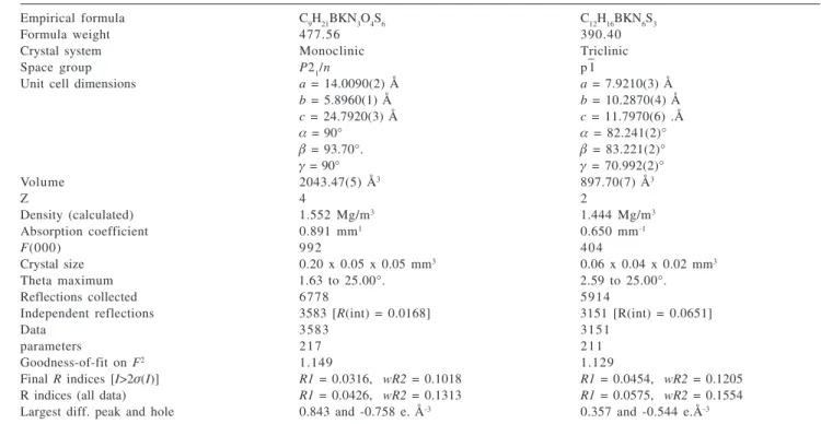
them is 85(3)o. In each unit the potassium cation is
coordinated to six water molecules. Two of them are
symmetrically dependent of other neighboring K(H2O)6
units. A view focusing on the potassium solvate sphere is
shown in Figure 2. The soft nature of the Mt- anion is
Table 1. Crystal data and structure refinement for compounds KMt.4H2O (1) and KTm (2)
Empirical formula C9H21BKN3O4S6 C12H16BKN6S3
Formula weight 477.56 390.40
Crystal system Monoclinic Triclinic
Space group P21/n p
– 1
Unit cell dimensions a = 14.0090(2) Å a = 7.9210(3) Å
b = 5.8960(1) Å b = 10.2870(4) Å
c = 24.7920(3) Å c = 11.7970(6) .Å
α = 90° α= 82.241(2)°
β = 93.70°. β= 83.221(2)°
γ = 90° γ = 70.992(2)°
Volume 2043.47(5) Å3 897.70(7) Å3
Z 4 2
Density (calculated) 1.552 Mg/m3 1.444 Mg/m3
Absorption coefficient 0.891 mm1 0.650 mm-1
F(000) 992 404
Crystal size 0.20 x 0.05 x 0.05 mm3 0.06 x 0.04 x 0.02 mm3
Theta maximum 1.63 to 25.00°. 2.59 to 25.00°.
Reflections collected 6778 5914
Independent reflections 3583 [R(int) = 0.0168] 3151 [R(int) = 0.0651]
Data 3583 3151
parameters 217 211
Goodness-of-fit on F2 1.149 1.129
Final R indices [I>2σ(I)] R1 = 0.0316, wR2 = 0.1018 R1 = 0.0454, wR2 = 0.1205
R indices (all data) R1 = 0.0426, wR2 = 0.1313 R1 = 0.0575, wR2 = 0.1554
Largest diff. peak and hole 0.843 and -0.758 e. Å-3 0.357 and -0.544 e.Å-3
Figure 1. ORTEP3 view of the anion and K(H2O)6 units of compound KMt.4H2O (1) showing the atom-labelling scheme. Displacement ellipsoids drawn at the 50% probability. H atoms are represented by circles of arbitrary size. (Symmetry Codes:#1 -x+1, -y, -z+1; #2 -x+1, -y+1,
indicated by the lack of interaction between its donor atoms and the potassium ion.
The geometry around the potassium cation can be described as a very distorted octahedron, better as a pentagonal pyramid, as observed before for other hydrotris
borates.14 The largest deviation from the 90o degree
octahedral geometry takes place in OW(1)-K-OW(2) and
OW(4)-K-OW(3) angles, with values of 120.8(1)o and
71.80(6)o, respectively, Table 2. The K-OW(1) bond
distance is the shortest one, 2.676(2)Å, and is shorter than
the sum of the K++ O2– ionic radii of 2.78Å.15 Compound 1
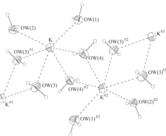
has a polymeric structure, with an infinite chain consisting
of K(H2O)n units that grows along the b axis, inside an
approximate hexagonal cavity that is formed by the packing of the anions, that has an average diameter of 10(1)Å, Figure 3. The equivalent bond distances in the three rings were found to have similar values and all of them agree with the expected values for the bonds involved.
The bond distances and angles found for compound 1,
are similar to the ones recently described in the literature for the TlTbz, Tbz = 2-mercaptobenzothiazoline,
complex.4 In this compound the Tl cations are coordinated
to the sulfur atoms and there are infinite one-dimensional polymeric chains, that exhibit a zigzag array of Tl and S atoms that are formed by the association of the monomeric units through the thione sulfur atoms.
The structure of compound 2, described here and
shown in Figure 4, exhibits similarities to the X-ray
structure previously reported for NaTm.4.5H20,3 such as bond
distances and angles, Table 3. Crystallographic parameters and data collection and refinement details are summarized in Table 1.
Figure 2. ORTEP3 view of potassium coordination environment in KMt.4H2O (1). Displacement ellipsoids drawn at the 50% proba-bility.(Symmetry Codes: #1 -x+1, -y, -z+1; #2 -x+1, -y+1, -z+1; #3 x,
y+1, z).
The counter-ion K+ of 2 was found to be coordinated
to two sulfur atoms of the hydrotris(methimazole)borate unit and to other three sulfur atoms, symmetrically dependent, belonging to neighboring KTm units, which is indicative of the soft nature of the ligand, and resulting in a pentacoordination environment around the cation. A better view of the environment around the potassium cation is shown in Figure 5.
The geometry around K+ can be described as distorted
square pyramidal. The K-S(1) bond, which is the longest one in the structure, forms the apex of the pyramid. The
K-S(2)#3 is the shortest K-S bond in the structure and
Figure 3. ORTEP3 view of KMt.4H2O (1) showing the approximate hexagonal cavity that is formed by the packing of the anions along the b and the infinite chain of K(H2O)n units.
Table 2. Selected bond distances (Å) and angles (°) for atoms and the potassium ion for KMt.4H2O (1) according to X-ray diffraction analysis
Symmetry Codes:#1 –x+1, -y, -z+1; #2 –x+1, -y+1, -z+1.
K-OW(3) 2.784(2) K-S(21) 4.945(2) K-S(22) 4.323(2) K-K#2 3.859(1)
K-K#1 4.077(1)
N(1)-C(13)-S(12) 113.7(2) S(11)-C(13)-S(12) 120.1(2) C(11)-N(1)-B 121.0(2) C(13)-N(1)-B 123.6(2) N-B-N (mean value) 108.6(3) N-B-H (mean value) 110(2) OW(1)-K-OW(3)#1 111.5(1)
OW(1)-K-OW(2) 120.8(1) OW(4)-K-OW(3) 71.80(6) OW(2)-K-OW(3) 75.84(6) OW(3)#1-K-OW(4)#2 78.43(6)
OW(4)#2-K-OW(3) 80.88(6)
OW(3)#1-K-OW(2) 83.67(6)
OW(3)#1-K-OW(3) 83.94(6)
OW(1)-K-OW(4)#2 86.06(7)
OW(1)-K-OW(4) 89.99(6) OW(4)#2-K-OW(4) 90.36(6)
OW(2)-K-OW(4) 96.97(6) C(11)-C(12) 1.521(4) N(1)-C(13) 1.316(3) N(1)-C(11) 1.484(3) N(2)-C(21) 1.477(4) N(2)-C(23) 1.318(3) N(3)-C(31) 1.475(3) N(3)-C(33) 1.313(4) S(11)-C(13) 1.697(3) S(12)-C(13) 1.750(3) S(12)-C(12) 1.813(3) S(21)-C(23) 1.687(3) S(22)-C(22) 1.809(3) S(22)-C(23) 1.756(3) S(31)-C(33) 1.699(3) S(32)-C(32) 1.810(3) S(32)-C(33) 1.747(3) B-H 0.96
N-B (mean value) 1.564(4) K-OW(1) 2.676(2) K-OW(3)#1 2.698(2)
K-OW(2) 2.723(2) K-OW(4)#2 2.730(2)
together with the K-S(3) are shorter than the sum of the K+
+ S2– ionic radius of 3.22Å.15 In this compound there are
infinite dimeric chains, along the a axes, that are held
together by the potassium atoms, Figure 6. The structure
of the sodium salt of Tm, NaTm.4.5H20, was shown to
consist of discrete Tm– anions having a distorted
one-dimensional chains of hydrated sodium cations.1
Conclusions
The potassium salts of the anions
hydrotris(2-mercaptothiazolyl)borate, Mt–, and hydrotris
Table 3. Selected bond distances (Å) and angles (º) for compound 2 according to X-ray diffraction analysis
N(11)-C(11) 1.362(4) C(13)-N(11)-B 127.3(3) N(11)-C(13) 1.394(4) C(11)-N(11)-C(13) 108.3(3) N(12)-C(11) 1.358(4) C(11)-N(12)-C(12) 109.5(3) N(12)-C(12) 1.378(4) C(11)-N(12)-C(14) 125.7(3) N(12)-C(14) 1.456(4) C(12)-C(13)-N(11) 107.8(3) S(1)-C(11) 1.709(3) C(12)-N(12)-C(14) 124.7(3) C(12)-C(13) 1.347(5) C(13)-C(12)-N(12) 107.4(3) H-B (mean value) 1.550(5) N(11)-C(11)-S(1) 127.1(3) N-H 0.96 N(11)-C(11)-N(12) 107.0(3) S(3)-K#2 3.270(1) N(12)-C(11)-S(1) 126.0(2)
S(2)-K#3 3.152(1) K#2-K-K#3 132.27(3)
S(1)-K#3 3.292(1) K#3-S(1)-K 74.54(3)
K-S(1) 3.301(1) K-S(3)-K#2 92.86(3)
K-S(1)#3 3.292(1) S(2)#3-K-S(1) 110.83(3)
K-S(2)#3 3.152(1) S(2)#3-K-S(1)#3 97.87(3)
K-S(3) 3.167(1) S(2)#3 -K-S(3) 140.39(3)
K-S(3)#2 3.270(1) S(2)#3 -K-S(3)#2 80.07(3)
K-K#3 3.992(2) S(3)#2 -K-S(1) 99.05(3)
K-K#2 4.664(2) S(3)#2-K-S(1)#3 154.34(3)
N-B-N (mean value) 109(2) S(3)-K-S(1)#3 78.23(3)
H-B-N (mean value) 110.4(3) S(3)-K-S(3)#2 87.14(3)
C(11)-N(11)-B 124.3(3) S(1)#3-K-S(1) 105.46(3)
Symmetry codes: #1 x+1,y,z; #2 –x, -y, -z; #3 –x+1, -y-z.
Figure 5. ORTEP3 view of the compound 2 showing in details the potassium coordenation environment. Displacement ellipsoids drawn at the 50% probability.(Symmetry Codes: #1 x+1, y, z; #2 -x, -y, -z; #3 -x+1, -y, -z).
Figure 4. ORTEP3 view of compound 2 showing the atom-label-ling scheme. Displacement ellipsoids drawn at the 50% probability. H atoms are represented by circles of arbitrary size.
(methimazolyl)borate, Tm–, were prepared in better yields
than previously described in the literature3,4 and were
characterized in the solid state and in solution. The solution of their structures confirms the softer nature of
the anion in 1, as compared to 2, indicated by the lack of
interaction of the anion with the cation, in 1, that is
surrounded by six water molecules. The X-ray diffraction analyses also determined that the salts have an extended structure.
Supplementary Material
Supplementary Crystallographic data have been deposit with the Cambridge Crystallographic Data centre
as supplementary publication no. CCDC 181571
(KMt.4H2O) and 181572 (KTm). Copies of available
material can be obtained free of charge, on application to the Director, CCDC, 12 Union Road, Cambridge, CB2 1EZ, UK (fax: +44123-336-033; e-mail: deposit@ ccdc.cam.ac.uk or http:www.ccdc.ac.uk ).
References
1. Lee, B. Y.; Moon, H.; Chung, Y. K.; Organometallics1993, 12, 3879; Cardoso, A. M.; Clark, R. J. H.; Moorhouse, S.;
J.Chem. Soc. Dalton 1980, 1156; Bunker, M. J.; De Cian, A.; Green, M. L. H.; Moreau, J. E.; Siganporia, N. J.; J. Chem. Soc. Dalton 1980, 2155; Hitchcock, P. B.; Lappert, M. F.; Milne, C. R. C.; J. Chem. Soc. Dalton 1981, 180; Lucas, C. R.; Labinger, J. A.; Schwartz, J.; Inorg. Synth. 1976, 16, 107. 2. Trofimenko, S.; J. Am. Chem. Soc.1967, 89, 3170. 3. Garner, M.; Reglinski, J.; Cassidy, I.; Spicer, M. D.; Kennedy,
A.R.; Chem. Comm.1996, 1975.
4. Ojo J.F.; Slavin, P. A.; Reglinski, J.; Garner, M.; Spicer, M.D.; Kennedy, A.R.; Teat, S.J.; Inorg. Chim. Acta 2001, 313, 15. 5. Soares, L.F.; Silva, R.M.; Inorg. Synth. 2002, 33, 200. 6. Enraf-Nonius COLLECT.; Nonius BV, Delft, The Netherlands,
1997-2000
7. Otwinowski, Z.; Minor, W. In Methods in Enzymology, 276; Carter Jr., C.W.; Sweet, R.M., eds., Academic Press: New York, 1997, pp. 307-326.
8. Blessing, R. H.; Acta Cryst. 1995, A51, 33.
9. Sheldrick,G.M.; SHELXS-97. Program for Crystal Structure Resolution. Univ. of Göttingen, Germany, 1997.
10. Sheldrick, G.M.; SHELXL-97. Program for Crystal Structures Analysis. Univ. of Göttingen, Germany, 1997.
11. Farrugia,L.; Wingx,J..; J. Appl. Cryst. 1999, 32, 837. 12. Farrugia, L. J.; ORTEP3 for Windows.; J. Appl. Cryst. 1997,
30, 565.
13. Santini , C.; Lobbia, G. G.; Pettinari, C; Pellei, M.; Inorg. Chem. 1998, 37, 890.
14. Weis, K.; Varenkamp, H.; Inorg. Chem. 1997, 36, 5589. 15. Huheey, J. E.; Inorganic Chemistry. Principles of Structure
and Reactivity, Harper Internacional SI Edition: New York, 1983, p.258.
Received: December 4, 2003
Published on the web: August 24, 2004