316
Brazilian Journalof otorhinolaryngology 74 (2) March/april 2008 http://www.rborl.org.br / e-mail: [email protected]
CASE REPORT
Ancient schwannoma of the vagus
nerve, resection with continuous
monitoring of the inferior laryngeal
nerve
Claudio Gilberto Yuji Nakano 1, Luiz Claudio Bosco
Massarollo 2, Erivelto Martinho Volpi 3, José Geraldo
Barbosa Junior 4, Vitor Arias 5, Rubens Yassuzo Ykko
Ueda 6
INTRODUCTION
Schwannomas (neurinomas, neurilem-momas) are benign, single, slow-growing en-capsulated tumors that originate in the sheath of cranial or spinal nerves,1 and that rarely undergo
malignant transformation.
Descriptions have shown that about 25% of cases occur in the head and neck;2 there are
only 95 references of vagus nerve involvement.3
These tumors appear mostly between the third and fifth decades of life; there is no sex predo-minance.4 The clinical picture usually consists
of a relatively pain-free bulge in the neck; the differential diagnosis should be made with other parapharyngeal tumors or neoplasms in the jugular foramen.3
The senile schwannoma (SS) is a rare variant that was first described by Ackrman and Taylor in 1951;2 its features are: wide areas of
hyalinized matrix, hypercellularity with nuclear polymorphism and cell hyperchromatism. A microscopic description of SS in serial and histological sections reveals two cell types: the Antoni type A or fasciculated type (elongated cells, arranged in intertwining bundles in various directions or in a spiral layout), and the Antoni type B or reticular type (polymorphic cells that define small vacuoles, giving the tumor a honeycomb aspect). Antoni type B cells pre-dominate in SS. Absence of mitosis is the main feature that differentiates a SS from a malignant schwannoma. Twelve cases of head and neck SSs have been described so far, of which one involved the vagus nerve.5
Surgery is the treatment of choice; there is a high rate of vagus nerve injury during this procedure.3 There are descriptions of resections
of vagus nerve schwannomas associated with neurostimulation3,6 and observation of
esopha-geal6 contractions or endoscopic visualization of the larynx.3 The current article is the first case
report of resection of a vagus nerve schwannoma under continuous electrophysiological monito-ring of the recurrent laryngeal nerve.
CASE REPORT
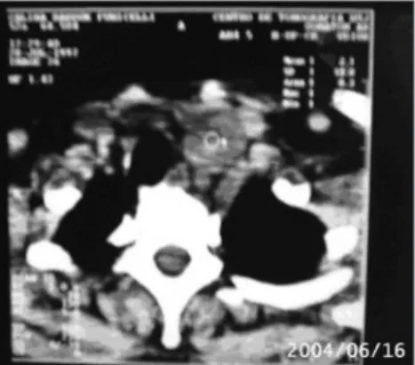
A female, 59-year-old patient reported a 10-year history of multinodular goiter and a palpable nodule in the left supraclavicular fossa. She complained of coughing upon flexing the neck and upon percussion of the
supraclavi-cular nodule, which was gradually worsening. Computed tomography revealed a nodule in the cervical-thoracic transition point, juxtaposed to the trachea and the left lower pole of the thyroid (Figure 1). Fine needle aspiration was done for cytology, which suggested a mesen-chymal tumor.
The patient was operated on 21 June 2004; the initial procedure was a total thyroi-dectomy and dissection with preservation of the recurrent laryngeal nerves. At this point an encapsulated tumor was found close to the lower pole of the left thyroid lobe, which extended retrosternally.
The vagus nerve tumor was completely removed under continuous monitoring (NIM-2® System); laryngeal innervation was preserved. The patient was discharged on the first posto-perative day with no intercurrences; direct la-ryngoscopy after the surgical procedure revealed normally functioning vocal folds. Three years after surgery there are no signs of recurrence or changes in phonation.
Histopathology showed areas of vacuo-lization, increased cellularity, pleomorphism and hyalinization. Immunohistochemistry was stron-gly reactive for vimentin and the S-100 protein, which confirmed the diagnosis of SS.
DISCUSSION
Primary tumors of the vagus nerve are uncommon. Schwannomas are infrequent and
the SS variant has been described previously only once.3
Surgery has a high rate of vocal fold in-jury and paralysis, particularly in tumors located close to the jugular foramen.2
Fujino6 (2000) described the
intracap-sular enucleation technique for vagus nerve tumors, which has become the standard surgical method - together with neurostimulation - for the treatment of these tumors.
Mevio2 (2003) reported vagus nerve
tumor resection with neurostimulation and endoscopic observation of the ipsilateral vo-cal fold. The use of electrodes together with endotracheal ventilation tubes for continuous intra-operative monitoring during thyroidectomy has been well described in the literature.7 This
system makes possible a simplified non-invasive technique that is just as sensitive as laryngeal muscle monitoring.7
This is the second report of a vagus nerve SS and the first report of a case in which continuous laryngeal nerve electrophysiological monitoring was used when resecting a primary vagus nerve tumor.
CONCLUSION
Schwannomas should be included in the differential diagnosis of vagus nerve tu-mors. Whenever possible, surgical removal of these tumors should include continuous intra-operative electrophysiological monitoring of the laryngeal nerve.
REFERENCES
1. Conley JJ. Neurogenic tumors in the neck. Arch Otolaryngol 1955; 61:167-80.
2. Ackerman LV, Taylor FH. Neurogenous tumors within the torax: a clinicopathological evaluation of 48 cases. Cancer 1951;4:669-91.
3. Mevio E, Gorini E et al. Unusual Cases of Cervical Nerves Schwannomas: Phrenic and Vagus Nerve Involvement. Auris Nasus Larynx 2003;30:209-13. 4. Park CS, Suh KW, Kim CK. Neurilemmomas of the
cervical vagus nerve. Head Neck 1991;13:439-41. 5. Saydam L, Kizilay A et al. Ancient Cervical Vagal
Neurilemmoma: A Case Report. Am Journal Oto-laryngology 2000;21(1):61-4.
6. Fujino K, Shinohara K et al. Intracapsular Enucleation of Vagus Nerve-Originated Tumors for Preservation of Neural Function. Otolaryngol Head Neck Surg 2000;123:334-6.
7. Eisele DW. Intraoperative Electrophysiologic Monito-ring Of The Recurrent Laryngeal Nerve. Laryngos-cope 1996;106:443-9.
Keywords: intraoperative eletrophysiologic monitoring, recurrent laryngeal nerve, vagus nerve, neurinnoma, ancient schwannoma.
1 Medical student, FCMSCSP.
2 Chief of the Head & Neck Surgery Unit, Sao Cristovao Hospital and Guarulhos Oncology Institute. 3 Assistant physician - Head & Neck Surgery Unit, HCFMUSP.
4 Assistant physician - Head & Neck Surgery Unit, Sao Cristovao Hospital. 5 Pathologist, Adolfo Lutz Institute and FMUSP.
6 Surgeon, Head & Neck Surgery Unit, Sao Cristovao Hospital.
Sao Cristovao Hospital and Guarulhos Oncology Institute.
Address for correspondence: Instituto de Oncologia de Guarulhos (IOG) - Rua dos Metalurgicos 7 Vila das Palmeiras Guarulhos 07013-131. Tel. (0xx11) 6468-0236/ 6408-5734
Paper submitted to the ABORL-CCF SGP (Management Publications System) on July 23th, 2006 and accepted for publication on November 17th, 2006. cod. 3289. Rev Bras Otorrinolaringol
2008;74(2):316.