412
Radiol Bras. 2017 Nov/Dez;50(6):405–415 Letters to the Editorhttp://dx.doi.org/10.1590/0100-3984.2016.0072 diaphragmatic paralysis, the diagnostic criteria for which are a
DTF below 20% in B-mode(5) and paradoxical breathing, char -acterized by a curve below the baseline in M-mode(6). At this writing, the patient is being monitored and is under conservative treatment, showing gradual clinical improvement.
REFERENCES
1. Ferrari G, De Filippi G, Elia F, et al. Diaphragm ultrasound as a new index of discontinuation from mechanical ventilation. Crit Ultrasound J. 2014;6:8.
2. Matamis D, Soilemezi E, Tsagourias M, et al. Sonographic evaluation of the diaphragm in critically ill patients. Technique and clinical applications. Intensive Care Med. 2013;39:801–10.
3. Francis CA, Hoffer JA, Reynolds S. Ultrasonographic evaluation of
diaphragm thickness during mechanical ventilation in intensive care
patient. Am J Crit Care. 2016;25:e1–8.
Rachel Zeitoune1, Ana Célia Baptista Koifman2, Marina Shu Fong1, Roberto Mogami1
1. Hospital Universitário Pedro Ernesto (HUPE), Rio de Janeiro, RJ, Brazil. 2. Hos-pital Universitário Gaffrée & Guinle (HUGG), Rio de Janeiro, RJ, Brazil. Mailing address: Dra. Rachel Zeitoune. Hospital Universitário Pedro Ernesto – Serviço de Radiologia. Boulevard 28 de Setembro, 77, Vila Isabel. Rio de Janeiro, RJ, Brazil, 20551-030. E-mail: [email protected].
4. Sarwal A, Walker FO, Cartwright MS. Neuromuscular ultrasound for evaluation of the diaphragm. Muscle Nerve. 2013;47:319–29.
5. Summerhill EM, El-Sameed YA, Glidden TJ, et al. Monitoring recovery from diaphragm paralysis with ultrasound. Chest. 2008;133:737–43. 6. Lloyd T, Tang YM, Benson MD, et al. Diaphragmatic paralysis: the use of
M mode ultrasound for diagnosis in adults. Spinal Cord. 2006;44:505– 8.
Malignant peripheral nerve sheath tumor of the vagus nerve: an uncommon cause of progressive dyspnea
Dear Editor,
A healthy, nonsmoking, 27-year-old male patient was re -ferred to our institution for investigation of a three-month history of progressive dyspnea. He reported that his dyspnea worsened on physical exertion and signiicantly limited his daily activities. He reported no cough, fever, night sweats, or weight loss; nor did he report any new lumps or masses during the last three months. Upon skin examination, multiple subcutaneous nodules and café-au-lait spots were noted, together with bi -lateral axillary freckles (Figure 1a). Collectively, those clinical indings met the criteria for a diagnosis of neuroibromatosis, which was so far undiagnosed. Pulmonary auscultation revealed diffuse wheezing in the right upper hemithorax. His biochemi -cal proile was unremarkable. The patient then underwent a
computed tomography (CT) scan of the chest with intravenous contrast administration, which revealed a 20-cm right cervico -thoracic mass presumably arising from the right vagus nerve (Figures 1b–d). Because of the background of neuroibroma -tosis, a hypothesis of malignant peripheral nerve sheath tumor (MPNST) was raised and further conirmed by incisional biopsy and histological analysis. Given the proximity to vital structures, the patient was treated with a chemotherapy protocol for soft tissue sarcomas in an attempt to reduce the tumor bulk preop-eratively. Because of a poor cellular response and recrudescence of the respiratory symptoms, the patient was deemed ineligible for any aggressive interventions.
MPNSTs are exceedingly rare sarcomas in the general pop -ulation, with a lifetime risk of less than 0.01%. Conversely, in association with neuroibromatosis, these tumors arise in higher frequency because of malignant transformation from preexisting plexiform neuroibromas(1). Overall, these tumors are associated
Figure 1. Findings on physical exam-ination and CT. a:Café-au-lait spots (curved arrows) and axillary freckles (arrowhead) upon skin inspection. b–d: Axial CT scans of the neck (b,d) and chest (c,d) showing the MPNST. Note the heterogeneous enhance-ment after contrast administration (b) and the stenosis of the right main bronchus lumen (c), which
413
Radiol Bras. 2017 Nov/Dez;50(6):405–415Letters to the Editor
http://dx.doi.org/10.1590/0100-3984.2016.0055
Felipe Welter Langer1, Daiane dos Santos1, Giordano Rafael Tronco Alves1, Gustavo Suertegaray1, Carlos Jesus Pereira Haygert1
1. Department of Radiology and Imaging Diagnosis, University Hospital of Santa Maria, Federal University of Santa Maria (UFSM), Santa Maria, RS, Bra-zil. Mailing address: Dr. Felipe Welter Langer. Department of Radiology and Imaging Diagnosis, University Hospital of Santa Maria, Federal University of Santa Maria. Avenida Roraima, 1000, Camobi. Santa Maria, RS, Brazil, 97105-900. E-mail: [email protected].
with high local invasion, rapid growth, and early distant metasta -sis unless they are excised in a timely manner(2). The most com -mon locations for MPNST in neuroibromatosis patients are the extremities, head, and neck. Thoracic involvement, however, is remarkably rare, few cases having been reported(3). According to the size and location of the intrathoracic tumor, compressive manifestations such as pain, dyspnea, dysphagia, and superior vena cava syndrome may be the presenting manifestations, as seen in our patient, who reported dyspnea as the sole symptom related to his MPNST(3,4).
The identiication of MPNST in neuroibromatosis patients may be troublesome for several reasons. First, the existence of multiple benign neuroibromas may delay the identiication of changes in plexiform neuroibromas. In addition, because su -pericial cutaneous neuroibromas do not undergo malignant transformation, MPNSTs often remain undetected until they reach a moderate size or cause compressive symptoms. Further-more, CT and magnetic resonance imaging might not be ac -curate enough to differentiate benign from malignant lesions with any degree of reliability in the very early stages, although advances have been made in the area of positron emission to-mography(4–6). Therefore, any suspicious lesions should gener -ally prompt histological sampling(7).
Although the mainstay of successful treatment of an MPNST is surgical excision after disease staging, neoadjuvant chemotherapy may be employed in order to reduce its dimen-sions beforehand, especially in patients with ledimen-sions surrounding vital organs. Radiotherapy might also delay recurrence, although it has not been shown to improve survival in MPNST patients(8).
REFERENCES
1. Hirbe AC, Gutmann DH. Neuroibromatosis type 1: a multidisciplinary approach to care. Lancet Neurol. 2014;13:834–43.
2. Porter DE, Prasad V, Foster L, et al. Survival in malignant peripheral nerve sheath tumours: a comparison between sporadic and neuroibroma
-tosis type 1-associated tumours. Sarcoma. 2009;2009:756395.
3. Chao BH, Stogner-Underwood KA, Kiev J, et al. Intrathoracic malignant peripheral nerve sheath tumour in neuroibromatosis 1. Journal of Clini
-cal Oncology. 2008;26:2216–8.
4. Grimer R, Judson I, Peake D, et al. Guidelines for the management of soft tissue sarcomas. Sarcoma. 2010;2010:506182.
5. Yap YS, McPherson JR, Ong CK, et al. The NF1 gene revisited – from bench to bedside. Oncotarget. 2014;5:5873–92.
6. Salamon J, Veldhoen S, Apostolova I, et al. 18F-FDG PET/CT for detec
-tion of malignant peripheral nerve sheath tumours in neuroibromatosis type 1: tumour-to-liver ratio is superior to an SUVmax cut-off. Eur Radiol. 2014;24:405–12.
7. Gutmann DH, Aylsworth A, Carey JC, et al. The diagnostic evaluation and multidisciplinary management of neuroibromatosis 1 and neuroibroma -tosis 2. JAMA. 1997;278:51–7.
8. Brems H, Beert E, de Ravel T, et al. Mechanisms in the pathogenesis of malignant tumours in neuroibromatosis type 1. Lancet Oncol. 2009; 10:508–15.
Burkitt-like lymphoma of the brain mimicking an intraventricular colloid cyst
Dear Editor,
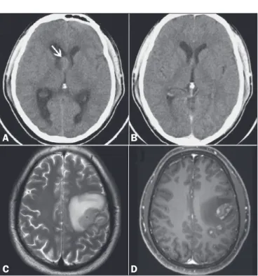
A 32-year-old male sought treatment, complaining of head -ache. Computed tomography (CT) of the brain revealed hyper -dense intraventricular nodule to the right of the foramen of Monro, highly suggestive of a colloid cyst (Figure 1A). The patient was using dexamethasone as pain therapy. In a CT scan of the brain obtained one month later, no nodules were observed (Figure 1B). Cervical and thoracoabdominal CT scans also showed no abnor -malities. At two months, the patient presented with convulsions. Magnetic resonance imaging (MRI) of the brain showed a cere -bral mass (Figures 1C and 1D). Histopathological and immuno -histochemical analysis of a biopsy sample revealed Burkitt-like lymphoma, which is one of the non-Hodgkin lymphomas. Ancil -lary examinations ruled out systemic disease and viral infection.
Lymphomas are designated primary when they originate at and are conined to a given site(1–3). Primary central nervous sys -tem (CNS) lymphomas account for up to 6% of brain neoplasms and 1–6% of extranodal lymphomas; approximately 90% of pri -mary CNS lymphomas are non-Hodgkin lymphomas of the dif-fuse large B-cell subtype(1–6). The incidence of CNS lymphoma is higher in the presence of certain immunodeiciencies, especially human immunodeiciency virus (HIV) infection(2). Among im-munocompetent individuals, the prevalence of CNS lymphoma is highest (60–67%) in men 45–75 years of age. In that group, CNS lymphomas present as a single homogeneous mass (in 62%), often in the supratentorial compartment (in 83%) and notably in the deep white matter (in 57%). The corpus callosum and regions surrounding the ventricles are typically affected. Perilesional
Figure 1. A: Non-contrast-enhanced CT scan of the brain, showing well-delineated, discretely hyperdense intraventricular nodule to the right of the foramen of Monro (arrow), promoting slight dilation of the lateral ventricles (obstructive hydrocephalus). B: Follow-up CT of the brain, obtained one month later, showing no such nodule. C,D: MRI of the brain after episodes of seizures, T2-weighted sequence (C) and paramagnetic contrast-enhanced T1-weighted sequence (D), showing an intra-axial frontoparietal mass in the left cerebral hemisphere, with intense perilesional vasogenic edema and heterogeneous enhancement.