909
IMAGES IN NEUROLOGY
DOI: 10.1590/0004-282X20130140
Perineurioma of the posterior interosseous
nerve: surgical treatment
Perineurioma do nervo interósseo posterior: tratamento cirúrgico
Djalma Felipe da Silva Menéndez, Roberto Sergio Martins, Mario Gilberto Siqueira, Igor Araújo Ferreira da Silva,
Lívia Barreira Cavalcante, Roberto Falzoni, Luciano Henrique Lopes Foroni, Manoel Jacobsen Teixeira
Intraneural perineurioma is a benign tumor that occurs
in less than 1% of peripheral nerve tumors; no more than 90
cases have been reported
1,2. Tumorous lesions of the posterior
interosseous nerve (PIN) have rarely been described
3–5.
An 18-year-old woman presented with a longstanding
his
tory of spontaneous progressive weakness in the PIN
dis tribution (Figure 1). Ultrasonography and MRI studies
(Figure 2) showed a nodular lesion in the PIN, measuring
1.0 cm at its greatest diameter. At surgical exploration a tumor
(Figure 3) involving all the nerve fascicles was entirely re sected.
he nerve was repaired by termino-terminal neurorrhaphy.
Figure 4 shows the histological examination.
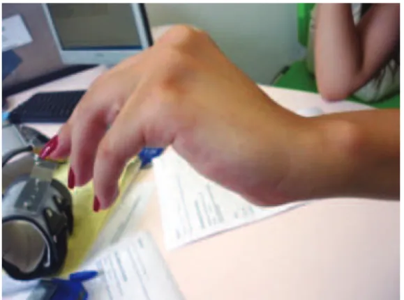
Figure 1.
Wrist extension partially preserved and deviation of
the hand to the radial side. There was no finger extension.
Figure 3.
Intraoperative view of the tumor involving all the
fascicles of the PIN. PIN: posterior interosseous nerve; RN:
radial nerve; SB: superficial branch of the radial nerve; T: tumor.
Grupo de Cirurgia de Nervos Periféricos, Divisão de Neurocirurgia Funcional do Hospital das Clínicas da Universidade de São Paulo, São Paulo SP, Brasil.
Correspondence:Djalma Felipe da Silva Menéndez; R. Teodoro Sampaio 498 / Apto. 121; 05406-000 São Paulo SP - Brasil; E-mail: [email protected]
Conflict of interest: There is no conflict of interest to declare.
Received 19 May 2013; Received in final form 01 June 2013; Accepted 10 June 2013.
Figure 2.
(A, B) MRI: heterogeneous oval lesion, with slight
post-contrast enhancement, located in the PIN, measuring 1.0
cm at its greatest diameter; (C) Ultrasonography: nodule (N)
located within the PIN.
A
B
C
Figure 4.
(A) Histological slide (H&E stain) showing diffuse
pseudo-onion-bulb leaflets and thinly myelinated fibres at the
centre of them; (B) Schwann cell preparation (S-100 protein) on
immunohistochemistry demonstrates reactivity of the myelinated
fibres at the centre, and absence of reactivity, of the surrounding
pseudo-onion bulbs. Furthermore, epithelial membrane antigen
(EMA) was positive, confirming perineurial origin. These findings,
considered together, are diagnostic of perineurioma
5.
910
Arq Neuropsiquiatr 2013;71(11):909-910References
1. Groeneweg AJM, Hartman EH, Fleischeuer R, Visser LH. An
unusual location of ulnar nerve pathology: a perineurioma of the ulnar nerve in the upper arm. Muscle Nerve 2011;44:593-596.
2. Heilbrun ME, Tsuruda JS, Townsend JJ, Heilbrun MP. Intraneural perineurioma of the common peroneal nerve. Case report and review of the literature. J Neurosurg 2011;94:811-815.
3. Mauermann ML, Amrami KK, Kuntz NL, et al. Longitudinal study of intraneural perineurioma – a benign, focal hypertophic neuropathy of youth. Brain 2009;132:2265-2276.
4. Lallemand RC, Weller RO. Intraneural neurofibromas involving the posterior interosseous nerve. J Neurol Neurosurg Psychiatry 1973;36:991-996. 5. Sachanandani NS, Brown JM, Zaidman C, Brown SS, Mackinnon