DOI: 10.1590/0004-282X20160080
VIEW AND REVIEW
A diagnostic approach for neurodegeneration
with brain iron accumulation: clinical features,
genetics and brain imaging
Uma orientação diagnóstica para neurodegeneração com acúmulo cerebral de ferro:
aspectos clínicos, genéticos e de neuroimagem
Rubens Paulo Araújo Salomão
1, José Luiz Pedroso
1, Maria Thereza Drumond Gama
1, Lívia Almeida Dutra
1,
Ricardo Horta Maciel
2, Clécio Godeiro-Junior
3, Hsin Fen Chien
4, Hélio A. G. Teive
5, Francisco Cardoso
2,
Orlando G. P. Barsottini
1Neurodegeneration with brain iron accumulation (NBIA)
represents a heterogeneous group of inherited
neurodegenera-tive diseases, characterized by excess iron accumulation,
partic-ularly in the basal ganglia, and to a lesser degree in substantia
nigra and adjacent areas
1. NBIA is considered to be a very rare
dis-ease group, with a prevalence of less than 1/1,000,000 in general
population
1. Common clinical features of NBIA include
move-ment disorders, particularly parkinsonism and dystonia,
cogni-tive dysfunction, pyramidal signs, and retinal abnormalities
1,2,3.
Hunt in 1917 described a case report of juvenile parkinsonism
associated with progressive atrophy of globus pallidus
4. In 1922,
Hallervorden and Spatz reported a family with ive afected sisters
1Universidade Federal de São Paulo, Departamento de Neurologia, Divisão de Neurologia Geral, São Paulo SP, Brasil;
2Universidade Federal de Minas Gerais, Clínica de Desordens do Movimento, Departmento de Neurologia, Belo Horizonte MG, Brasil; 3Universidade Federal do Rio Grande do Norte, Unidade de Transtornos do Movimento, Departamento de Medicina Integrada, Natal RN, Brasil; 4Universidade de São Paulo, Instituto de Ortopedia e Traumatologia, São Paulo SP, Brasil;
5Universidade Federal do Paraná, Hospital de Clínicas, Unidade de Desordens do Movimento, Curitiba PR, Brasil.
Correspondence: Orlando G. P. Barsottini; Departamento de Neurologia, Universidade Federal São Paulo; Rua Pedro de Toledo 650; 04041-002 São Paulo SP, Brasil; E-mail: orlandobarsottini@gmail.com
Conflict of interest: There is no conlict of interest to declare. Received 10 April 2016; Accepted 26 April 2016.
ABSTRACT
Neurodegeneration with brain iron accumulation (NBIA) represents a heterogeneous and complex group of inherited neurodegenerative
diseases, characterized by excessive iron accumulation, particularly in the basal ganglia. Common clinical features of NBIA include movement
disorders, particularly parkinsonism and dystonia, cognitive dysfunction, pyramidal signs, and retinal abnormalities. The forms of NBIA
described to date include pantothenase kinase-associated neurodegeneration (PKAN), phospholipase A2 associated neurodegeneration
(PLAN), neuroferritinopathy, aceruloplasminemia, beta-propeller protein-associated neurodegeneration (BPAN), Kufor-Rakeb syndrome,
mitochondrial membrane protein-associated neurodegeneration (MPAN), fatty acid hydroxylase-associated neurodegeneration (FAHN),
coenzyme A synthase protein-associated neurodegeneration (CoPAN) and Woodhouse-Sakati syndrome. This review is a diagnostic
approach for NBIA cases, from clinical features and brain imaging indings to the genetic etiology.
Keywords: neurodegeneration with brain iron accumulation; NBIA; clinical features; brain imaging; genetics.
RESUMO
A neurodegeneração com acúmulo cerebral de ferro (sigla em inglês NBIA) representa um grupo heterogêneo e complexo de doenças
neurodegenerativas hereditárias, caracterizada pelo acúmulo cerebral de ferro, especialmente nos núcleos da base. O quadro clínico das NBIAs
em geral inclui distúrbios do movimento, particularmente parkinsonismo e distonia, disfunção cognitiva, sinais piramidais e anormalidades da
retina. As formas de NBIA descritas até o momento incluem neurodegeneração associada a pantothenase kinase (PKAN), neurodegeneração
associada a phospholipase A2 (PLAN), neuroferritinopatia, aceruloplasminemia, neurodegeneração associada a beta-propeller protein (BPAN),
síndrome de Kufor-Rakeb, neurodegeneração associada a mitochondrial membrane protein (MPAN), neurodegeneração associada a “fatty acid
hydroxylase” (FAHN), neurodegeneração associada a coenzyme A synthase protein (CoPAN) e síndrome de Woodhouse-Sakati. Esta revisão é uma
orientação para o diagnóstico das NBIAs, partindo das características clínicas e achados de neuroimagem, até a etiologia genética.
with neuropathological conirmation of lesions of globus pallidus
and substantia nigra
5. Davidson described in 1954, a case series of
ive patients presenting with progressive parkinsonism, dystonia,
and spasticity, associated with pyramidal and pallidal lesions, and
created the term pallidopyramidal degeneration (PPD)
6. However,
this disease was worldwide known as Hallervorden-Spatz
syn-drome (HSS)
3. Julius Hallervorden and Hugo Spatz were German
physicians, who performed several neuropathological studies on
brain specimens of mental retardation persons, executed during
the hird Reich euthanasia program (Aktion–T-4)
1,2,3. After conir
-mation of Hallervorden and Spatz’s involvement in the
euthana-sia program of the Nazi regime in Germany, and the recent
neu-roimaging and genetic discoveries, this syndrome was renamed
NBIA
1,2. In 2010, Horstink et al. suggested that PPD was a
misno-mer and conclude that the existence of PPD as a distinct
noso-logical entity is doubtful
7. In 2013, Kara et al. argued that the use
of the term NBIA is not ideal and suggested the term
pallidopyra-midal syndromes (PPS), however NBIA is the most known
world-wide term
2. To date ten forms of NBIA has been described, eight
with autosomal recessive inheritance, one autosomal dominant
form, and one with X-linked dominant inheritance
1,2. he most
common forms are pantothenase kinase-associated
neurodegen-eration (PKAN) (30-50% of NBIA cases), due to mutations in the
PANK2
gene, followed by phospholipase A2 associated
neurode-generation (PLAN) due to
PLA2G6
gene mutations,
mitochon-drial membrane protein-associated neurodegeneration (MPAN)
due to
c19orf12
mutations, and beta-propeller protein-associated
neurodegeneration (BPAN) causing SENDA syndrome (static
encephalopathy of childhood with neurodegeneration in
adult-hood) (gene
WDR45
, chromosome Xp11.23). Probably most of
the case published in the literature as HSS were PKAN
1. Other
less common forms are fatty acid hydroxylase-associated
neuro-degeneration (FAHN), coenzyme A synthase protein-associated
neurodegeneration (CoPAN), Kufor-Rakeb syndrome (PARK9),
Woodhouse-Sakati syndrome, neuroferritinopathy and
acerulo-plasminemia
1,2. he forms of NBIA described to date and the re
-spective gene mutations are listed in Table 1.
Pantothenase kinase-associated neurodegeneration
(PKAN)
PKAN is an autosomal recessive disorder characterized
by mutations in the gene encoding a mitochondrial
panto-thenate kinase (
PANK2
) at locus 20p13-p12.3
8. It is the most
common disorder of the NBIA group
9,10,11. he classic clini
-cal presentation of PKAN is characterized by early-onset
(mean age is 14 years - range from 1 to 28y) and rapidly
pro-gressive course. he afected child presents gait impairment
and movement disorders (particularly dystonia and
par-kinsonism). Spasticity and brisk tendon relexes are com
-mon. Cognition is frequently impaired
1,12,13,14. Retinitis pig
-mentosa may occur, associated or not with acanthocytes
in blood cells
15,16. he majority of individuals (85%) become
wheelchairbound within 15 years after the beginning of
symptoms
13,14. Speech and swallowing are afected with dis
-ease progression. Death is usually secondary to respiratory
infections, cardiorespiratory complications, malnutrition
state and, rarely,
status dystonicus
. Atypical phenotypes with
slowly progressive course have a late onset. Neuropsychiatric
symptoms are common and may be early signs. hey include
mood lability, impulsivity, non-speciic behavioral changes,
and obsessive-compulsive features
1,14.
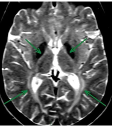
Brain magnetic resonance imaging (MRI) plays an im
-portant role in the investigation of PKAN since it shows the
‘
eye-of- the-tiger
’ sign, deined as a medial area of hyperintense
signal within hypointense signal in globus pallidus bilaterally and
best demonstrated on T2WI and SWI (Figure 1). Hypointense
signal may also be observed in the substantia nigra
15.
here is no current speciic therapy to stop disease progres
-sion. Treatment is supportive and intend to relief associated
symptoms
1,13,14. Dystonia and spasticity are usually managed
with anticholinergic drugs, benzodiazepines, botulinum toxin,
oral baclofen and intrathecal baclofen in severe cases. he role
of the brain iron accumulation in the pathophysiology of the
disease remains under discussion, and iron chelation therapy
has been investigated as a disease modifying approach
1,14,17.
Phospholipase A2 associated neurodegeneration
(PLAN)
PLAN is an autosomal recessive form of NBIA. he dis
-ease is caused by failure in the ubiquitously expressed
PLA2G6
gene, which maps to chromosome 22q13.1
10,18. his
gene encodes phospholipase A2 group VI, which may
dis-rupt membrane homeostasis, involved in free fatty acids and
lysophospholipids synthesis, resulting in
neurodegenera-tion, atrophy, brain iron accumulaneurodegenera-tion, gliosis and
degener-ation of the optic pathways
9,19. he majority of PLAN cases
have early-onset of symptoms, with beginning in childhood.
PLA2G6
-associated diseases have variable syndromes and
may include: classic infantile neuroaxonal dystrophy (INAD),
atypical neuroaxonal dystrophy (aNAD) of childhood-onset
and
PLA2G6
-related dystonia-parkinsonism with late onset
in adulthood (PARK14)
1,9,13.
Table 1.
Forms of NBIA described to date and the respective
gene mutations.
NBIA subtype
Gene mutation
PKAN
PANK2
PLAN
PLA2G6
Neuroferritinopathy
FTL1
Aceruloplasminemia
Ceruloplasmin
BPAN
WDR45
Kufor-Rakeb syndrome
ATP13A2
(PARK9)
MPAN
C19orf12
FAHN
FA2H
CoPAN
CoASY
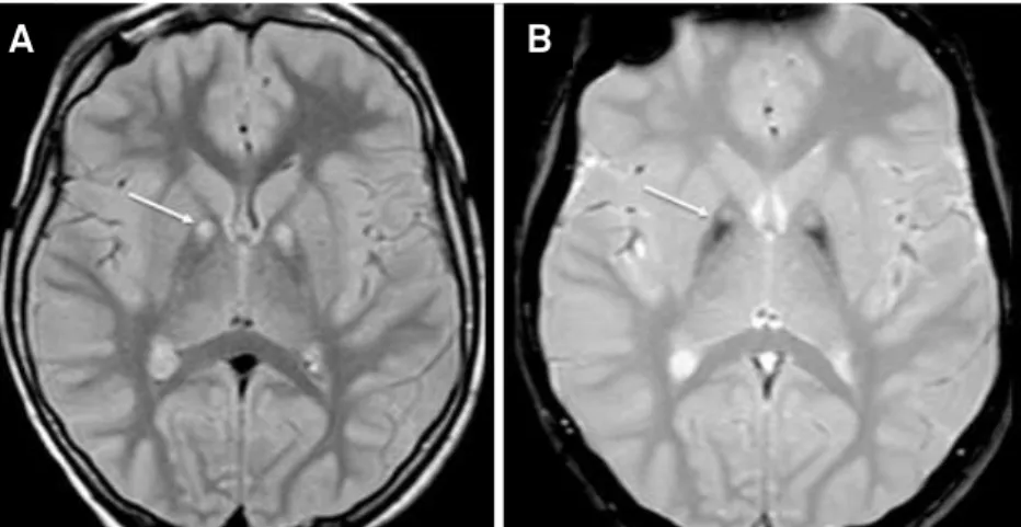
INAD is characterized by early-onset mental
developmen-tal delay, ataxia with cerebellar atrophy, neuropathy and optic
atrophy. Patients usually have hypotonia, kyphoscoliosis and
limb contractures. he symptoms usually occurs before 2 years
old and the progression of the disease is rapid leading to death
in the irst decade
9,20. An estimated 50% of patients have
abnor-mal iron accumulation in brain MRI, evolving globus pallidus,
dentate nuclei and substantia nigra (Figure 2)
10. Optic pathway
atrophy is a relevant clue for the disease
9.
aNAD was previously described as Karak syndrome. his
disease is less aggressive than classic INAD. he typical clinical
presentation of aNAD include early-onset (older than INAD)
ataxia and dysarthria, hypotonia, arelexia, dystonia and cog
-nitive impairment
1,20. Children with aNAD may develop optic
atrophy
19. Brain MRI usually have similar features observed in
INAD: abnormal iron accumulation in globus pallidus, dentate
nuclei and substantia nigra, and cerebellar atrophy
20.
Adult-onset PLAN with dystonia and parkinsonism,
described as PARK14, has onset in young adulthood. Clinical
features include parkinsonism-dystonia syndrome with
vari-able response to dopaminergic medications and
neuropsy-chiatric symptoms
9. Brain MRI may be normal or may dis
-close iron accumulation in globus pallidus, substantia nigra
and striatum
21.
here is no speciic treatment for PLAN. Symptomatic treat
-ment for spasticity, dystonia and parkinsonism should be tried
1.
A levodopa course for parkinsonism may improve symptoms
13.
Neuroferritinopathy
Neuroferritinopathy is a rare autosomal dominant NBIA
of adult-onset related to a mutation in the ferritin light chain
gene
FTL1,
on chromosome 19q13.3
19. he onset of symptoms
is predominantly described in young adulthood or middle
age
1. he clinical manifestation includes psychiatric symptoms
(psychosis, anxiety and depression), frontal lobe dysfunction,
dystonia, choreoathetosis, rigidity and spasticity. Other
ab-normalities described in patients with neuroferritinopathy
in-clude lingual dyskinesia, blepharospasm, cerebellar symptoms,
Figure 1.
Brain MRI of a patient with pantothenase kinase-associated neurodegeneration (PKAN). Axial FLAIR- (A) and
GRE T2-weighted (B) MRI discloses ‘
eye-of- the-tiger
’ sign, deined as a medial area of hyperintense signal within an hypointense
signal in globus pallidus bilaterally.
A
B
parkinsonism and palatal tremor
10,22. Blood tests show low
lev-els of serum ferritin, typically ≤20 μg/dl, which may reinforce
the diagnosis
22. Brain MRI indings include iron accumulation
in globus pallidus, caudate, substantia nigra, red nuclei and
putamen. In late stages of the disease cystic necrosis in basal
ganglia and globus pallidus may occur
22-24. Pathological
stud-ies have demonstrated ubiquitin and tau positive neuroaxonal
spheroids and neuroilaments, and ferritin-positive inclusions
in putamen and cerebellum
23. Treatment is symptomatic and
supportive, and dystonia may beneit with botulinum toxin.
No curative treatments are available
22,24.
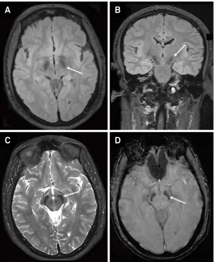
Aceruloplasminemia
Aceruloplasminemia is an autosomal recessive diseased
caused by mutation in the ceruloplasmin gene. Ceruloplasmin
is a copper-bound ferroxidase essential to normal iron
metab-olism, including cell iron elux and oxidation of ferrous iron
to ferric iron
25. When ceruloplasmin activity is severely
de-creased, iron overload occurs and tissue iron deposition and
degeneration follows. Iron accumulates primarily in the brain,
retina and pancreas, giving rise to the classical presentation of
the disease, a triad consisting of neurological symptoms,
reti-nal degeneration and diabetes mellitus. he prevalence of ac
-eruloplasminemia is higher in Japan
26.
Symptoms onset in aceruloplasminemia usually occur in the
fourth decade. Typically, the irst manifestation of the disease is
diabetes mellitus accompanied by microcytic anemia with low
serum iron, elevated ferritin and hepatic iron deposition
with-out ibrosis or cirrhosis
27. Retinal macular degeneration is usual
-ly asymptomatic
28,29. Neurological symptoms develop later in the
disease, around the sixth decade of life. Movement disorders are
the most common neurological features in aceruloplasminemia
and include ataxia, cranio-facial dyskinesias (torticollis,
blepha-rospasm, facial grimacing and tongue dystonia), parkinsonism
and dysarthria
30,31. Dementia and other neuropsychiatric
symp-toms are frequent in aceruloplasminemia
30,32.
Diagnosis of aceruloplasminemia is based on the absence
or very low serum ceruloplasmin. Brain MRI usually disclos
-es severe iron accumulation in caudate, pallidus, putamen,
dentate nuclei, red nuclei, substantial nigra, thalamus,
infe-rior and supeinfe-rior colliculi and cortex (Figure 3). his wide
-spread pattern of involvement helps distinguish
aceruloplas-minemia from other NBIA, in which iron deposition is either
more limited (e.g. conined to the pallidus) or associated
with other features (e.g. cavitation, cerebellar atrophy or thin
corpus callosum)
30,33. White matter T2 hyperintensities can
also occur
34. Heterozygous cases can present with an
incom-plete clinical picture, generating further diagnostic diicul
-ties
32,35,36. Genetic testing conirms the diagnosis.
A number of iron chelating agents have been tried with
mixed results and data on eicacy is scarce
37. Deferasirox
re-duces hepatic but not brain iron overload. Sporadic reports
have demonstrated that deferasirox and deferiprone may
de-lay neurological symptoms
36,38,39.
Beta-propeller protein-associated
neurodegeneration (BPAN)
BPAN is a X-linked dominant form of NBIA caused by
mu-tations in
WDR45
gene, a
β
-propeller scafolding protein, as
-sociated with disruption of autophagosome maturation,
ac-cumulation of aberrant autophagic structures and damaged
cellular components
40,41.
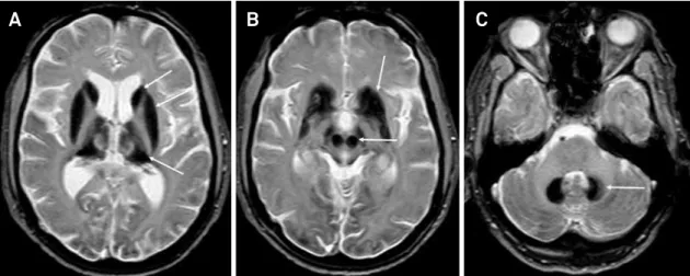
Clinically, BPAN is characterized by two phases of
dis-ease: an early neurological disorder with rapid progression
and a static encephalopathy of childhood. he early on
-set phenotype includes neurological symptoms similar to
Rett-like syndrome: epilepsy, sleep disorders and global de
-velopmental delay, followed by a rapid progressive onset of
parkinsonism, dystonia, and dementia
14,42. he second form
is a static encephalopathy of childhood with
neurodegenera-tion in adulthood (SENDA syndrome), which typically
pres-ents with hyperintense signal of the substantia nigra with a
central band of hypointense signal in brain MRI (Figure 4)
43.
Figure 3.
Brain MRI of a patient with aceruloplasminemia MRI discloses global and severe iron accumulation in caudate, pallidus,
thalamus, putamen (A), red nuclei, substantial nigra (B) and dentate nuclei (C).
Neuropathological studies have disclosed large axonal
spher-oids, siderophages, reactive astrocytes, severe neuronal loss
and abundant tau-positive neuroibrillary tangles, suggesting
the occurrence of a tauopathy
40,43.
Kufor-Rakeb syndrome - PARK 9
he mutation of the gene
ATP13A2
(PARK9) results in a rare
autosomal recessive form of juvenile parkinsonism, previously
known as Kufor-Rakeb syndrome
44. he irst cases described
presented early parkinsonism, pyramidal signs, upgaze
pal-sy and mental retardation
45. However, it is recognized that the
wide-ranging abnormalities in the gene leads to loss of function
mutations, which impact the pathophysiological function of
ATP13A2 protein resulting in considerable clinical heterogene
-ity
46. Moreover, the mutations in heterozygous state have been
found in parkinsonian patients suggesting that heterozygous
carriers may increase risk for development of the disease
45.
he onset of Kufor-Rakeb syndrome is mostly before the
age of 20, with variable disease progression. he clinical man
-ifestations comprise levodopa-responsive parkinsonism,
dys-tonia, pyramidal signs, facial-inger mini-myoclonus, supra
-nuclear gaze palsy and cognitive impairment
47. Brain MRI
usually shows global atrophy. he disease is included in NBIA
group since T2* weighted sequence discloses iron accumula
-tion in basal ganglia in some patients
48.
Mitochondrial membrane protein-associated
neurodegeneration (MPAN)
MPAN is an autosomal recessive disease caused by
mu-tations in
C19orf12
gene. Pathological studies have
demon-strated increased iron accumulation in the globus pallidus
and substantia nigra, gliosis, neuronal loss, and eosinophilic
spheroidal structures in the globus pallidus. he characteris
-tic peripheral axonal spheroids of PLAN may be seen in some
patients. MPAN is considered a synucleinopathy, with Lewy
bodies and Lewy neurites in basal ganglia and neocortex
19,49.
he disease may manifest from the irst decade of life
(3-16 years) to adulthood (until 30 years). he most common
features are early lower limb spasticity with extensor plantar
response, dysarthria, dystonia (involving hands and feet), optic
atrophy, neuropsychiatric abnormalities and cognitive decline.
he presentation in adults is more variable, with prominent
neuropsychiatric symptoms, parkinsonism and gait disorders.
Others clinical indings that support the diagnosis of MPAN
are dysphagia, axonal motor neuropathy and bladder and/or
Figure 4.
Brain MRI of a patient with beta-propeller protein-associated neurodegeneration (BPAN) or SENDA. There is a marked
bilateral hypointense signal in substantia nigra observed in axial and coronal FLAIR (A and B), axial T2 (C) and axial SWI (D) sequences.
A
B
bowel dysfunction. he disease progression is slow and the
lifespan is long in most of the cases
19,49.
he diagnosis of MPAN is made by detection of biallelic
pathogenic variants in
C19orf12
gene
.
Brain MRI shows iron
accumulation in substantia nigra and globus pallidus on T2*
and GRE sequences, normally without the eye of the tiger
sign, which is typically found in PKAN. Cortical and
cerebel-lar atrophy may be seen in advanced disease
19,49,50.
Similar to
others NBIA, there is no curative treatment and the
manage-ment of patients relies on rehabilitation and symptomatic
medications: anti-spastic agents, anticholinergics,
dopami-nergic agents and botulinum toxin.
Fatty acid hydroxylase- associated
neurodegeneration (FAHN)
FA2H mutations were previously known to cause
leukodys-trophy and a form of hereditary spastic paraplegia (HSP), which
was classiied as SPG35
51,52,53. FA2H produces 2-hydroxylated
fatty acids for incorporation into 2-hydroxydihydroceramide
and 2-hydroxyceramide. hese ceramide species serve as
precursors for the synthesis of galactosylceramides and
sul-fatides, essential lipid components of normal myelin
54,55.
Phenotypically, afected patients demonstrated features simi
-lar to those observed in INAD
55. he clinical phenotype is char
-acterized by childhood-onset spastic paraplegia, ataxia and
dystonia. here are prominent ophthalmologic features such
as acquired strabismus, nystagmus and optic atrophy. Intellect
is usually spared in FAHN patients. Seizures may be present
51.
Brain MRI in FAHN usually shows bilateral globus pallidus
T2 hypointense signal, characterizing iron accumulation, pon
-tocerebellar atrophy and cortical atrophy (Figure 5). Conluent
periventricular T2 white matter hyperintense signal were also
observed along with thinning of the corpus callosum
51,55. It is
a matter of discussion if FAHN should be included in one or
more of the 3 following groups: NBIA, HSP or leukodystrophy.
CoA synthase protein associated neurodegeneration
(CoPAN)
Coenzyme A synthesis (
CoASY
) is a cofactor in all living
organisms and is involved in several enzymatic reactions.
Patients with
CoASY
mutations present a clinical picture
similar to those with PKAN. CoPAN phenotype is
charac-terized by early-onset spastic-dystonic paraparesis with a
later appearance of parkinsonian features, cognitive
im-pairment and pronounced obsessive-compulsive disorder.
The disease has a slow progression with loss of ambula
-tion during adolescence and adulthood
56. Brain MRI usu
-ally shows bilateral “eye-of-the-tiger” sign. CT scan shows
bilateral calcifications and corresponding to the central
spot visible on MRI
21.
Woodhouse-Sakati syndrome and SCPx deficiency
Woodhouse–Sakati syndrome is a rare autosomal re
-cessive disorder caused by a mutation in the
C2orf37
gene,
manifesting with hypogonadism, deafness, alopecia,
diabe-tes mellitus and progressive dystonia, chorea, dysarthria and
cognitive impairment. Brain MRI discloses iron accumula
-tion in the substantia nigra and globus pallidus, and white
matter lesions. However, some patients may have only subtle
white matter abnormalities
57,58.
Finally, sterol carrier protein x (SCPx) deiciency has been
associated with NBIA in a patient with adult-onset
spinocere-bellar ataxia, slow ocular saccades, and deafness. 3T MRI brain
revealed abnormal T2 signal and susceptibility-weighted se
-quences suggested increased mineral deposition in the basal
ganglia. Potentially pathogenic mutations were identiied in
SCP2. SCPx is a peroxisomal enzyme with thiolase activity
required for the breakdown of branched chain fatty acids and
the pathogenic efects are likely to be mediated by the accu
-mulation of branch chain fatty acids, as in other
peroxisom-al disorders. Patients with SCP2 mutation may have
abnor-mal fatty-acid acyl-CoA metabolism, which has emerged as a
common disease mechanism in NBIA. his suggests that the
brain iron accumulation is secondary to an underlying
meta-bolic defect, questioning the role of iron chelation as a
treat-ment in all forms of NBIA
59.
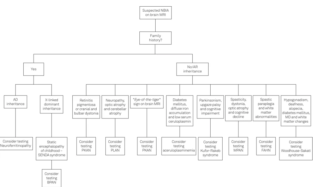
Diagnostic approach to neurodegeneration with
brain iron accumulation
To diagnose the NBIA subtype is a challenge. Careful
attention to clinical phenotype and neuroimaging features
is crucial. Family history in order to determine inheritance
is relevant since mendelian inheritance forms may direct
investigation to neuroferritinopathy. Neuroimaging
fea-tures may disclose key findings for different NBIA
sub-types (Table 2). Altogether, clinical features including
non-neurological manifestation, associated movement
disorders, age at onset, family history and detailed
neuro-imaging characteristics should guide to the genetic
test-ing investigation (Figure 6).
593
R
ubens P
a
ul
o Ar
a
újo Sal
omã
o e
t al
. Neur
odeg
ener
a
tion with br
ain ir
on accumula
tion
Figure 6.
A guidance with clinical and neuroimaging tips to better require speciic genetic testing in NBIA patients.
NBIA: neurodegeneration with brain iron accumulation; MRI: magnetic resonance imaging; AD: autosomal dominant; AR: autosomal recessive; MD: movement disorders; BPAN: beta-propeller protein-associated neurodegeneration; PKAN: pantothenase kinase-associated neurodegeneration; PLAN: phospholipase A2 associated neurodegeneration; MPAN: mitochondrial membrane protein-associated neurodegeneration; FAHN: fatty acid hydroxylase-associated neurodegeneration.
Suspected NBIA
on brain MRI
Family
history?
Yes
No/AR
inheritance
Retinitis
pigmentosa
or cranial and
bulbar dystonia
Neuropathy,
optic atrophy
and cerebellar
atrophy
Diabetes
mellitus,
diffuse iron
accumulation
and low serum
ceruloplasmin
Parkinsonism,
upgaze palsy
and cognitive
impairment
Spasticity,
dystonia,
optic atrophy
and cognitive
decline
Spastic
paraplegia
and white
matter
abnormalities
Hypogonadism,
deafness,
alopecia,
diabetes mellitus,
MD and white
matter changes
Static
encephalopathy
of childhood –
SENDA syndrome
Consider
testing
PKAN
Consider
testing
Kufor-Rakeb
syndrome
Consider
testing
Woodhouse-Sakati
syndrome
AD
inheritance
X-linked
dominant
inheritance
Consider
testing
PKAN
Consider
testing
PLAN
Consider
testing
MPAN
Consider
testing
FAHN
Consider
testing
BPAN
Consider
testing
aceruloplasminemia
Consider testing
Neuroferritinopathy
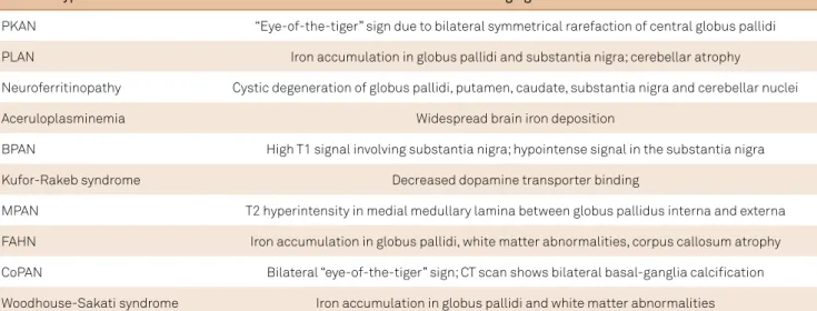
Table 2.
Key imaging indings for different NBIA subtypes.
NBIA subtype
Neuroimaging features
PKAN
“Eye-of-the-tiger” sign due to bilateral symmetrical rarefaction of central globus pallidi
PLAN
Iron accumulation in globus pallidi and substantia nigra; cerebellar atrophy
Neuroferritinopathy
Cystic degeneration of globus pallidi, putamen, caudate, substantia nigra and cerebellar nuclei
Aceruloplasminemia
Widespread brain iron deposition
BPAN
High T1 signal involving substantia nigra; hypointense signal in the substantia nigra
Kufor-Rakeb syndrome
Decreased dopamine transporter binding
MPAN
T2 hyperintensity in medial medullary lamina between globus pallidus interna and externa
FAHN
Iron accumulation in globus pallidi, white matter abnormalities, corpus callosum atrophy
CoPAN
Bilateral “eye-of-the-tiger” sign; CT scan shows bilateral basal-ganglia calciication
Woodhouse-Sakati syndrome
Iron accumulation in globus pallidi and white matter abnormalities
References
1. Hogarth P. Neurodegeneration with brain iron accumulation: diagnosis and management. J Mov Disord. 2015;8(1):1-13. doi:10.14802/jmd.14034
2. Kara E, Hardy J, Houlden H. The pallidopyramidal syndromes: nosology, aetiology and pathogenesis. Curr Opin Neurol. 2013;26(4):381-94. doi:10.1097/WCO.0b013e3283632e83 3. Haylick SJ, Westaway SK, Levinson B, Zhou B, Johnson MA,
Ching KH et al. Genetic, clinical, and radiographic delineation of Hallervorden-Spatz syndrome. N Engl J Med. 2003;348(1):33-40. doi:10.1056/NEJMoa020817
4. Hunt J. A system disease of the paralysis agitans type, characterized by atrophy of the motor cells of the corpus striatuma contribution to the functions of the corpus striatum. Brain. 1917;40(1):58-148. doi:10.1093/brain/40.1.58 First published title: Progressive atrophy of the globus pallidus.
5. Hallervorden J, Spatz H. Eigenartige erkrankung im extrapyramidalen system mit besonderer beteiligung des globus pallidus und der substantia nigra: Ein beitrag zu den beziehungen zwischen diesen beiden zentren. Z Gesamte Neurol Psychiatr. 1922;79(1):254-302. doi:10.1007/BF02878455
6. Davison C. Pallido-pyramidal disease. J Neuropathol Exp Neurol. 1954;13(1):50-9. doi:10.1097/00005072-195401000-00007 7. Horstink MW, Dekker MC, Montagna P, Bonifati V, van De Warrenburg
BP et al. Pallidopyramidal disease: a misnomer? Mov Disord. 2010;25(9):1109-15. doi:10.1002/mds.23118
8. Kruer M. The neuropathology of neurodegeneration with brain iron accumulation. Int Rev Neurobiol. 2013;110:165-94. doi:10.1016/B978-0-12-410502-7.00009-0
9. Kurian MA, McNeill A, Lin JP, Maher ER. Childhood disorders of neurodegeneration with brain iron accumulation (NBIA). Dev Med Child Neurol. 2011;53(5):394-404. doi:10.1111/j.1469-8749.2011.03955.x
10. Levi S, Finazzi D. Neurodegeneration with brain iron accumulation: update on pathogenic mechanisms. Front Pharmacol. 2014;5:99. doi: 10.3389/fphar.2014.00099
11. Levi S, Rovida E. Neuroferritinopathy: from ferritin structure modiication to pathogenetic mechanism. Neurobiol Dis. 2015; 81:134-43. doi:10.1016/j.nbd.2015.02.007
12. Lee CH, Lu CS, Chuang WL, Yeh TH, Jung SM, Huang CL et al. Phenotypes and genotypes of patients with pantothenate
kinase-associated neurodegeneration in Asian and Caucasian populations: 2 cases and literature review. ScientiicWorldJournal. 2013;2013:860539. doi:10.1155/2013/860539
13. Tonekaboni SH, Mollamohammadi M. Neurodegeneration with brain iron accumulation: an overview. Iran J Child Neurol. 2014;8(4):1-8.
14. Meyer E, Kurian MA, Haylick SJ. Neurodegeneration with brain iron accumulation: enetic diversity and pathophysiological mechanisms. Annu Rev Genomics Hum Genet. 2015;16(1):257-79. doi:10.1146/annurev-genom-090314-025011
15. Amaral L, Gaddikeri S, Chapman PR, Roy R, Gaddikeri RS, Marussi Vh et al. Neurodegeneration with brain iron accumulation: clinicoradiological approach to diagnosis. J Neuroimaging. 2015;25(4):539-51. doi:10.1111/jon.12195
16. Pedroso JL, Proveti P, Teixeira LF, Sallum JM, Barsottini OG. Retinitis pigmentosa in pantothenate kinase-associated neurodegeneration. Arq Neuropsiquiatr. 2014;72(10):816-7. doi:10.1590/0004-282X20140122
17. Cossu G, Abbruzzese G, Matta G, Murgia D, Melis M, Ricchi V et al. Eficacy and safety of deferiprone for the treatment of pantothenate kinase-associated neurodegeneration (PKAN) and neurodegeneration with brain iron accumulation (NBIA): results from a four years follow-up. Parkinsonism Relat Disord. 2014;20(6):651-4. doi:10.1016/j.parkreldis.2014.03.002
18. Khateeb S, Flusser H, Oir R, Shelef I, Narkis G, Vardi G et al. PLA2G6 mutation underlies infantile neuroaxonal dystrophy. Am J Hum Genet. 2006;79(5):942-8. doi:10.1086/508572
19. Schneider SA, Dusek P, Hardy J, Westenberger A, Jankovic J, Bhatia KP. Genetics and pathophysiology of neurodegeneration with brain iron accumulation (NBIA). Curr Neuropharmacol. 2013;11(1):59-79. doi:10.2174/157015913804999469
20. Gregory A, Polster BJ, Haylick SJ. Clinical and genetic delineation of neurodegeneration with brain iron accumulation. J Med Genet. 2009;46(2):73-80. doi:10.1136/jmg.2008.061929
22. Levi S, Rovida E. Neuroferritinopathy: from ferritin structure modiication to pathogenetic mechanism. Neurobiol Dis. 2015;81:134-43. doi:10.1016/j.nbd.2015.02.007
23. Hautot D, Pankhurst QA, Morris CM, Curtis A, Burn J, Dobson J. Preliminary observation of elevated levels of nanocrystalline iron oxide in the basal ganglia of neuroferritinopathy patients. Biochim Biophys Acta. 2007;1772(1):21-5. doi:10.1016/j.bbadis.2006.09.011
24. Keogh MJ, Morris CM, Chinnery PF. Neuroferritinopathy. Int Rev Neurobiol. 2013;110:91-123.
doi:10.1016/B978-0-12-410502-7.00006-5
25. Miyajima H. Aceruloplasminemia. Neuropathology. 2015;35(1):83-90. doi:10.1111/neup.12149
26. Miyajima H, Kohno S, Takahashi Y, Yonekawa O, Kanno T. Estimation of the gene frequency of aceruloplasminemia in Japan. Neurology. 1999;53(3):617-9. doi:10.1212/WNL.53.3.617
27. Bosio S, De Gobbi M, Roetto A, Zecchina G, Leonardo E, Rizzetto M et al. Anemia and iron overload due to compound heterozygosity for novel ceruloplasmin mutations. Blood. 2002;100(6):2246-8. doi:10.1182/blood-2002-02-0584
28. Tai M, Matsuhashi N, Ichii O, Suzuki T, Ejiri Y, Kono S et al. Case of presymptomatic aceruloplasminemia treated with deferasirox. Hepatol Res. 2014;44(12):1253-8. doi:10.1111/hepr.12292
29. Vroegindeweij LH, Beek EH, Boon AJ, Hoogendoorn M, Kievit JA, Wilson JH et al. Aceruloplasminemia presents as Type 1 diabetes in non-obese adults: a detailed case series. Diabet Med. 2015;32(8):993-1000. doi:10.1111/dme.12712
30. McNeill A, Birchall D, Hayflick SJ, Gregory A, Schenk JF, Zimmerman EA et al. T2* and FSE MRI distinguishes four subtypes of neurodegeneration with brain iron accumulation. Neurology. 2008;70(18):1614-9. doi:10.1212/01.wnl.0000310985.40011.d6
31. Miyajima H, Kono S, Takahashi Y, Sugimoto M. Increased lipid peroxidation and mitochondrial dysfunction in aceruloplasminemia brains. Blood Cells Mol Dis. 2002;29(3):433-8. doi:10.1006/bcmd.2002.0561
32. Miyajima H, Kono S, Takahashi Y, Sugimoto M, Sakamoto M, Sakai N. Cerebellar ataxia associated with heteroallelic ceruloplasmin gene mutation. Neurology. 2001;57(12):2205-10. doi:10.1212/WNL.57.12.2205
33. Kruer MC, Boddaert N. Neurodegeneration with brain iron accumulation: a diagnostic algorithm. Semin Pediatr Neurol. 2012;19(2):67-74. doi:10.1016/j.spen.2012.04.001
34. Grisoli M, Piperno A, Chiapparini L, Mariani R, Savoiardo M. MR imaging of cerebral cortical involvement in aceruloplasminemia. AJNR Am J Neuroradiol. 2005;26(3):657-61.
35. Kuhn J, Bewermeyer H, Miyajima H, Takahashi Y, Kuhn KF, Hoogenraad TU. Treatment of symptomatic heterozygous aceruloplasminemia with oral zinc sulphate. Brain Dev. 2007;29(7):450-3. doi:10.1016/j.braindev.2007.01.001 36. Rusticeanu M, Zimmer V, Schleithoff L, Wonney K, Viera
J, Zimmer A et al. Novel ceruloplasmin mutation causing aceruloplasminemia with hepatic iron overload and diabetes without neurological symptoms. Clin Genet. 2014;85(3):300-1. doi:10.1111/cge.12145
37. Pan PL, Tang HH, Chen Q, Song W, Shang HF. Desferrioxamine treatment of aceruloplasminemia: long-term follow-up. Mov Disord. 2011;26(11):2142-4. doi:10.1002/mds.23797
38. Finkenstedt A, Wolf E, Höfner E, Gasser BI, Bösch S, Bakry R et al. Hepatic but not brain iron is rapidly chelated by deferasirox in aceruloplasminemia due to a novel gene mutation. J Hepatol. 2010;53(6):1101-7. doi:10.1016/j.jhep.2010.04.039
39. Bove F, Fasano A. Iron chelation therapy to prevent the manifestations of aceruloplasminemia. Neurology. 2015;85(12):1085-6. doi:10.1212/WNL.0000000000001956 40. Haylick SJ, Kruer MC, Gregory A, Haack TB, Kurian MA,
Houlden HH et al. β-Propeller protein-associated neurodegeneration: a new X-linked dominant disorder with brain iron accumulation. Brain. 2013;136(6):1708-17. doi:10.1093/brain/awt095
41. Long M, Abdeen N, Geraghty MT, Hogarth P, Hayflick S, Venkateswaran S. Novel WDR45 mutation and pathognomonic BPAN imaging in a young female with mild cognitive delay. Pediatrics. 2015;136(3):714-7.
doi:10.1542/peds.2015-0750
42. Ichinose Y, Miwa M, Onohara A, Obi K, Shindo K, Saitsu H et al. Characteristic MRI indings in beta-propeller protein-associated neurodegeneration (BPAN). Neurol Clin Pract. 2014;4(2):175-7. doi:10.1212/01.CPJ.0000437694.17888.9b
43. Paudel R, Li A, Wiethoff S, Bandopadhyay R, Bhatia K, Silva R et al. Neuropathology of Beta-propeller protein associated neurodegeneration (BPAN): a new tauopathy. Acta Neuropathol Commun. 2015;3(1):39.
doi:10.1186/s40478-015-0221-3
44. Ramirez A, Heimbach A, Gründemann J, Stiller B, Hampshire D, Cid LP et al. Hereditary parkinsonism with dementia is
caused by mutations in ATP13A2, encoding a lysosomal type 5 P-type ATPase. Nat Genet. 2006;38(10):1184-91. doi:10.1038/ng1884
45. Di Fonzo A, Chien HF, Socal M, Giraudo S, Tassorelli C, Iliceto G et al. ATP13A2 missense mutations in juvenile parkinsonism and young onset Parkinson disease. Neurology. 2007;68(19):1557-62. doi:10.1212/01.wnl.0000260963.08711.08
46. Lees AJ, Singleton AB. Clinical heterogeneity of ATP13A2 linked disease (Kufor-Rakeb) justiies a PARK designation. Neurology. 2007;68(19):1553-4. doi:10.1212/01.wnl.0000265228.66664.f4 47. Park JS, Blair NF, Sue CM. The role of ATP13A2 in Parkinson’s
disease: clinical phenotypes and molecular mechanisms. Mov Disord. 2015;30(6):770-9. doi:10.1002/mds.26243 48. Chien HF, Bonifati V, Barbosa ER. ATP13A2-related
neurodegeneration (PARK9) without evidence of brain iron accumulation. Mov Disord. 2011;26(7):1364-5. doi:10.1002/mds.23514
49. Schulte EC, Claussen MC, Jochim A, Haack T, Hartig M, Hempel M et al. Mitochondrial membrane protein associated
neurodegeneration: a novel variant of neurodegeneration with brain iron accumulation. Mov Disord. 2013;28(2):224-7. doi:10.1002/mds.25256
50. Skowronska M, Kmiec T, Kurkowska-Jastrzębska I, Czlonkowska A. Eye of the tiger sign in a 23 year patient with mitochondrial membrane protein associated neurodegeneration. J Neurol Sci. 2015;352(1-2):110-1. doi:10.1016/j.jns.2015.03.019
51. Pedroso JL, Handfas BW, Abrahão A, Kok F, Barsottini OG, Oliveira AS. Fatty acid 2-hydroxylase deiciency: clinical features and brain iron accumulation. Neurology. 2015;84(9):960-1. doi:10.1212/WNL.0000000000001316 52. Schneider SA, Bhatia KP. Three faces of the same gene: FA2H links
neurodegeneration with brain iron accumulation, leukodystrophies, and hereditary spastic paraplegias. Ann Neurol. 2010;68(5):575-7. doi:10.1002/ana.22211
53. Dick KJ, Eckhardt M, Paisán-Ruiz C, Alshehhi AA, Proukakis C, Sibtain NA et al. Mutation of FA2H underlies a complicated form of hereditary spastic paraplegia (SPG35). Hum Mutat. 2010;31(4):E1251-60. doi:10.1002/humu.21205
55. Kruer MC, Paisán-Ruiz C, Boddaert N, Yoon MY, Hama H, Gregory A et al. Defective FA2H leads to a novel form of neurodegeneration with brain iron accumulation (NBIA). Ann Neurol. 2010;68(5):611-8. doi:10.1002/ana.22122
56. Dusi S, Valletta L, Haack TB, Tsuchiya Y, Venco P, Pasqualato S et al. Exome sequence reveals mutations in CoA synthase as a cause of neurodegeneration with brain iron accumulation. Am J Hum Genet. 2014;94(1):11-22. doi:10.1016/j.ajhg.2013.11.008
57. Alazami AM, Al-Saif A, Al-Semari A, Bohlega S, Zlitni S, Alzahrani F et al. Mutations in C2orf37, encoding a nucleolar protein, cause hypogonadism, alopecia, diabetes mellitus, mental retardation,
and extrapyramidal syndrome. Am J Hum Genet. 2008;83(6):684-91. doi:10.1016/j.ajhg.2008.10.018 58. Alazami AM, Schneider SA, Bonneau D, Pasquier L,
Carecchio M, Kojovic M et al. C2orf37 mutational spectrum in Woodhouse-Sakati syndrome patients. Clin Genet. 2010;78(6):585-90.
doi:10.1111/j.1399-0004.2010.01441.x