Arquivos Brasileiros de Cardiologia - Volume 85, Nº 3, September 2005
Cardiac Papillary Fibroelastoma. Experience of
an Institution
Primary intracardiac tumors are rare, with prevalence between 0.0017% and 0.19% from non-selected autopsy studies. Approximately 75% are benign and almost half of them are myxomas. The remaining tumors are divided among rabdomyomas, lipomas and fibroelastomas. Myxomas are the most common intracardiac tumors in adult age and rabdomyomas the most common among pediatric population.
Papillary fibroelastoma (PFE) is a relative rare benign heart tumor, corresponding to approximately 8% of intracardiac tumors. They most commonly manifested in cardiac valves1. In the past, they either consisted of
M a i l i n g a d d r e s s : C h a r l e s M a d y • A v . D r . E n é a s d e C a r v a l h o A g u i a r , 4 4 - 0 5 4 0 3 - 0 0 0 - S ã o P a u l o , S P - B r a z i l
E-mail: charles.mady@incor.usp.br Received on 09/09/04 • Accepted on 03/04/05
Case Report
Case Report
Case Report
Case Report
Case Report
Case Report
Suzelle F. de M. Oliveira, Ricardo Ribeiro Dias, Fábio Fernandes, Noedir A. G. Stolf,
Charles Mady, Sérgio Almeida de Oliveira
IInstituto do Coração do Hospital das Clínicas (FMUSP) - São Paulo, SP - Brazil
C
ASE
R
EPORTS
Case 1 - A 27-year-old female patient, under Turner
Syndrome follow-up, sent due to echocardiographic finding of tumoral, pedunculate, movable in tricuspid valve topography image. She did not show symptoms and her physical exam was normal, except for bodily marks from the syndrome itself. Echocardiogram evidenced a round echodense movable image, located at right atrium, adhered to apical portions of tricuspid valve septal leaflet, with signs of left ventricular filling obstructions. The 1.8 cm x 1.2 cm mass was submitted to surgical exeresis (fig. 1). Microscopic exam confirmed papillar y fibroelastoma diagnosis.
Case 2 - Female patient, 67 years old, clinically
asymptomatic, submitted to physical examination in which discreet systolic cardiac murmur was detected. Transesophageal echocardiogram showed pedunculate movable mass in aortic valve, which moved towards the aorta (fig. 2). Magnetic nuclear resonance imaging evidenced the referred mass in aortic valve leaflet (fig. 3). She was submitted to tumor exeresis. Diagnostic confirmed papillary fibroelastoma.
Case 3 - A 63-year-old female diabetic patient, with
mitral commissurotomy history due to rheumatic mitral stenosis and chronic atrial fibrillation. She made use of oral anticoagulant with coumarinic and showed recurring episodes of transitor y ischemic attack (TIA).
Arquivos Brasileiros de Cardiologia - Volume 85, Nº 3, September 2005
Echocardiogram evidenced mitral stenosis with valve area of 1.4 cm2 and anomalous echoes of 1.8 cm x 1.8 cm in
tendinous chord, suggestive of vegetation. There were left atrium thrombus signs. She was sent to surgical treatment and, as a finding, a characteristic tumoral formation with a diameter of, 2 cm, which extended to the papillary muscle, was evidenced in mitral valve anterior leaflet. The exchange of mitral valve through biological prosthesis and tumor exeresis were performed. Microscopic exam confirmed papillary fibroelastoma diagnosis (fig. 4).
Case 4 - Male patient, 59 years old, with history severe
mitral regurgitation due to myxomatous degeneration, made use of coumarinic in force of chronic atrial fibrillation and showed a TIA episode. He was under a follow-up due to functional class III heart failure (NYHA). He was submitted to surgical treatment, in which chord rupture was evidenced and quadrangular resection of posterior cuspid was carried out, followed by raffia and posterior annuloplasty with a bovine pericardium strip. Besides chronic valvopathy with fibrosis and myxoid material deposit, tumorgenicity compatible with papillar y fibroelastoma was evidenced in microscopic exam.
Case 5 - A 49-year-old male patient, with history of
rheumatic mitral lesion, under follow-up due to functional class III (NYHA). He was submitted to mitral valve exchange for biological prosthesis. Histopathological finding of material sent for study was compatible with papillary fibroelastoma, located in one of tendinous chords.
Patients evolved without immediate complications and free from reincidences in all five cases.
D
ISCUSSION
Papillary fibroelastoma is a low-prevalence benign tumor, with tendency to valvar involvement2,4,5.
Concerning frequency, it corresponds to the third most common primary intracardiac tumor, preceded by myxomas and lipomas6. It represents less than 10% from
all primary intracardiac tumors, either those studied in autopsy or after resection1,7,8.
Approximately 90% of PFE undertakes cardiac valves, usually as a single lesion, on the atrial face of atrioventricular valves or on any one of the sides of semilunar valves3,4.
They rarely occur as multiple lesions1,6.
Approximately 44% of PFE is found in aortic valves, followed by the undertaking of mitral valve in 35% of the cases, in 15%, in tricuspid valves and in 8%, in pulmonary ones9. Repor ts of cases on those tumors have
demonstrated undertaking of all endocardial surfaces, including papillary muscles, tendinous chords, the septum or free walls from any of cardiac chambers1,5,8,10.
The size of described PFE varied from 0.1 to 4 cm, and most of them were smaller than 1cm of diameter4.
Their genesis remains controversial. Real neoplasias until
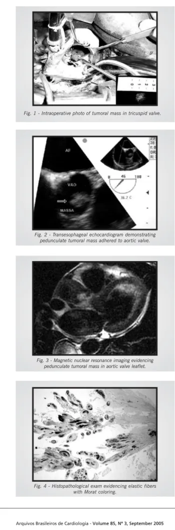
Fig. 3 - Magnetic nuclear resonance imaging evidencing pedunculate tumoral mass in aortic valve leaflet.
Fig. 4 - Histopathological exam evidencing elastic fibers with Morat coloring.
Fig. 1 - Intraoperative photo of tumoral mass in tricuspid valve.
Fig. 2 - Transesophageal echocardiogram demonstrating pedunculate tumoral mass adhered to aortic valve.
Arquivos Brasileiros de Cardiologia - Volume 85, Nº 3, September 2005
hamartomas, organized thrombi, reactive responses to mechanic trauma, to surgical or radiotherapy damage1,3,4,8,9
have been considered. Prevalence is unknown due to the group of non-diagnosed silent tumors4.
The age of patients is variable, from cases in neonates to well-advanced age patients1,4,7. Most of it is described
in adults, over 50 years of age, and there is no difference between sexes1,4,9.
PFE is an incidental finding in most cases, although among symptomatic patients the clinical presentation is variable and dependent on location, motility and size of tumor4,9. As most of it originates from left chambers (more
than 95% of the cases), the most feared complication is the systemic embolization, especially for cerebral or coronary circulation4,9. It is not clear whether the embolus
is tumor- or platelet-origin and if systemic anticoagulation could prevent from such events2,4,9. The most common
clinical presentation described was stroke or transitory ischemic attack (TIA). Other described manifestations were: angina, myocardial infarction, sudden death, heart failure, syncope, pulmonary embolism, blindness, peripheral embolism of renal infarction9. In aortic valve
tumor patients, sudden death and myocardial infarction were the most common manifestations. In rebuttal, stroke was the prevailing presentation9 in mitral valve tumors.
Tumoral motility is the only independent mortality and non-fatal embolization predictor9.
Electrocardiographic findings are non-specific, as atrial arrhythmias may occur. Thoracic radiography may demonstrate signs of increase of cardiac chambers, pulmonary hypertension or congestion, especially if the tumor is occluding the mitral valve. Transthoracic echocardiogram is the ideal method for tumor diagnosis and characterization, as it usually demonstrates the mass with its varied proportions, motile or not, well-delimited, pedunculate or sessile, of round, oval or irregular shape2.
They are mostly small (99% smaller than 2.0 cm)2. In a
case-control study, the sensitivity and specificity of echocardiogram was 88.9% and 87.8%, respectively2.
Magnetic resonance imaging demonstrates the mass in valve leaflet or cardiac chamber, and the presence of enhancement with gadolinium in tumoral mass, increases the suspicion level9. Cardiac catheterization does not
contribute for the diagnosis. In coronary angiography it is possible to visualize total occlusions of coronary arteries, as well as aneurysmatic dilatations and secondary narrowing to tumoral emboli1,9.
PFE has characteristic appearance, resembling a sea anemone, with multiple ramifications held by a pedicle to endocardium. At histological exam, it consists of an endothelium coat, which covers a connective tissue matrix with variable amounts of collagen, smooth muscle cells and elastic fibers2,4.
For symptomatic patients, surgical exeresis is the choice for treatment, by trying to always preserve valve tissue and its function7. Among asymptomatic individuals, surgical
procedure is controversial, as tumoral motility is the determining factor of surgical indication, for being an independent embolization and death predictor. Surgery is curatory and there is no report on reincidences4,9.
Follow-up of asymptomatic patients who were not submitted to surgery must include anticoagulation, although its efficacy in protection against embolic phenomena is controversial2.
Management before a left side isolated lesion includes surgical removal, when the mass is big and/or movable or in the presence of patent ovale foramen due to the possibility of paradoxical embolism2.
In the last years, PFE has progressed from an autopsy incidental finding to an in vivo diagnosed disease with potential injurious complications, which require proper diagnosis and treatment. Due to its rareness, data on the treatment and follow-up are mostly derived from accounts from patients and experiences as those described in our work.
1. Pacini D, Farneti PA, Leone O, Galli R. Cardiac papillar y fibroelastoma of the mitral valve chordae. Eur J Cardiothoracic Surg 1998; 13: 322-4.
2. Sun JP, Asher CR, Yang XS et al. Clinical and echocardiographic characteristics of papillary fibroelastomas. Circulation 2001; 103: 2687-93.
3. Kurup AN, Tazelaar, HD, Edwards WD et al. Iatrogenic cardiac papillary fibroelastoma: A study of 12 Cases (1990 to 2000). Hum Pathol 2002; 33: 1165-9.
4. Shahian DM, Labib SB, Chang G. Cardiac papillary fibroelastoma. Ann Thorac Surg 1995; 59: 538-41.
5. Gologorsky E, Gologorsky A. Aortic valve fibroelastomas as an incidental intraoperative transesophageal echocardiographic finding. Anesth Analg 2002; 95: 1198-9.
R
EFERENCES
6. Tanaka H, Narisawa T, Mori T et al. Double Primary Left Ventricular and Aortic Valve Papillary Fibroelastoma. Circ J 2004; 68: 504-6.
7. Di Mattia DG, Assaghi A, Mangini A et al. Mitral valve repair for anterior leaflet papillary fibroelastoma: two case descriptions and a literature review. Eur J Cardiothoracic Surg1999; 15: 103-7.
8. Georghiou GP, Erez E, Vidne BA, Aravot D. Tricuspid valve papillary fibroelastoma: an unusual cause of intermittent dyspnea. Eur J Cardiothoracic Surg2003; 23: 429-31.
9. Gowda RM, Khan IA, Nair CK et al. Cardiac papillary fibroelastoma: A comprehensive analysis of 725 cases. Am Hear t J 2003; 146: 404-10.
10. Gowda RM, Khan IA, Mehta NJ et al. Cardiac Papillary Fibroelastoma originating from pulmonary vein – a case report. Angiology 2002; 53: 745-8.