419
Original Article
Myocardial Contrast Echocardiography in Patients
with Suspected or Known Coronary Artery Disease.
Comparison with Myocardial Nuclear Scintigraphy
Marcia Carrinho, Alvaro Moraes, Fernando Morcerf, Caio Medeiros, Márcia Castier,
Luis José Martins Romeo
Rio de Janeiro, RJ - Brazil
ECOR Ecocardiografia and Universidade Federal Fluminense Mailing address: Márcia Carrinho - Rua Barata Ribeiro, 432/201 Cep 22040-020 - Rio de Janeiro, RJ, Brazil
E-mail: [email protected] Received for publication: 9/24/2003 Accepted for publication: 07/04/2004 English version by Stela Maris Costalonga
Objective
To compare myocardial contrast echocardiography (MCE) using PESDA and adenosine in bolus (ADN) with myocardial nuclear scintigraphy (NS) in patients (pts) undergoing routine investigation with a high probability of having coronary artery disease.
Methods
This study comprised 125 pts (85 men) with 58.4 ± 10.6 years, who underwent MCE and NS within 4 weeks. MCE was performed with PESDA in a continuous infusion at rest and after administration of an adenosine bolus. The LV walls was divided into 3 territories related to the coronary arteries, in a total of 375 territories. MCE was normal when an increase in contrast intensity occurred after ADN. The reduction in contrast intensity at rest or after ADN was defined as an abnormal MCE result. NS was perfor-med according to classical protocols. When compared per patient, both examinations were considered concordant when they were normal or abnormal, independent of its location. The comparison by territory was considered concordant when perfusion defects existed or not in the same territory. The chi-square test was used to determine the significance of concordance.
Results
In 106/125 pts, MCE and NS were concordant (84.8% -P < 0.001). Concordance occurred in 342/375 territories (91.2% - P < 0.001). For the LAD territory, concordance was 87.2%; for the RCA, 93.6%; and for the CX, 92.8% (P < 0.001).
Conclusion
An excellent concordance exists between MCE and NS in assessing pts for coronary artery disease; therefore, MCE may represent a good alternative for assessing myocardial perfusion.
Key words
echocardiography, myocardial perfusion, echocardiographic contrast medium, coronary artery disease, adenosine,myo-cardial nuclear scintigraphy
The primary objective of perfusion studies is to assess ischemia and viability of myocardial cells. The 3 most frequently used me-thods are stress echocardiography, pharmacological stress echo-cardiography, and myocardial nuclear scintigraphy. Of these 3 methods, only scintigraphy directly assesses perfusion. Myocardial contrast echocardiography is an alternative method for scintigraphy because it provides reproducibility, real-time image assessment, in addition to being noninvasive and relatively inexpensive 1. All
comparative studies between both methods have shown an ac-ceptable concordance, validating the use of contrast echocardio-graphy as an alternative for myocardial nuclear scintiechocardio-graphy 2-5.
This study aimed at comparing myocardial contrast echocar-diography using perfluorocarbon-exposed sonicated dextrose albumin (PESDA) and adenosine in bolus with myocardial nuclear scinti-graphy in patients with a high probability of having coronary artery disease and undergoing routine diagnostic investigation.
Methods
This is a longitudinal, prospective study of 125 consecutive patients with suspected or known coronary artery disease, who underwent myocardial contrast echocardiography indicated by their attending physician from July 1997 to October 2001. The clinical characteristics of the patients are shown in table I.
Myocardial nuclear scintigraphy was performed in 2 large nu-clear medicine centers in Rio de Janeiro at a maximum interval of 4 weeks from myocardial contrast echocardiography. The patients who had any coronary event between these 2 examinations were excluded from the study. Both examinations were assessed inde-pendently and qualitatively.
All patients provided formal consent for undergoing myocardial contrast echocardiography after oral and written clarification, ac-cording to the research protocol approved by the National Com-mittee on Ethics and Research of the Health Ministry.
The contrast agent PESDA was prepared according to the method previously reported by Porter et al 6.
The equipment used is commercially available (HDI 3000 and HDI 5000, ATL, Bothell, WA, USA). Image preparation and con-trast administration were performed according to the protocol of Morcerf et al 7, who use continuous infusion of PESDA with second
420
The echocardiographic images were obtained with apical 4-chamber and 2-4-chamber views. Image acquisition began right after optimization of the equipment controls and PESDA dripping, and immediately before adenosine injection, and continued until disappearance of its effect on the myocardium. The visual gradation of the contrast in the myocardium at rest and its intensification or reduction after adenosine bolus injection was performed by 2 in-dependent reviewers based on videotape or optical disk images 7.
Each reviewer assessed the contrast distribution and homo-geneity in the left ventricular walls in all echocardiographic views at rest. After adenosine injection, the increase in, reduction in, or maintenance of contrast intensity in the left ventricular walls were assessed with the maximum effect of the drug, allowing the de-finition of normal and abnormal (fixed or reversible defects) patterns of myocardial perfusion 7.
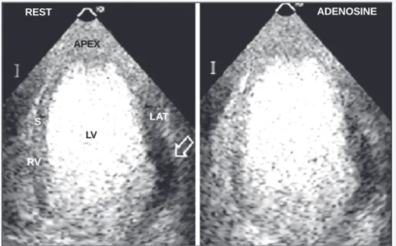
Myocardial perfusion was normal when a marked intensification of the contrast occurred in all segments of the left ventricular walls after adenosine injection (fig. 1). Myocardial perfusion was abnormal when a reduction in contrast intensity occurred. If this was observed only after adenosine injection, the defect was con-sidered reversible (fig. 2); if observed already in the evaluation at rest, with no alteration with adenosine injection, the defect was considered fixed (fig. 3).
In each patient, the left ventricular walls were divided into 3 coronary territories or beds. The anterior wall and the anterior por-tion of the interventricular septum, as well as the apical 2/3 of its posterior portion, were considered part of the territory of the anterior descending artery (AD). The lateral wall was considered to belong to the territory of the circumflex artery (CX). The posterolateral and posterior walls, and the basal posterior portion of the interventricular septum were considered territories of the right coronary artery (RC). The scintigraphic examination used in all patients was 99m
Tc-sestamibi SPECT associated with dipyridamole stress. The data were obtained at rest and after venous infusion of dipyridamole at the dosage of 0.56 mg/kg diluted in 20 mL of saline solution in 4 minutes. After 2 minutes, an additional dose of 0.28 mg/kg diluted in 10 mL was administered. For SPECT, 99mTc-sestamibi was
in-jected 6 minutes after infusion of dipyridamole, with image acqui-sition after 1 hour. Tomographic cuts were performed in the 3 major cardiac axes (minor vertical, major vertical, and horizontal). All tests were considered effective.
When compared per patient, the examinations were considered concordant when both were normal or both had perfusion altera-tions independent of their locaaltera-tions.
The comparison between myocardial contrast echocardiogra-Table I - Clinical characteristics of the patients
Age 58.4 ± 10.6 years
Sex: men/women 85 (68%) /40 (32%)
Typical OR suggestive chest pain 71 (56.8%)
Diabetes 19 (15.2%)
Systemic arterial hypertension 68 (54.4%) Previous myocardial infarction 26 (20.8%)
Previous revascularization 34 (27.2%)
CLBBB 10 (8%)
With cine coronary angiography 39 (31.2%)
n (%) - number of cases and percentages; CLBBB - complete left bundle-branch block.
REST ADENOSINE
S
RV LV
LAT
Fig. 1 - Apical 4-chamber view showing the normal pattern of myocardial perfusion with great intensification of the contrast medium observed in all segments after adenosine injection. LAT - lateral wall; S - interventricular septum; RV - right ventricle; LV - left ventricle.
Fig. 3 - Apical 4-chamber view showing an evident perfusion abnormality at rest, maintained after adenosine administration, in the basal region of the lateral wall (white arrow), characterizing a fixed perfusion defect. LAT - lateral wall; LV - left ventricle; RV - right ventricle; S - interventricular septum.
REST ADENOSINE
APEX
RV S
LV
LAT
Fig. 4 - Example of concordance between the methods in a 54-year-old male patient with chest pain suggestive of angina. Cine coronary angiography with important obstructive lesion (85%) in the circumflex artery (A and B are examples of MCE, where A is the image without treatment, B is the image coded for colors, and C is an example of MS). All images were obtained during the peak stress, showing the reversible perfusion defect in the basal region of the lateral wall (white arrows). AP - LV apical region; MS - myocardial nuclear scintigraphy; MCE - myocardial contrast echocardiography; LAT - lateral wall; S - interventricular septum.
AP
S LAT
AP
S > < LAT
Fig. 2 - Apical 2-chamber view showing the normal perfusion pattern at rest with evident abnormality after adenosine administration in the left ventricular apex (black arrow), characterizing a reversible perfusion defect. ANT - anterior wall; INF - inferior wall; LV - left ventricle.
REST ADENOSINE
INF LV
421
phy and myocardial nuclear scintigraphy per territory of coronary perfusion was considered concordant whether a perfusion defect existed or not in the same territory in both examinations, inde-pendent of the perfusion pattern.
The analysis of concordance between the 2 methods was perfor-med per patient and per coronary perfusion territory in regard to the presence or absence of perfusion defect. The territories of coronary perfusion were analyzed both as a whole, as well as individually.
The chi-square test was used for determining the significance of the distribution of concordance. The Student t test was used for comparing the values of concordance for each coronary territory. Variability lower than 0.05 was considered significant.
Results
The overall analysis of the 125 patients studied showed con-cordant myocardial contrast echocardiography and myocardial nu-clear scintigraphy in 106 patients (84.8%, χ2 = 58.579; P<0.001). Forty-five of these examinations were normal, and 61 were abnormal (39 reversible defects and 22 fixed defects). In 19 patients (15.2%), the examinations were discordant.
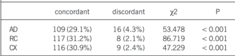
In the 125 patients, 375 coronary territories were analyzed. The overall concordance in these territories was 91.2% (342 in 375 territories, χ2 = 210.905; P < 0.001). The distribution of the normal and abnormal examinations is shown in table II.
The assessment of concordance for each territory (AD, RC, and CX) separately is briefly shown in table III.
Although the concordance for the alterations in the anterior descending artery territory was smaller in terms of percentage, the comparison of the 3 territories using the Student t test showed no significance (P > 0.05).
The form of adenosine administration posed neither compli-cations nor additional risks to the examinations. The mean dose of adenosine used per examination was 3.9 ampules. The side effects related to the administration of adenosine (facial warmth sensation, chest discomfort, headache, and transient complete atrioventricular block) were reported in less than 10% of the examinations.
Discussion
For 2 decades, assessment of myocardial perfusion through scintigraphy was considered the gold-standard for diagnosing co-ronary artery disease and the result of the procedures of reperfusion, for studying myocardial viability, and for stratifying the risk 8.
In the past 10 years, several studies reported that contrast echocardiography produces myocardial opacification when the contrast is intravenously administered 9, and, mainly, may identify
the presence of perfusion defects at rest and during pharmacological stress 10.
The study of myocardial perfusion performed with echocar-diography and peripheral infusion of contrast and the study performed with scintigraphy were first compared by Kaul et al 2, who studied
30 patients with suspected or known coronary artery disease by using the FS-069 second-generation contrast and dipyridamole as the stressor agent. When the analysis was performed per patient, concordance between the 2 methods was 86%, and, when the analysis was performed in each of the 3 coronary territories, con-cordance was 90% (88% for the territories with abnormal perfusion and 91% with normal perfusion). These promising values contri-buted to continuation of the research for the development of con-trast echocardiographic techniques.
Porter et al 11 compared contrast echocardiography with PESDA
at rest and after stress with sestamibi SPECT using videointensity for analysis (quantitative). They found 92% sensitivity and 84% specificity for myocardial contrast echocardiography, with excellent concordance in regard to location and extension of the perfusion defects.
Although the techniques of the 2 examinations are analogous, significant differences exist: 1) the location of the tracer is different in each technique; the contrast medium used for echocardiography is restricted to the vascular space. When injected through the intravenous route, the gas microbubbles mix with blood, and their concentration in a myocardial region reflects the blood volume in this region 12. Myocardial nuclear scintigraphy involves the
intra-cellular uptake of the tracer concentrated in the mitochondria 13.
Contrast echocardiography studies the integrity of the microvas-culature, while the nuclear technique involves the need for intact muscle cell; 2) the difference relates to the characteristic of the image. The spatial resolution of echocardiography is 1 mm, while that of scintigraphy is 1 cm. Therefore, conditions in which a reduction in the thickness of the left ventricle and abnormal motion of its walls occur (akinesia, complete left bundle-branch block) may create or emphasize the perfusion defects on myocardial nuclear scintigraphy. On the other hand, myocardial contrast echo-cardiography can identify perfusion defects independent of the alterations in contractility, as already reported by Moraes et al 14,
even in patients with previous myocardial infarction 15 or with
complete left bundle-branch block 16.
Because it is a recent technique, myocardial contrast echo-cardiography is still difficult to interpret, mainly because of atte-nuations and shadows that usually relate to the basal segments of the inferior and lateral walls, or to the greater destruction of the contrast medium by the ultrasound in the apical region. The ul-trasound reflection in the microbubbles is not homogeneous, re-Table II - Analysis of the results in 375 coronary territories
Normal MCE Abnormal MCE
normal MS 272 (72.5%) 16 (4.3%)
abnormal MS 17 (4.5%) 70 (18.7%)
χ2 = 210.905 – p < 0.001
n (%) - number of cases and percentages; MS - myocardial nuclear scintigraphy; MCE - myocardial contrast echocardiography.
Table III - Assessment of concordance for each coronary territory
concordant discordant χ2 P
AD 109 (29.1%) 16 (4.3%) 53.478 < 0.001 RC 117 (31.2%) 8 (2.1%) 86.719 < 0.001 CX 116 (30.9%) 9 (2.4%) 47.229 < 0.001
422
sulting in a decrease in signal intensity and amplitude, more intense in the distal field. Different types of contrast media, as well as the administration in the form of bolus injection may lead to different degrees of attenuation.
Independently of the type of echocardiographic contrast medium used and of its route of administration, researchers have emphasized the need for staff training for acquisition and analysis of the echo-cardiographic images, according to each protocol, aiming at mi-nimizing the incidence of artifacts to increase the sensitivity of myo-cardial contrast echocardiography, since specificity is very satisfactory4.
The choice of the stressor agent is an important factor for the greater or lower degree of accuracy of perfusion echocardiography. The blood vessels distal to an obstructive lesion are maximally dilated at rest, while the proximal areas are normal. This indicates that the myocardial region distal to the obstruction has a smaller coronary flow reserve, defined as the maximum possible increase in flow that can occur in 1 single coronary artery undergoing stress. Coronary flow reserve can be studied with physical exercise, inotropic agents, and vasodilating drugs, such as adenosine and dipyridamole. Vasodilators are the preferred agents for contrast echocardiography.
Ronderos et al 17 used dipyridamole during myocardial contrast
echocardiography to study 35 patients diagnosed with perfusion defects on myocardial nuclear scintigraphy. Those authors reported that 32 of those patients also had perfusion defects on echocar-diography.
As in the present study, Heinle et al 18 used adenosine for
assessing coronary flow reserve. Adenosine is a potent coronary vasodilator. Its peripheral injection increases coronary flow 3 to 4 times due to a decrease in coronary resistance, resulting in an increase in blood volume 19, which causes a variation in the intensity
of myocardial contrast.
Although the overall concordance between myocardial contrast echocardiography and myocardial nuclear scintigraphy was similar in our study and that of Heinle et al18 – 84.8% and 81%,
respec-tively – significant differences are observed in the studies in regard to the analysis of each coronary territory.
Heinle et al18 reported the following concordances for the
territories of the anterior descending (AD), right coronary (RC), and circumflex (CX) arteries: 81%, 76%, and 72%, respectively. In our study, the concordances were 87.2% (109/125 territories), 93.6% (117/125 territories), and 92.8% (116/125 territories), respectively (tab. III). The differences are mainly based on the protocol used for myocardial contrast echocardiography.
Heinle et al 18 used power Doppler as the image modality. It
more frequently generates artifacts in the distal fields, causing “fixed defects” mainly in the lateral wall, which are not seen on scintigraphy. This can explain the lower concordance of this study for the territory of the CX. In addition, power Doppler is very sensitive to cardiac movements, which, in this study, were inten-sified by the use of adenosine, which is known to cause tachypnea as a reflex mechanism.
This study applied the protocol developed by Morcerf et al 7,
which uses the administration of adenosine bolus. This protocol has a sensitivity of 91.7% and specificity of 98.6%, with excellent intra- and interobserver concordance (k = 0.94, and k = 0.91, respectively). The continuous infusion of the contrast medium
provides longer time for the adjustment of the initial image, in an attempt to reduce, as much as possible, the artifacts inherent to the echocardiographic study with contrast medium. In addition, the effect of the use of adenosinebolus is felt within a short time, facilitating the maintenance of the same adjustments of the equip-ment, the same velocity of contrast infusion, and a rapid comparison with the baseline image. The characteristic that differentiates this protocol and that could justify the high concordance in the RC and CX territories (93.6% and 92.8%, respectively) is the existence of an expected normality pattern for the administration of adenosine. This pattern is the great increase in the intensity of the contrast medium, which can be visually detected in the areas without obstruction or with noncritical obstruction. Therefore, even when heterogeneity of perfusion exists at rest, due to the presence of artifacts, with an increase in contrast intensity after adenosine administration, perfusion is considered normal because coronary flow reserve is preserved.
Based on these considerations, concordance with myocardial nuclear scintigraphy was detected in 106 (84.8%) of the 125 patients studied. The statistical study showed that this concordance was highly significant (P < 0.001). The same significance was observed when the analysis was performed by perfusion bed. Overall concordance was 91.2% (342/375 territories).
Although without statistical significance, a slight reduction in concordance among the examinations was observed in regard to the AD perfusion bed (tab. III). This may be due to the greater incidence, among the discordances in this territory, of patients with complete left bundle-branch block, which is a known factor of false-positive results in scintigraphic examinations. Of the 16 discordant examinations in the AD territories, 5 had complete left bundle-branch block.
A better understanding of the discordances is important. Ini-tially, the results of myocardial contrast echocardiography are compared with those of myocardial nuclear scintigraphy, considered the gold standard. However, it is not possible to know which method has the correct information, because scintigraphy also has false-positive results.
In this study, discordance occurred in 19 examinations as follows: MIBI was the abnormal examination in 13 patients, and myocardial contrast echocardiography was abnormal in 6. Of the 13 patients with alteration only in MIBI, 6 had complete left bundle-branch block, 2 had previous myocardial infarction, and 1 patient had already undergone myocardial revascularization sur-gery. Of the 6 patients with alteration only on myocardial contrast echocardiography, 2 had previous myocardial infarction, and 3 had already undergone revascularization (2 patients with angioplasty and 1 patient with surgery). These data point to the need for correlating with another method, maybe coronary anatomy as-sessment (cine coronary angiography), aiming at improving the comprehension of the possibilities of myocardial contrast echo-cardiography in assessing myocardial perfusion.
423
1. Porter TR, Li S, Kricsfeld D, et al. Detection of myocardial perfusion in multiple echocardiographic windows with one intravenous injection of microbubbles using transient response second harmonic imaging. J Am Col Cardiol 1997;29:791-9. 2. Kaul S, Senior R, Dittrich H, et al. Detection of coronary artery disease with
myo-cardial contrast echocardiography: comparison with 99mTc-sestamibi single-pho-ton emission computed tomography. Circulation. 1997;96:785-92.
3. Porter TR, Cwajg J, Li S, et al. Correlation of myocardial contrast enhancement de-termined with accelerated intermittent imaging with dual isotope SPECT imaging during exercise and dobutamine stress. J Am Soc Echocardiogr 1999;12:350. 4. Marwick TH, Brunken R, Meland N, et al. Accuracy and feasibility of contrast
echocardiography for detection of perfusion defects in routine practice: Compari-son with wall motion and Technetium-99m sestamibi single-photon emission computed tomography. J Am Coll Cardiol 1999;32:1260-9.
5. Jucquois I, Nihoyannopoulos P, D’hont AM, et al. Comparison of myocardial con-trast echocardiography with NC 100100 and Tc Sestamibi Spect for detection of resting myocardial perfusion abnormalities in patients with previous myocardial infarction. Heart 2000;83:518-24.
6. Porter TR, Xie F. Transient myocardial contrast following initial exposure to diag-nostic ultrasound pressures with minute doses of intravenously injected microbub-bles: demonstration and potential mechanisms. Circulation 1995;92:2391-5. 7. Morcerf F, Moraes A, Carrinho M, et al. Estudo da reserva de fluxo coronariano
com uso endovenoso de microbolhas (Ecocardiografia com contraste) e adenosi-na. Apresentação de protocolo para apresentação clínica em pacientes com sus-peita de doença arterial coronariana. Arq Bras Cardiol2002;78:281-9. 8. Ladenheim ML, Pollock BH, Rozanki A, et al. Extent and severity of myocardial
hypoperfusion as predictors of prognosis in patients with suspected coronary ar-tery disease. J Am Coll Cardiol 1986;7:464-71.
9. Villanueva F, Glasheen WP, Sklenar J, et al. Successful and reproducible myocardial opacification during two-dimensional echocardiography from right heart injection of contrast. Circulation 1992;85:1557-64.
References
10. Xie F, Porter TR. Acute myocardial ischemia and reperfusion can be visually iden-tified non-invasively with intravenous perfluoropropane-enhanced sonicated al-bumin ultrasound contrast. Circulation 1994;90:1-555.
11. Porter TR, Cwajg J, Li S, et al. Correlation of myocardial contrast enhancement de-termined with accelerated intermittent imaging with dual isotope SPECT imaging during exercise and dobutamine stress. J Am Soc Echocardiogr 1999;12:350. 12. Kaul S. Clinical applications of myocardial contrast echocardiography. In:
Braun-wald E (Eds.) Heart Disease: A Textbook of Cardiovascular Medicine (Update 1). Philadelphia: W.B Saunders 1997;304-13.
13. Dahlberg ST, Leppo JA. Physiologic properties of myocardial perfusion tracers. Cardiol. Clin 1994;12:169-85.
14. Moraes A, Morcerf F, Carrinho M et al. The ability of myocardial contrast echocar-diography to identify perfusion defects is independent of rest wall motion abnor-malities. Circulation 1998;98(supp I):502.
15. Morcerf F, Moraes A, Carrinho M et al. Intravenous infusion contrast echocardio-graphy to assess myocardial revascularization procedures. Circulation 1998;98 (supp I):19.
16. Carrinho M, Moraes A, Morcerf F, et al. Eco de Contraste Miocárdico com micro-bolhas e adenosina em pacientes com bloqueio completo do ramo esquerdo. Rev SOCERJ 1999;XII (suplemento A):23.
17. Ronderos RE, Boskis M, Namsik C, et al. Correlation between myocardial perfu-sion abnormalities detected with intermittent imaging using intravenous perfluo-rocarbon microbubbles and radioisotope imaging during high-dose dipyridamole stress echo Clin Cardiol 2002;25:103.
18. Heinle SK, Noblin J, Goree-Best P, et al. Assessment of myocardial perfusion by harmonic power Doppler imaging at rest and during adenosine stress. Comparison with 99m Tc-Sestamibi SPECT imaging. Circulation 2000;102:55-60. 19. Belardinelli L, Linden J, Berne RM. The cardiac effect of adenosine. Progress in
Cardiovac Dis 1989; 17: 73-97.
is known to favor the diagnostic accuracy of the method; 2) in regard to the protocol of myocardial contrast echocardiography used, it is worth noting that its reproducibility was adequately tested in few diagnostic centers; 3) the qualitative and nonquan-titative analysis could interfere with the results; 4) unlike myocardial contrast echocardiography, the scintigraphic examinations were performed in 2 different laboratories. Therefore, although the exa-minations were interpreted by cardiologists with certification in
nuclear cardiology, who followed the same standard protocol for MIBI with dipyridamole, variation in the diagnostic criteria may have occurred.