Orthodontic camouflage of skeletal Class III
malocclusion with miniplate: a case report
Marcel Marchiori Farret1, Milton M. Benitez Farret2, Alessandro Marchiori Farret3
Introduction: Skeletal Class III malocclusion is often referred for orthodontic treatment combined with orthognathic surgery. However, with the aid of miniplates, some moderate discrepancies become feasible to be treated without surgery.
Objective: To report the case of a 24-year-old man with severe skeletal Angle Class III malocclusion with anterior cross-bite and a consequent concave facial profile. Methods: The patient refused to undergo orthognathic surgery; therefore, orthodontic camouflage treatment with the aid of miniplates placed on the mandibular arch was proposed. Results: Af-ter 18 months of treatment, a Class I molar and canine relationship was achieved, while anAf-terior crossbite was corrected by retraction of mandibular teeth. The consequent decrease in lower lip fullness and increased exposure of maxillary incisors at smiling resulted in a remarkable improvement of patient’s facial profile, in addition to an esthetically pleasing smile, respectively. One year later, follow-up revealed good stability of results.
Keywords: Angle Class III malocclusion. Orthodontic anchorage procedures. Orthodontic appliance design.
1 Professor, Centro de Estudos Odontológicos Meridional (CEOM), Graduate
Program in Orthodontics, Passo Fundo/RS, Brazil; and Fundação para Reabilitação das Deformidades Crânio-faciais (FUNDEF), Lajeado/RS, Brazil.
2 Professor, Universidade Federal de Santa Maria (UFSM), Santa Maria/RS,
Brazil.
3 Private practice, Santa Maria/RS, Brazil.
DOI: http://dx.doi.org/10.1590/2177-6709.21.4.089-098.oar
How to cite this article: Farret MM, Farret MMB, Farret AM. Orthodontic camouflage of skeletal Class III malocclusion with miniplate: a case report. Dental Press J Orthod. 2016 July-Aug;21(4):89-98.
DOI: http://dx.doi.org/10.1590/2177-6709.21.4.089-098.oar
Submitted: November 26, 2015 - Revised and accepted: February 11, 2016
» The authors report no commercial, proprietary or financial interest in the products or companies described in this article.
» Patients displayed in this article previously approved the use of their facial and in-traoral photographs.
Contact address: Marcel Marchiori Farret E-mail: [email protected]
Introdução: a má oclusão de Classe III esquelética é frequentemente tratada com associação entre Ortodontia e Cirurgia Ortognática. No entanto, com o auxílio das miniplacas, discrepâncias moderadas tornaram-se passíveis de serem tratadas sem a necessidade de cirurgia ortognática. Objetivo: relatar o caso de um paciente de 24 anos, com má oclusão de Classe de III Angle e Classe III esquelética, mordida cruzada anterior e consequente perfil facial côncavo. Métodos: o paciente recusou-se a realizar a cirurgia ortognática, assim, foi proposta a compensação da má oclusão com o auxílio de miniplacas na arcada inferior. Resultados: após 18 meses de tratamento, foram obtidas relações de molares e caninos em Classe I, e a mordida cruzada anterior foi corrigida, por meio da distalização de todos os dentes inferiores. A consequente redução na projeção do lábio inferior, associada à maior exposição dos incisivos superiores no sorriso, resultou em um perfil facial consideravelmente mais agradável e um sorriso esteticamente melhor, respectivamente. A análise um ano após o trata-mento revelou boa estabilidade dos resultados obtidos.
tional and esthetic results.3,4,5 However, several patients refuse surgery. In such situations, orthodontic camou-lage treatment may be an alternative, particularly if
dis-crepancy is slight or moderate.3,4,6
The introduction of skeletal anchorage has increased the number of patients with skeletal problems who can be treated by mechanical orthodontic treatment only, thereby avoiding the need for complementary orthognathic
sur-gery.2,7 Mini-implants are preferred for patients with slight
discrepancies because of less invasive insertion and removal
procedures.8,9 However, in patients with moderate skeletal
and dental discrepancies, miniplates are the treatment of choice to improve anchorage and eliminate the possibility of contact between implant screws and tooth roots during
tooth movement, as it can occur with mini-implants.2,8,10,11
In the present study, we report the case of a 24-year-old man with severe skeletal Angle Class III malocclusion who was treated by orthodontic camoulage treatment with miniplate anchorage.
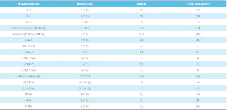
terior teeth and decreased visibility of maxillary anterior teeth at smiling. Extraoral examination revealed a con-cave facial proile (Fig 1). Clinical manipulation in cen-tric relation demonstrated that there was no mandibular anterior deviation during bite closing. Intraoral exami-nation and analysis of dental casts revealed Angle Class III malocclusion, Class III canine relationship, anterior crossbite, and maxillary incisor crowding, with a nega-tive discrepancy of 4 mm (Figs 2 and 3). Furthermore, Bolton analysis revealed 1-mm excess for maxillary pos-terior teeth and 2-mm excess for mandibular anpos-terior teeth. Cephalometric analysis revealed skeletal Class III (ANB = −5°) malocclusion, a hypodivergent facial pat-tern (SN-GoGn = 20°, FMA = 9° and Y-axis = 44°), se-vere maxillary incisor proclination, and uprighted man-dibular incisors (Fig 4).
Figure 2 - Pretreatment intraoral photographs.
Figure 3 - Pretreatment dental casts.
crowding; and (4) improve facial esthetics by straight-ening the facial proile and increasing maxillary incisor exposure at smiling.
Treatment alternatives
The irst treatment option for this patient was or-thognathic surgery for maxillary advancement, which would certainly improve facial esthetics and simplify subsequent mechanical orthodontic treatment; how-ever, the patient refused to undergo surgery. The sec-ond option was mechanical orthodontic treatment with Class III elastics and a sliding jig on the mandibular arch. This would require prolonged use of elastics with extremely good patient compliance and could result in some undesirable efects, such as counterclockwise occlusal plane rotation, with less maxillary incisor and greater mandibular incisor exposure. The third option was the use of mini-implants as anchorage unit; which was disregarded because the required tooth movement was extensive and the mini-implant would require re-moval and relocation at some point during treatment. Eventually, camoulage orthodontic treatment with miniplate anchorage was proposed and the patient agreed with this option. In this planning, treatment would be started with alignment and leveling of lower and upper arches, except for maxillary incisors, thus avoiding further proclination. Ater alignment and lev-eling of the upper arch, stripping was considered from second molar to irst premolar on each side, so as to gain space for incisors alignment. On the lower arch, ater alignment and leveling, miniplates would be inserted on each side of the posterior mandible, so to be used as the anchorage unit to retract all mandibular teeth. During anterior crossbite correction, a posterior bite plate was also planned to be used, so as to avoid interferences be-tween maxillary and mandibular incisors.
and 0.016-in, 0.018-in, and 0.020-in stainless steel archwires. The archwires were not inserted for inci-sors to avoid proclination and premature contact be-tween maxillary and mandibular incisors. Stripping from the mesial surface of the maxillary second molar to the mesial surface of the maxillary first premolar was performed on both sides, followed by distaliza-tion of all maxillary posterior teeth. Mandibular pos-terior teeth were aligned and leveled up to a 0.020-in stainless steel archwire, and at this point in treatment, miniplates were placed on the external oblique ridge. Subsequently, a 0.019 × 0.025-in stainless steel arch-wire was set with hooks between the canine and first premolar on both sides and connected to the mini-plates by means of elastomeric chains, thus resulting in a load of 400 g/f on each side (Fig 5).
Figure 5 - Intraoral photographs at the beginning of mandibular dentition distalization.
Figure 6 - Intraoral photographs when maxillary and mandibular incisors were included on the mechanics to correct anterior crossbite.
Figure 7 - Intraoral photographs after anterior crossbite correction.
Treatment results
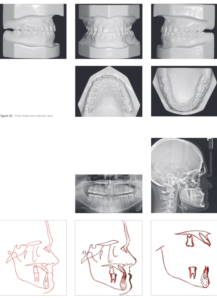
Patient’s treatment was complete after 18 months. His facial profile remarkably improved with an es-thetically pleasing smile (Fig 8). Intraoral examina-tion and dental casts analysis revealed a Class I molar and canine relationship on both sides, with excel-lent intercuspation (Figs 9 and 10). Due to anterior Bolton discrepancy, spaces were kept unchanged be-tween maxillary lateral incisors and canines, which would be filled with composite resin. Anterior cross-bite was successfully corrected and adequate overjet
Figure 8 - Post-treatment facial photographs.
Figure 10 - Post-treatment dental casts.
Figure 13 - 1-year post-treatment intraoral photographs.
Table 1 - Cephalometric measurements.
Measurements Norms (SD) Initial Post-treatment
SNA 82° (3) 86 85
SNB 80° (3) 91 90
ANB 2° (2) -5 -5
Facial convexity (NA.APog) 0° (2) -13 -14
Facial angle (PoOr.NPog) 87° (3) 103 102
Y-axis 59° (6) 44 45
SN.GoGn 32° (3) 20 21
1-NA (°) 22° 44 41
1-NA (mm) 5 mm 6 8
1-NB (°) 25° 13 5
1-NB (mm) 5 mm 3 0
Inter-incisal angle 131° (5) 126 139
Ul-S line 0 mm (2) -5 -4
Ll-S line 0 mm (2) -1 -3
IMPA 90° (4) 85 74
FMA 25° (3) 9 11
FMIA 65° (4) 86 95
DISCUSSION
The present article reported the case of a 24-year-old man with severe skeletal Angle Class III malocclusion. The patient was treated by orthodontic camoulage treat-ment with miniplate anchorage. In the last few years, only slight skeletal discrepancies in adult patients were usually
treated without orthognathic surgery.9 Treatment options
included the use of Class III elastics alone or in combination with a sliding jig or even headgears, stripping, and tooth
extraction.3,6,10 Unfortunately, all these options were
asso-ciated with complications, such as counterclockwise
rota-tion of the occlusal plane,2,4,12,13 patient’s noncompliance
with elastics or headgears,14,15 patient’s refusal to undergo
extraction, and the creation of Bolton discrepancy in cases of stripping. The advent of skeletal anchorage increased the reliability of results because it does not require patient compliance and it is associated with minimal or no side ef-fects. In this context, miniplates represent the best option for simultaneous multiple tooth movement because of the increased stability generated by multiple screws instead of a single screw as with implants. Conventionally, mini-plates are inserted at two sites in Class III patients: on the external oblique ridge with the active end positioned at the mesial or distal surface of the irst molar or on the lower border of the mandible with the active end positioned at the
mesial surface of the irst molar.10 For the presented case,
the surgeon faced some diiculty during the procedure and had to ix right and let miniplates with their active ends around the mesial and distal surfaces of the irst molar, re-spectively, with no mechanical issues thereater.
In patients with moderate skeletal Class III malocclu-sion, one question must always be addressed by orthodon-tists: is it possible to camoulage this malocclusion? There are several parameters inluencing this decision. First, the extent of compromise on facial esthetics, and whether compromise is a big concern for the patient must be
judged.4,5,13 In the present study, the patient was not hugely
concerned about his facial esthetics, and proile concavity was moderate. Certainly, if the patient’s chief complaint was facial esthetics, orthognathic surgery, and not cam-oulage treatment alone, would be necessary. The second parameter is the anteroposterior position and angulation of maxillary and mandibular incisors. In patients with an edge-to-edge anterior bite or a slight anterior crossbite, correction can be achieved ater judging the extent of maxillary incisor proclination and mandibular incisor ret-roclination. Our patient showed severe maxillary incisor
proclination; however, mandibular incisors were not ret-roclined, thereby facilitating orthodontic camoulage by means of incisor retraction. The third parameter is thick-ness of mandibular symphysis, which should be adequate
to allow extensive incisor retraction.3 Fortunately, in our
patient, the anteroposterior dimension of the symphysis was adequate. Finally, the last parameter is the degree of anteroposterior discrepancy. Even if facial esthetics is ac-ceptable, the symphysis is thick enough, and mandibular incisors are slightly proclined, camoulage is not possible if anteroposterior discrepancy is too severe. Considering that anteroposterior discrepancy was moderate in the patient reported herein, orthodontic camoulage was selected.
One major concern for orthodontists is stability of camoulage treatment ater mandibular incisor retraction
in patients with Class III malocclusion.5,14 Considering
that the entire arch is retracted by 4–5 mm, the tongue has less space ater treatment, thus resulting in extreme tongue pressure on mandibular incisors and consequent relapse with premature contact between incisors and
abnormal spacing between mandibular teeth.10,16 Some
alternatives to improve stability in such cases include
achieving an ideal overjet, overbite, and intercuspation;2,5
maintenance of mandibular posterior teeth in an upright position ater distalization because distal tipping tends cause them to return to their original position according
to their root apices;10,14,15 using a 3 × 3 bonded mandibular
retainer for an undetermined period of time;13
myofunc-tional therapy to eliminate tongue interposition during swallowing and rest; and to position the tip of the tongue at the incisive papilla during swallowing and in the
poste-rior region of the oral cavity at rest.17
Superimpositions at follow-up revealed excessive re-modeling of the symphysis because of mandibular inci-sor retraction. Inciinci-sors centered on the symphysis at the beginning of treatment maintained the centers at the end of treatment, thus avoiding gingival recession in the
long-term and improving stability.5 In the case presented herein,
analysis one year ater treatment revealed excellent stability of results. The patient will remain under post-treatment follow-up once a year.
CONCLUSION
2013 Sept;40(3):256-63.
3. Choi JY, Lim WH, Chun YS. Class III nonsurgical treatment using indirect skeletal anchorage: A case report. Korean J Orthod. 2008;38(1):60-7.
4. Lin J, Gu Y. Preliminary investigation of nonsurgical treatment of severe skeletal Class III malocclusion in the permanent dentition. Angle Orthod. 2003 Aug;73(4):401-10.
5. Moullas AT, Palomo JM, Gass JR, Amberman BD, White J, Gustovich D. Nonsurgical treatment of a patient with a Class III malocclusion. Am J Orthod Dentofacial Orthop. 2006;129(4 Suppl):S111-8.
6. Kuroda Y, Kuroda S, Alexander RG, Tanaka E. Adult Class III treatment using a J-hook headgear to the mandibular arch. Angle Orthod. 2010 Mar;80(2):336-43. 7. Freire-Maia B, Pereira TJ, Ribeiro MP. Distalization of impacted mandibular
second molar using miniplates for skeletal anchorage: case report. Dent Press J Orthod. 2011;16(4):132-6.
8. Kuroda S, Tanaka E. Application of temporary anchorage devices for the treatment of adult Class III malocclusions. Semin Orthod. 2011;17(2):91-7. 9. Sugawara Y, Kuroda S, Tamamura N, Takano-Yamamoto T. Adult patient with
mandibular protrusion and unstable occlusion treated with titanium screw anchorage. Am J Orthod Dentofacial Orthop. 2008 Jan;133(1):102-11.
12. Baek S-H, Yang I-H, Kim K-W, Ahn H-W. Treatment of Class III malocclusions using miniplate and mini-implant anchorage. Semin Orthod. 2011;17(2):98-107. 13. Sobral MC, Habib FA, Nascimento AC. Vertical control in the Class III
compensatory treatment. Dental Press J Orthod. 2013 Mar-Apr;18(2):141-59. 14. Chung KR, Kim SH, Choo H, Kook YA, Cope JB. Distalization of the mandibular
dentition with mini-implants to correct a Class III malocclusion with a midline deviation. Am J Orthod Dentofacial Orthop. 2010 Jan;137(1):135-46. 15. Sakai Y, Kuroda S, Murshid SA, Takano-Yamamoto T. Skeletal Class lll
severe openbite treatment using implant anchorage. Angle Orthod. 2008 Jan;78(1):157-66.
16. Saito I, Yamaki M, Hanada K. Nonsurgical treatment of adult open bite using edgewise appliance combined with high-pull headgear and Class III elastics. Angle Orthod. 2005 Mar;75(2):277-83.