243
J Vasc Bras. 2013 Jul.-Set.; 12(3):243-246 http://dx.doi.org/10.1590/jvb.2013.033
C A S E
R E P O R T
Acute limb ischemia secondary to radiation-induced arteritis:
case report
Insuficiência arterial aguda secundária a arterite induzida por radiação: relato de caso
José Emerson dos Santos Souza1, Leonardo Pessoa Cavalcante1, Marcos Velludo Bernardes1, Marcos Henrique Parisati1, Patrícia de Souza Lacerda1, Raquel Magalhães Pereira2
Abstract
Radiation-induced arteritis is a rare but well-known complication of radiotherapy. his report describes the case of a 34-year-old woman with uterine cervical cancer who was diagnosed with left iliofemoral deep vein thrombosis (DVT) 2 years after radiotherapy, and 2 months later, during the treatment of DVT with efective anticoagulation, developed an episode of acute arterial ischemia of the left lower limb secondary to a long subocclusive lesion of the external iliac artery. he patient was treated with angioplasty and stenting of the lesion and recovered uneventfully after the endovascular procedure.
Keywords: radiotherapy; constriction, pathologic; angioplasty.
Resumo
A arterite induzida por radiação é uma rara mas bem documentada complicação da radioterapia. O presente relato descreve o caso de uma mulher de 34 anos, diagnosticada com neoplasia de colo do útero, a qual, dois anos após sessões de radioterapia desenvolveu trombose venosa profunda (TVP) iliofemoral esquerda; dois meses depois, durante tratamento para TVP com devida anticoagulação, a paciente apresentou quadro de insuiciência arterial aguda do membro inferior esquerdo secundária a uma longa lesão suboclusiva da artéria ilíaca externa. A paciente foi tratada com angioplastia transluminal percutânea e implantação de stent autoexpansível, recuperando-se sem intercorrências após o procedimento endovascular.
Palavras-chave: radioterapia; constrição patológica; angioplastia.
1 Hospital Universitário Francisca Mendes, Vascular and Endovascular Surgery Service, Manaus, AM, Brazil. 2 Universidade Federal do Amazonas – UFAM, Manaus, AM, Brazil.
Financial support: None.
Conlicts of interest: No conlicts of interest declared concerning the publication of this article. Submitted on: 11.02.12. Accepted on: 04.13.13.
Radiation-induced acute limb ischemia
244 J Vasc Bras. 2013 Jul.-Set.; 12(3):243-246
With the aid of road mapping, the left femoral artery was punctured, and the EIA lesion was easily crossed with a hydrophilic 0.035-in, 180-cm stiff
shaft and an angled loppy-tip guide wire; after
that, the lesion was pre-dilated using a 6-mm × 40-mm non-compliant percutaneous transluminal angioplasty (PTA) balloon. Subsequently, 2
self-expandable nitinol stents were deployed: irst, an
8-mm × 80-mm stent distally, and then a 9-mm × 80-mm stent proximally, with an overlapping zone of about 3 cm. For post-dilatation, a 7-mm × 80-mm non-compliant PTA balloon was used (Figure 2).
Both balloons had to be inlated to nearly their burst
pressure to open the lesion adequately. Pressure was measured across the stented region, but no pressure
gradient was identiied. The completion angiogram
showed a satisfactory angiographic result (Figure 3).
On the irst post-procedure day, she had normal
pedal pulses, and color Doppler US detected normal
triphasic low down to the foot on both left pedal
arteries. Clinically, there was remission of the left foot numbness and her left foot movements were preserved. She was discharged from the hospital on the third post-procedure day with a referral for outpatient anticoagulation and INR control.
DISCUSSION
Radiation-induced stenosis has been described in various organs after radiotherapy for several types of tumors. According to Teixeira et al.4,
radiation-induced proctitis chronically leading to rectal stenosis is a complication seen in about 1% to 20%
INTRODUCTION
External-beam radiation for malignancies may
cause inlammation and ibrosis in adjacent large arteries and lead to clinically signiicant stenosis.
These lesions are often indistinguishable from
atherosclerosis; however, location and coninement
to an area previously irradiated indicates that radiation was the cause1,2.
The most affected vascular beds are: cervical and cranial arteries, secondary to radiotherapy
for esophageal squamous cell carcinoma; visceral arteries, secondary to radiotherapy for lymphoma;
and the iliac arteries, secondary to radiotherapy for cervical cancer2,3.
The diagnoses is usually made on clinical grounds,
and conirmed by noninvasive and invasive test
results. This study describes a case of external iliac artery (EIA) radiation-induced subocclusive lesion that led to a rare clinical presentation of acute arterial
insuficiency in a patient already on anticoagulation
for the treatment of deep vein thrombosis (DVT) of the same limb.
CASE REPORT
A 34-year-old woman with no risk factors for atherosclerosis was diagnosed with advanced cervical cancer in 2008 and underwent chemotherapy, radiotherapy (57.6 Gy radiation in 32 fractions) and brachytherapy (4 sessions) in the same year.
In June 2011, the patient presented with sudden left lower extremity swelling, calf pain and tightness, but pedal pulses were palpable. A color Doppler ultrasound (US) scan revealed left iliofemoral DVT. She was hospitalized, treated with sequential parenteral and oral anticoagulation (warfarin), and discharged with a stable international normalized ratio (INR) of 2.0 to 3.0.
In August 2011, she returned to the emergency department because of intense pain of the left lower thigh and calf, walking impairment and left foot numbness. Physical examination revealed absence of left femoral, popliteal and pedal pulses and palpable normal pulses in the contralateral limb. The skin temperature on the whole left limb was low, and the left foot was cyanotic. Laboratory tests were normal, except for an INR of 2.3.
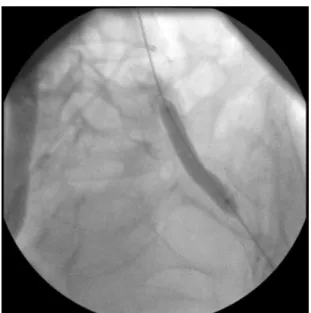
The patient was then taken to an endovascular suite and underwent digital angiography, which revealed a long, uniform subocclusive lesion of the left EIA (Figure 1). Filling was delayed in all the distal arteries, but their angiographic appearance was normal.
José Emerson dos Santos Souza, Leonardo Pessoa Cavalcante et al.
245
J Vasc Bras. 2013 Jul.-Set.; 12(3):243-246
of patients that undergo pelvic region radiotherapy. Novais et al.5 found that esophageal
radiation-induced stenosis affects 25% to 67% of patients irradiated for the treatment of esophageal primary
tumors and 1% to 20% for the treatment of adjacent
tumors (breast, lung, thyroid).
Although still rarely seen in oncology practice, arterial radiation-induced stenosis is becoming more
common due to the increased use of radiotherapy and prolonged survival of cancer patients1,6. The vessels
involved are directly associated with cancer location and the irradiation received.
Arterial occlusive disease pathogenesis begins with direct cellular damage due to radiation or by free radicals produced by its action7. Cell damage leads
to ibrosis, intimal thickening, elastic middle layer degeneration, adventitia ibrosis and vasa vasorum
damage3,6. After that, in addition to narrowing, there
may be thrombus formation, ulceration and distal embolization1.
Although well documented, histological changes are nonspecific and virtually identical to those observed in atherosclerosis1,2. Some authors suggest
that vasa vasorum injury and the consequent arterial
wall ischemia are among the few morphological characteristics that separate radiation-induced lesions from spontaneous atherosclerosis8. It has also
been suggested that the presence of atherosclerosis risk factors further promotes lesion development after radiotherapy2,6. However, the predisposition
to atherosclerosis only accelerates vascular injury
after irradiation, because the absence of lesions at uninvolved areas indicates that radiotherapy is the primary etiology1,2,6.
Therefore, the treatment of cervical cancer was
the main cause of arterial injury in our patient, as
arteriography showed that the disease was restricted to the left EIA, and all the other pelvic and left lower limb vessels had a normal angiographic appearance. Besides that, age and the absence of other atherosclerosis risk factors confirmed the radiation-induced etiology of the arterial disease. The radiation dose reported in the literature to be associated with iliac and femoral artery stenosis is 39.5 to 80 Gy9.
In our patient, acute arterial insufficiency symptoms appeared about 2 years after radiotherapy. In the literature, the time between irradiation and signs and symptoms of damage varies: Piedbois et al.2
described a range of 1 to 10 years (mean 6 years); Dorresteijn et al.10, reported a mean 13 years;
Hassen-Khodja et al.11 found a mean10 years between
cervical irradiation and carotid stenosis treatment. The clinical signs and symptoms of radiation-induced arterial stenosis are not different from other chronic arterial blockages. Therefore, in the arterial involvement of the lower extremities,
varying degrees of arterial insuficiency may occur,
from exertion muscular ischemia (intermittent claudication) to rest pain and tissue loss12.
Our patient presented with an uncommon clinical
picture of acute arterial insuficiency. Although
Figure 2. Post-dilatation of two self-expandable nitinol stents with a 7-mm × 80-mm non-compliant percutaneous translu-minal angioplasty balloon.
Radiation-induced acute limb ischemia
246 J Vasc Bras. 2013 Jul.-Set.; 12(3):243-246
and Review of the Literature. Cardiovasc Intervent Radiol. 2006;29:1144-7. PMid:16845557. http://dx.doi.org/10.1007/ s00270-005-0230-x
7. Brown KR, Rzucidlo E. Acute and chronic radiation injury. J Vasc Surg. 2011;53(15S):15S-21S. PMid:20843630. http://dx.doi. org/10.1016/j.jvs.2010.06.175
8. Zhou W, Bush RL, Lin PH, Lumsden AB. Radiation-associated venous stenosis: Endovascular treatment options. J Vasc Surg. 2004;40(1):179-82. PMid:15218482. http://dx.doi.org/10.1016/j. jvs.2004.03.039
9. Himmel PD, Hassett JM. Radiation-induced chronic arterial injury. Semin Surg Oncol. 1986;2:225-47. http://dx.doi.org/10.1002/ ssu.2980020405
10. Dorresteijn LDA, Vogels OJM, Leeuw FE, et al. Outcome of carotid artery stenting for radiation-induced stenosis. Int. J. Radiation Oncology Biol. Phys 2010;77(5):1386-90. PMid:20116932. http:// dx.doi.org/10.1016/j.ijrobp.2009.06.045
11. Hassen-Khodja R, Sala F, Declemy S, Lagrange JL, Bouillane PJ, Batt M. Surgical Management of Atherosclerotic Carotid Artery Stenosis after Cervical Radiation herapy. Ann Vasc Surg. 2000;14(6):608-11. http://dx.doi.org/10.1007/s100169910110 12. Pedron C, Ristow A, Cury JM Fº, Martin HS, Peixoto CC, Fonseca
LMB. Tratamento endovascular da oclusão das artérias Ilíacas. Radiol Bras. 2001;34(5):261-5. http://dx.doi.org/10.1590/ S0100-39842001000500004
13. Donas KP, Schwindt A, Pitoulias GA, Schönefeld T, Basner C, Torsello G. Endovascular treatment of internal iliac artery obstructive disease. J Vasc Surg. 2009;49(6):1447-51. PMid:19497505. http://dx.doi.org/10.1016/j.jvs.2009.02.207 14. Sullivan TM, Childs MB, Bacharach JM, Gray BH, Piedmonte ML.
Percutaneous transluminal angioplasty and primary stenting of the iliac arteries in 288 patients. J Vasc Surg. 1997;25(5):829-39. http://dx.doi.org/10.1016/S0741-5214(97)70212-X
Correspondence
Leonardo Pessoa Cavalcante Hospital Universitário Francisca Mendes – Serviço de Cirurgia Vascular/Endovascular Av. Camapuã, 108 - Cidade Nova II CEP 69097-720 - Manaus (AM), Brazil E-mail: [email protected]
Author information
LPC is chief, Endovascular Surgery Service, Hospital Universitário Francisca Mendes. MVB is chief, Vascular Surgery Service, Hospital Universitário Francisca Mendes. MHP is vascular surgeon, Vascular Surgery Service, Hospital Universitário Francisca Mendes. JESS and PSL are resident physicians (Vascular Surgery), Vascular Surgery Service, Hospital Universitário Francisca Mendes. RMP is medical student, Universidade Federal do Amazonas (UFAM).
Author’s contributions
Conception and design: JESS, LPC, RMP Analysis and interpretation: MHP, MVB Data collection: JESS, PSL Writing the article: JESS, LPC, RMP Critical revision of the article: LPC, MVB, MHP Final approval of the article*: JESS, LPC, MVB, MHP, PSL, RMP Statistical analysis: N/A Overall responsibility: JESS Obtained funding: None.
*All authors should have read and approved of the inal version of the article submitted to J Vasc Bras.
she had a chronic arterial lesion that was probably asymptomatic, iliofemoral DVT may have led to an imbalance of the arterial blood supply to the limb, previously compensated through collateral arteries. These arteries may explain why the pulses were palpable during physical examination two months before acute ischemia.
The clinical diagnosis of chronic arterial insufficiency is usually confirmed using color Doppler US and physiological tests (such as ABI)13,14.
Digital subtraction arteriography is usually reserved for surgical planning1,2,10. In the case described
here, urgent arteriography was performed after the clinical diagnosis of acute arterial occlusion, and the endovascular treatment was administered at the same time.
Although other treatment options have been described (drug therapy, endarterectomy, extra-anatomic bypass)1,2, PTA with stent implantation
is increasingly becoming the treatment of choice in similar cases because of its low invasiveness and high success rates, as described in the literature12,14.
PTA associated with stent implantation is extremely effective in the treatment of iliac arterial stenosis of any etiology14. Patients usually have a good
postoperative recovery, and clinical improvement is evident, as shown in this case and in the literature12,14.
CONCLUSION
This case demonstrated that PTA with implantation of self-expandable stents is a good therapeutic option for the treatment of radiation-induced EIA stenosis with clinical symptoms in patients treated for advanced pelvic cancer.
REFERENCES
1. Baerlocher MO, Rajan DK, Douglas J, Rubin BB. Primary stenting of bilateral radiation-induced external iliac stenoses. J Vasc Surg. 2004;40:1028-31. PMid:15557921. http://dx.doi.org/10.1016/j. jvs.2004.08.031
2. Piedbois P, Becquemin JP, Blanc I, et al. Arterial occlusive disease after radiotherapy: a report of fourteen cases. Radiother Oncol. 1990;17:133-40. http://dx.doi.org/10.1016/0167-8140(90)90101-2 3. Warrington KJ, Cooper Jr LT. Vasculitis and Other Arteriopathies. In: Cronenwett JL, Johnston KW, editors. Rutherford’s Vascular Surgery. Philadelphia: WB Saunders; 2010. p. 1156-68. http:// dx.doi.org/10.1016/B978-1-4160-5223-4.00076-7
4. Teixeira FV, Pilon B, Marchioni R. Tratamento da retite actínica hemorrágica com o uso de solução de formalina intra-retal: Relato de caso. Rev Bras Coloproct. 2002;22(3):184-9.
5. Novais P, Lemme E, Equi C, Medeiros C, Lopes C, Vargas C. Estenoses benignas de esôfago: abordagem endoscópica com velas de Savary-Gilliard. Arq Gastroenterol. 2008;45(4):290-4. PMid:19148356. http://dx.doi.org/10.1590/S0004-28032008000400006 6. Farrugia M, Gowda KMS, Cheatle TR, Ashok TP.