Case 3/2011 - Male, Smoker, Hypertensive and Alcohol Consumer,
with Rapidly Progressive Heart Failure
Fábio Figueirêdo Costa, Fernando Côrtes Remisio Figuinha, Luiz Alberto Benvenuti
Instituto do Coração (InCor) HC-FMUSP, São Paulo, SP - BrazilMailing address: Vera D. Aiello •
InCor - Av. Dr. Enéas de Carvalho Aguiar, 44 - 05403-000 - São Paulo, SP E-mail: [email protected]
Keywords
Heart failure; risk factors; alcoholism; hypertension; cardiomyopathy, dilated.
Editor: Alfredo José Mansur ([email protected])
Associate Editors: Desidério Favarato ([email protected]) Vera Demarchi Aiello ([email protected])
JV, male, 42 years old, bar owner, from Canto do Buriti (PI); referred for surgical treatment of heart failure.
A year before, there was onset of dyspnea on moderate exertion, and a few months before, there were episodes of paroxysmal nocturnal dyspnea; more recently, there was onset of orthopnea and lower limb edema. He denied chest pain and palpitations.
He smoked 20 cigarettes daily for 20 years, and consumed alcohol. His parents were alive, and the father hypertensive. He used 0.25 mg of digoxin; 60 mg of furosemide; 25 mg of spironolactone; and 10 mg of enalapril on a daily basis.
Physical examination (November 18, 2009) showed weight of 57.1 kg; height, 1.71 m; body mass index of 19.5 kg/ m²; heart rate, 100 bpm; blood pressure, 100 x 80 mmHg; increased jugular pressure; auscultation without changes; heart examination: normal rhythmic sounds with SS++ mitral area; abdomen showed no abnormalities; no edema and peripheral pulses were symmetrical.
Laboratory tests showed hemoglobin, 14.1 g/dl; hematocrit, 41%; creatinine, 1.04 mg/dl; sodium 143 mEq/l and potassium 4.1 mEq/l. Serology for Chagas’ disease was negative.
Chest radiography revealed pronounced cardiomegaly. The initial ECG revealed sinus rhythm with heart rate of 114 bpm; first degree AV block (PR = 240 ms); QRS duration 100 ms; left chamber overload; intraventricular conduction disturbance of the stimulus; anterior and inferior electrically inactive area; with elevated ST segment of V2 to V4 (Fig. 1).
Echocardiography of seven months before (April 13, 2009) revealed: left atrium of 39 mm; 8 mm septum; posterior wall, 7 mm; left ventricle was dilated with diastolic diameter of 85 mmHg and systolic diameter of 77 mm; and ejection fraction of 19% for pronounced diffuse hipokynesia. There was no valve change.
Abdomen ultrasound (September 03, 2009) showed fatty liver and gallbladder calculus.
Coronary angiography (Jan. 2010) revealed no coronary obstruction.
Digoxin was reduced to 0.125 mg; furosemide increased to 80 mg, enalapril increased to 20 mg and 12.5 mg of carvedilol were added.
In evolution, the patient was hospitalized for decompensated heart failure and outpatient visit (May 12, 2010), complained of dyspnea on mild exertion.
Blood pressure was 76 x 60 mmHg; heart rate 92 bpm; jugular venous hypertension; pulmonary auscultation was normal; cardiac auscultation revealed normal heart sounds and systolic murmur ++ in the mitral area; abdominal examination was normal and there was mild edema in lower limbs.
Cardiac transplantation was indicated.
The patient sought medical attention on June 15, 2010 due to worsening lower limb edema, dry cough and worsening dyspnea accompanied by hemoptysis.
On examination, he was conscious, tolerating the supine position well, with cold extremities, heart rate was 90 bpm; blood pressure, 110 x 70 mmHg; pulmonary auscultation revealed crackling rales at the bases; heart examination revealed a third sound and mitral systolic murmur ++/4+. The liver was palpated 3 cm below the costal margin and there was edema ++/4+ to half of the legs.
The patient was hospitalized and intravenous dobutamine and furosemide and subcutaneous enoxaparin were administered. Laboratory tests (June 14, 2010) showed hemoglobin, 17.8 g/dl; hematocrit 55%; leukocytes, 10.700/mm³ (neutrophils 75%, lymphocytes 16%, monocytes 9%); platelets 112.000/ mm³; urea , 72 mg/dl; creatinine 1.33 mg/dl; AST 52 U/l; ALT 47 U/l; gammaGT, 366 U/l; FA, 127 U/l; total bilirubin, 2.26 mg/dl; direct bilirubin, 1.78 mg/dl; sodium 142 mEq/l; potassium 4.8 mEq/l; TP (INR) 1.7; APTT, (rel) 1.19; D-dimer, 3553 ng/ml; lactate, 18 mg/dl; CK-MB, 7.8 ng/ml; and troponin I, 0.24 ng/ml. Arterial blood gas revealed pH 7.53; pO2, 84.7 mmHg; pCO2, 28.9 mmHg; O2 saturation, 97.4%; bicarbonate 24.2 mEq/l; and base excess, 3 mEq/l.
The initial ECG (June 14, 2010) was similar to the 1st
Figure 1 -ECG - sinus rhythm, irst degree AVB, left chamber overload, intraventricular conduction disturbance of the stimulus, anterior and inferior electrically inactive area with ST-segment elevation from V2 to V4.
Table 1 - Echocardiographic evolution
Cardiac
pre-transplantation Post-transplantation
Apr. 2009 June 15, 2010
August 18, 2010
September 1, 2010
Aorta (mm) - 31 29 29
Left atrium (mm) 39 49 46 45
Septum (mm) 8 8 13 13
LV wall (mm) 7 8 11 13
LV (D/S) (mm) 85/77 95/88 44/30 47/30
LVEF (%) 19 16 60 66
Mitral failure Mild Pronounced None None
sPAP (mmHg) - 57 36 41
The echocardiogram revealed diffuse and severe impairment of both ventricles; severe mitral regurgitation and pulmonary arterial hypertension (table 1).
Venous ultrasound of lower limbs (June 18, 2010) showed signs of superficial femoral vein thrombosis bilaterally; there were no signs of thrombosis in deep femoral, popliteal, tibial and fibular veins.
Right heart catheterization (July 1, 2010) revealed pulmonary artery pressure (S/D; M) 22/12/15 mmHg; pulmonary capillary, 12 mmHg; and right ventricle (S/diast initial;/diast final), 22/0/5 mmHg. Pulmonary arteriography revealed no signs suggestive of pulmonary thromboembolism. The patient evolved in need of dobutamine, even after administration of levosimendan.
There were episodes of bradycardia, ECG showed 2nd
degree atrioventricular block Mobitz 1.
The patient underwent orthotopic heart transplantation by the bicaval technique (November 08, 2010).
After the surgery, the patient developed hemodynamic stability and was intubated a few hours after arriving at the ICU for cardiac recovery.
Endomyocardial biopsy of August 20, 2010 showed acute cellular rejection degree R 2 (moderate, intermediate degree).
On day 11 post-transplant, the patient showed abdominal distension; there was worsening of renal function with creatinine, 2.23 mg/dl; and urea, 97 mg/dl; sodium 145 mEq/l; potassium 1.22; hemoglobin, 8.7%, and hematocrit 26%; leukocytosis, 24,000/mm3; and platelets, 313,000/mm3.
A few days later pleural effusion in the left hemithorax was found, which was drained with output of 1,250 ml of hematic fluid; however, a right pleural effusion was found and drained 400 ml.
There was no improvement in abdominal distension, there was drainage of “liquid bile” by nasogastric tube and the appearance of diarrhea. Stool culture was negative for Clostridium difficile and colonoscopy was negative for pseudomembranous colitis. Serology for cytomegalovirus was also negative.
One month after transplantation, there was respiratory failure requiring intubation, sepsis, multiple organ failure, anemia (hemoglobin 8 g/dl and thrombocytopenia 18,000/ mm³), and the patient died in asystole (October 9, 2010).
Clinical aspects
The young patient presented symptoms and signs suggestive of a syndrome of heart failure (HF). He was from endemic areas of Chagas’ disease1 and his cardiovascular risk factors
Before the physical examination and complementary examinations, differential diagnosis of chronic obstructive pulmonary disease was important; but the presence of jugular stasis and echocardiographic findings make structural heart disease more likely to be the cause of symptoms in the picture above. The absence of crackles in patients with chronic heart failure (HF) does not preclude the possibility of water retention, and is therefore a physical examination of low positive predictive value. The main cause for the absence of this finding is usually the employment of diuretic therapy2.
Biochemical and hematological laboratory tests often requested in patients under HF investigation serve both to assist in the diagnosis of HF, for example: BNP, and to detect the presence of diseases or risk factors associated and even the etiology, e.g.: thyroid hormones, serology for Chagas Disease, lipid dosage.
The electrocardiogram revealed anterior inactive area, which may suggest an ischemic etiology for heart disease. There was conduction disturbance in the left branch and LAHB associated, which, close to the anterior inactive zone are strong predictors of systolic dysfunction (DS)2. Due to the presence
of LAHB, it would be necessary to perform vectorcardiogram for further characterization of an inferior inactive zone.
The echocardiogram is the test most commonly used for the documentation of ventricular dysfunction, which correlates with prognosis in patients with HF2. The 1st examination
showed severe dilation of left ventricular chamber with severe DS associated. As the presence of diffuse hypokinesis without segmental impairment of ventricular function is a nonspecific finding, it does not allow us to rule out any type of heart disease as the cause of HF, even those which usually occur predominantly with segmental involvement, such as ischemic heart failure.
The dilated form of cardiomyopathy (CMP) represents about 90% of cardiomyopathies3 and is characterized by dilation and
impaired contraction of left ventricle (LV) or both ventricles. This definition fits perfectly the case of this patient. It is a broad and heterogeneous group of diseases that may represent the final various common stage of forms of cardiac aggression3. It can
be idiopathic, familial/genetic, viral and/or immune, alcoholic/ toxic, or may be associated with known cardiovascular disease. Histology is nonspecific. Dilated CMP usually evolves with progressive HF. Thromboembolism, arrhythmias and sudden death can occur at any stage of the process3.
In our case, we could think of alcoholic cardiomyopathy, once the patient worked in a bar and was alcoholic. The consumption of more than 90 g/day of alcohol by men for more than five years may lead to the development of structural heart disease4. Ischemic disease, hypertension (HTN), myocarditis,
valve impairment, congenital heart diseases and other cardiomyopathies should be dismissed. The treatment used to control the HF does not differ from that used for other causes2.
Cardiomyopathy has to be included in the diffential diagnosis despite initial coronary angiography with normal coronary arteries. The patient was a smoker, and could have presented previous embolic or thrombotic events that could lead to segmental ventricular dysfunction at first, but at an advanced stage could present as marked diffuse hypokinesia.
During evolution, the decompensation of HF could be attributed to coronary ischemia of embolic source, a not very rare complication reported in patients with advanced dilated CMP3.
Despite normal blood pressure on initial physical examination, advanced hypertensive cardiomyopathy could also be remembered, already with significant ventricular dilatation and dysfunction. Rarer causes such as Beriberi heart disease due to thiamine deficiency could be considered, due to association with alcoholism. The classic presentation is predominantly right HF and high cardiac output with warm extremities, peripheral edema, sinus tachycardia, wide pulses, B3 and apical systolic murmur; but in advanced stages it can evolve with reduced cardiac output, peripheral vasoconstriction and progressive worsening of HF, as in our case. There is rapid improvement of clinical picture with thiamine3 replacement.
The diagnosis of idiopathic dilated CMP was eventually done, since structural and functional cardiac changes could not be attributed to any associated disease. This presents no reliable statistics3 of its incidence in Brazil, but there are reports
that this form may be responsible for up to 37% of cases of dilated CMP5. It typically affects individuals aged between
18 and 50 and can occur in children and in the elderly. It is more common in men and blacks, and at least 25% of cases have genetic transmission of the disease. It is believed that genetic factors associated with changes in immune and infectious factors would act synergistically in the development of structural changes and subsequent development of clinical manifestations. It is estimated that between 10%-20% of cases of idiopathic CMP are caused by previous viral infection sequel6. The prognosis of idiopathic dilated CMP, as well as
other forms of dilated CMP, is determined mainly by the degree of ventricular dysfunction and the clinical manifestations (NYHA functional class, arrhythmias, thromboembolism, and others)3. The treatment of HF, ventricular dysfunction,
arrhythmias and other events is similar to the treatment of HF by other cardiomyopathies, because the lack of etiological definition prevents the specific treatment of this disease2.
At outpatient review, after therapy adjustment, the patient was hypotensive with low pulse pressure (BP of 76 x 60 mmHg), which prevented the increase in doses of drugs for HF. The patient maintained class III/IV despite optimized therapy to maximal tolerance. The patient had no indication of other types of therapy for advanced HF, such as cardiac resynchronization therapy, since QRS was around 100 ms. Other surgical treatments alternative to transplantation have not been considered for having questionable indication or were under study for patients with HF refractory to the standard treatment optimized2. The use of ventricular
assistance devices, despite the increasing number of publications demonstrating its benefits7 both for destination
therapy and for bridge to transplantation, has a restricted use in our environment due to its high cost.
Thromboembolism (PTE), since initial screening examination - D-dimer - had suggestive values. Despite screening evidence suggesting the diagnosis proposed, ultrasound of the lower limbs revealed no deep vein thrombosis and pulmonary arteriography did not demonstrate any signs suggesting PTE, strengthening the possibility, previously discussed, of worsening embolization to the coronary circulation and consequent myocardial infarction, which can be corroborated by the presence of increased markers of necrosis and worsening of echocardiographic parameters.
The persistent presence of HF class III/IV and the development of refractory HF with vasoactive drug addiction presented by the patient were two objective indications for cardiac transplantation8, which was eventually performed
during hospitalization of decompensation.
Patient’s difficulty of weaning from dobutamine after surgery may be initially explained by the occurrence of acute cellular rejection 2R (no findings of humoral rejection), even with echocardiography two days prior to biopsy without evidence of significant changes. Despite advances in immunosuppressive therapy, rejection remains a major cause of mortality in the first year after heart transplantation, acute cellular rejection being the most frequent one8. Most episodes
of cellular rejection are asymptomatic in its early stage or may have nonspecific symptoms. Manifestations of heart failure with signs of congestion, low cardiac output and systolic dysfunction in the ECO take longer and usually indicate severe rejection with high potential for graft failure. In the treatment of cellular rejection, besides the histological findings, risk factors for rejection, pretransplant immunological data, parameters of systolic and diastolic function on echocardiography and side effects of more aggressive therapies more must be taken into account8. Post-pulse therapy endomyocardial biopsy and
dose adjustment of immunosuppressive revealed that the dose adjustment was efficient.
The evolution of the patient after endomyocardial biopsy revealed data that make us think of another type of frequent complication in these individuals undergoing powerful immunosuppressive therapies - infection8. Infectious
complications after the transplant may be different in nature, both with regard to the etiological agent and in relation to the site of infection. Rare causes of serious infection in immunocompetent patients may be the cause of death in immunosuppressed transplant patients8. The clinical
abdominal picture presented makes us think about the possibility of CMV infection, with pseudomembranous colitis as the main differential diagnosis.
In about 30% of cases of gastrointestinal tract disease by CMV, antigenemia may result negative, whereas CRA is generally positive8. Because of the potential severity of the
disease in heart-lung transplantation, it is suggested, at least fortnightly, to observe the infection with CRA or antigenemia and treatment whenever the test is positive. The drug of choice for treatment is intravenous ganciclovir in cases of higher severity8, as it was done in our case.
Besides the abdominal picture, pleural effusion in the left hemithorax, and subsequently on the right, was also found. Pleural effusion could be attributed to the postoperative
period in which the patient was found. Nevertheless, because of immunosuppression, the hypothesis of an infectious complication should always be considered and investigated with thoracentesis and biochemical and microbiological analysis of the liquid.
Even with broad-spectrum antibiotic therapy used for the case, the patient evolved with refractory septic shock and multiple organ failure, and died about two months after transplantation.
Dr. Fábio Figueirêdo Costa, Dr. Fernando Côrtes Remisio Figuinha
Differential diagnoses
Advanced idiopathic dilated cardiomyopathy complicated by embolic myocardial infarction before transplantation and multiple organ failure due to abdominal focus refractory septic shock - gastrointestinal tract infection by CMV - and/ or pulmonary focus of bacterial etiology.
Dr. Fábio Figueirêdo Costa, Dr. Fernando Côrtes Remisio Figuinha
Pathological examination
The patient’s heart, removed during the transplantation, was sent for histopathological analysis, and weighed 632 g after fixation. It had a globular shape and increased volume, being devoid of both atria. The epicardium was smooth and shiny, with focal milk spots. The sections presented eccentric hypertrophy of both ventricles, with marked dilatation of the left ventricle. The myocardium was soft and greyish-brown, with extensive areas of thinning and transmural fibrous replacement of the left ventricular wall extending to the ventricular apex (Fig. 2). The endocardium showed whitish and slightly thickened areas at the right ventricular apex and at the left ventricular outflow, which showed an area of endocardial thrombosis under organization (Fig. 2). The epicardial cardiac and coronary valves had no abnormalities. Histological examination revealed diffuse hypertrophy of cardiomyocytes and transmural fibrous replacement of left ventricular wall (Fig. 3). Additionally, it confirmed endocardial thrombosis under organization in the left ventricle and endocardial thrombosis areas arranged in correspondence to the areas of endocardial whitish thickening. The examination of the coronary arteries showed incipient atherosclerotic lesions, normal for the age, no obstructive lesions (Fig. 4). The patient did not undergo necropsy.
Dr. Luiz Alberto Benvenuti
Anatomical-pathological diagnoses
Dilated cardiomyopathy; transmural infarction in the left ventricle, extensive, located on the sidewall; outbreaks of organized ventricular endocardial thrombosis and under organization; normal coronary arteries.
Dr. Luiz Alberto Benvenuti
Comments
Figure 2 -Four-chamber section of the heart showing eccentric hypertrophy with marked left ventricular dilation, which shows extensive transmural infarction with thinning of the lateral wall (arrows); endocardial thrombosis under organization at the left ventricular outlow (asterisk).
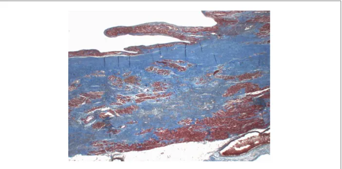
Figure 3 -Histological section of the left ventricular wall showing myocardial replacement by ibrosis, which is stained in blue, extending across the ventricular wall thickness. Masson’s trichrome, X 1.
Figure 4 -Histological section of epicardial coronary artery, which displays ample light without obstructive atherosclerotic lesions. HE, X 1.
examination of his heart, taken at the time of transplantation, revealed dilated cardiomyopathy with extensive area of old transmural infarction located on the left ventricular wall. The coronary arteries were normal without obstructive lesions, corroborating the results of coronary angiography examination performed five months before the transplant. The occurrence of acute myocardial infarction in the evolution of dilated cardiomyopathy with normal coronary arteries has been reported in previous studies, and may
greatly worsen the patient’s hemodynamic condition, eventually resulting in his death9. An interesting previous
report described a mechanical complication of infarction in these conditions, with rupture of the posteromedial papillary muscle of the mitral valve10. The most accepted
pathogenesis for infarction in dilated cardiomyopathy with normal coronary arteries is coronary embolism from recent thrombi or under organization located in the left heart chambers11,12, frequent in dilated cardiomyopathy
in the examination of the coronary arteries, as it may have occurred in this case. Finally, the etiology of dilated cardiomyopathy in our case is disputable. Due to the
patient’s history of alcoholism, it is possible that it is related to excessive alcohol consumption.
Dr. Luiz Alberto Benvenuti
References
1. Dias JPC, Coura JR. Epidemiologia. In: Dias JPC, Coura JR. (editores) Clínica e terapêutica da doença de Chagas: uma abordagem prática para o clínico geral. Rio de Janeiro: FIOCRUZ; 1997.p.33-66.
2. Bocchi EA, Marcondes-Braga FG, Ayub-Ferreira SM, Rohde LE, Oliveira WA, Almeida DR, et al / Sociedade Brasileira de Cardiologia. III Diretriz brasileira de insuficiência cardíaca crônica. Arq Bras Cardiol. 2009;93(1 supl.1):3-70.
3. Matsubara BB, Zanati SG, Okoshi K. Cardiomiopatias dilatadas. In: Nobre F, Serrano Jr CV. (editores). Tratado de cardiologia SOCESP. São Paulo: Manole; 2005.p.819-32.
4. Matsubara LS, Ferreira ALA. Cardiomiopatias tóxicas. In Nobre F, Serrano Jr CV. (Eds). Tratado de cardiologia SOCESP. São Paulo: Manole; 2005.p.845-57.
5. Markus MSP, Freitas HFG, Chizzola PR, Silva GT, Lima AC, Mansur AJ. Massa ventricular esquerda em portadores de insuficiência cardíaca. Arq Bras Cardiol. 2004;83(3):227-31,232-6.
6. Babonian C, Treasure T. Meta-analysis of the association of enteroviroses with human heart disease. Heart. 1997;78(6):539-43.
7. Slaughter MS, Rogers JG, Milano CA, Russel SD, Conte JV, Feldman D, et al. Advanced heart failure treated with continuous-flow left ventricular assist device. N Engl J Med. 2009;361(23):2241-51.
8. Bacal F, Souza-Neto JD, Fiorelli AI, Mejia J, Marcondes-Braga FG, Mangini S, et al. / Sociedade Brasileira de Cardiologia. II Diretriz brasileira de transplante cardíaco. Arq Bras Cardiol. 2009;94(1supl.1):e16-76.
9. Simões MV, Félix PR, Marin-Neto JA. Acute myocardial infarction complicating the clinical course of dilated cardiomyopathy in childhood. Chest. 1992;101(1):271-2.
10. Aiello VD, Mansur AJ, Favaratto D. Rupture of posteromedial papillary muscle as a mechanism of death in dilated cardiomyopathy. Int J Cardiol. 1996;54(1):73-5.
11. Canali G, Girardi P, Barbieri E. Coronary embolus and acute myocardial infarction in a patient with dilated cardiomyopathy and chronic atrial fibrillation. G Ital Cardiol. 2006;7(5):365-8.