Keywords
Heart failure/therapy; stroke volume; ventricular function; demographic aging.
Heart Failure with Preserved Ejection Fraction: Fighting
Misconceptions for a New Approach
Ricardo Fontes-Carvalho
1,2and Adelino Leite-Moreira
1,3Serviço de Fisiologia da Faculdade de Medicina do Porto1; Serviço de Cardiologia do Centro Hospitalar de Vila Nova de Gaia2; Centro de Cirurgia Torácica do Hospital de São João3, Porto - Portugal
Mailing address: Ricardo Fontes-Carvalho •
Serviço de Fisiologia da Faculdade de Medicina do Porto - Al. Prof. Hernâni Monteiro 4200 - 319 Porto - Portugal E-mail: [email protected], [email protected]
Manuscript received December 01, 2009; revised manuscript received February 17, 2010; accepted March 24, 2010.
Abstract
Over the last decades, heart failure with preserved ejection fraction (HFpEF) has received less attention by the medical and scientific communities, which led to the emergence of a number of misconceptions concerning its characteristics, diagnostic and therapeutic approach.
In recent years, new studies have changed the concepts traditionally associated with HFpEF, contributing to a new view towards this disease. This review is intended to discuss the latest evidence on HFpEF and to fight the main misconceptions associated with it in order to improve its diagnostic and therapeutic approach.
Today we have several data showing that HFpEF is a condition that requires a different clinical approach from that used in systolic heart failure (SHF). HFpEF is no longer seen as a “benign” disease because it is associated with a poor prognosis and high prevalence. Its pathophysiology is complex and not fully clarified. In addition to diastolic dysfunction, we now know that other cardiac and extracardiac factors are also involved in its onset and progression. Using recent consensus guidelines we have objective criteria for its diagnosis, especially by using the new echocardiographic parameters for assessing diastolic function, including the E/e’ ratio obtained by tissue Doppler. Finally, treatment of HFpEF remains unknown, because no therapeutic strategy has been shown to improve HFpEF prognosis. Thus, in this review we will also discuss the potentially new therapeutic targets for HFpEF.
Introduction
Heart failure (HF) represents a major and growing public health problem, affecting 2% - 3% of adults in developed countries1.
Patients with heart failure are classically divided into two groups: those with HF with preserved ejection fraction
(HFpEF), also called diastolic HF (DHF) and those with HF and reduced ejection fraction (HFrEF), better known as systolic HF (SHF)1.
In recent decades, HFpEF has received much less attention from medical and scientific communities, a situation that is finally starting to change. Such lack of attention resulted in the gradual emergence, within the medical community, of a series of misconceptions and dogmas concerning the epidemiology, diagnosis, pathophysiology and treatment of HFpEF.
With this review, we intend to explore and tackle the major misconceptions associated with HFpEF. We will discuss the latest evidence concerning HFpEF, providing a new view on this complex syndrome, in order to improve its clinical and therapeutic approach.
Frequent misconceptions in heart failure
with preserved ejection fraction
Misconception 1: HFpEF is a benign condition
Until recently, HFpEF had been considered an essentially “benign” disease associated with a better prognosis. Epidemiological studies have shown that the prognosis for these patients is as bad as those who have systolic HF (SHF)2,3. Patients with HFpEF have mortality rates of 29% after one year (versus 32% in patients with systolic HF), and 65% after five years (versus 68%)3.
The morbidity of HFpEF is also very high, requiring frequent admissions and a significant consumption of resources4,5. Once admitted due to HF, these patients have a high rate of readmission of 50% after one year5.
Equally worrying is the evidence showing that the survival of patients with HFpEF has not been improving in recent decades, unlike what has been observed in patients with systolic HF6. Such observation is probably related to the fact that the management and treatment of these patients are not producing the desired effects, probably due to various misconceptions concerning HFpEF.
from 38% to 54% out of cases of HF, a proportion that will continue to rise due to the progressive aging of population and expected increase in the prevalence of hypertension, obesity and diabetes.
Misconception 3: diastolic HF and systolic HF are the same condition
The classical separation of HF in HFpEF is questioned by several authors who argue that these relate to the same condition, albeit with different phenotypes9,10.
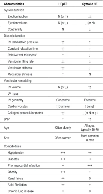
However, there are many demographic, epidemiological, histological, molecular and structural arguments, as well as some relating to ventricular function and even therapeutic effectiveness, which seem to clearly indicate that these two conditions are quite different (Table 1)6,11.
Regarding the characteristics of the population, patients with HFpEF are older, often female, and have a high prevalence of hypertension, diabetes and obesity, as well as various comorbidities such as atrial fibrillation, renal failure and anemia2-4,7,12,13 (Table 1).
The hearts of patients with systolic HF and HFpEF also have significant differences in terms of structure and ventricular function (Table 1). The hearts of patients with SHF present an eccentric ventricular modeling with increased diastolic volumes and the main anomaly occurs in LV systolic properties14 (Figure 1). By contrast, patients with HFpEF present as concentric remodeling, the volumes are normal or even reduced, and the main change occurs in the diastolic properties, with delayed relaxation and/or increased ventricular stiffness14-16 (Figure 1 and Table 1).
Other recently published studies have also shown differences at histological and molecular level. For example, analysis of endomyocardial biopsies revealed that cardiomyocytes of patients with HFpEF are structurally different, with larger diameters, greater stiffness and increased density of myofilaments, compared to patients with ICS17. Significant differences were also discovered at the molecular level. Titin is a molecule found inside the sarcomere which, given its elastic properties, is the main determinant of the stiffness of cardiomyocytes. It was found that in patients with HFpEF there is a change in the expression of the isoforms of this molecule - with increased expression of the stiffer isoform - or its degree of phosphorylation, which contributes to the increase in ventricular stiffness observed in these patients17,18. Patients with HFpEF and systolic HF also have significant differences in fibrosis and extracellular collagen matrix, due to distinct patterns of extracellular matrix metalloproteinases (MMP) and tissue inhibitors of such metalloproteinases (TIMP) activation. While in HFpEF there is a decreased degradation of extracellular matrix (resulting in increased ventricular stiffness), in dilated cardiomyopathy there is an increased matrix degradation19,20. In the HFpEF, diastolic dysfunction ca occur due to changes in the passive properties of the ventricle - particularly increased ventricular stiffness - or due to alterations in myocardial relaxation. The delay in myocardial realaxation seen in patients with HFpEF is caused by changes in calcium kinetics, especially by reduced activity
Table 1 – Comparison of characteristics of patients with systolic HF and HFpEF
Characteristics HFpEF Systolic HF
Systolic function
Ejection fraction N (or ↑) ↓↓
Ejection volume N (or ↓) ↓ (or N)
Contractility N ↓
Diastolic function
LV telediastolic pressure ↑↑ ↑
Constant relaxation time ↑↑ ↑
Relative wall thickness1 ↑ ↓
Ventricular illing rate ↓↓ ↓
Ventricular stiffness ↑↑ ↓
Myocardial stiffness ↑ N
Ventricular remodeling
LV volume N (or ↓) ↑↑
LV mass ↑ ↑
LV geometry Concentric Eccentric
Cardiomyocytes ↑ Diameter ↑ Length
Collagen extracellular matrix ↑↑ ↓ (or N or ↑)
BNP ↑ ↑↑
Age Often elderly All ages,
typically 50-70
Sex Often women More common
in men
Comorbidities
Hypertension +++ ++
Diabetes +++ ++
Prior myocardial infarction + +++
Obesity +++ +
Renal failure ++ 0
Atrial ibrillation ++ +
Chronic lung disease ++ 0
Abbreviations: LV - left ventricle, N - normal; ↑ - increased; ↓ - decreased; BNP -
brain natriuretic peptide; The relative wall thickness describes the left ventricular geometry and is deined as the ratio between the left ventricular thickness and the left ventricular cavity diameter.
of SERCA2, the main protein responsible for the reuptake of calcium back into the sarcoplasmic reticulum21.
Finally, strong arguments related to the response to pharmacological therapy justify the separation of these two conditions. Few clinical trials performed to date on HFpEF reveal that these patients do not respond as well to therapy commonly used in systolic HF, suggesting that different pathophysiological mechanisms operate in these two conditions.
Misconception 4: diastolic dysfunction is the only abnormality involved in HFpEF
The patophysiology of HFpEF is not totally understood, mainly because HFpEF affects an heterogeneous group of patients where different pathophysiological mechanisms may have a different relative importance.
Diastolic dysfunction plays a central role in the pathophysiology of this condition, since most patients present delayed myocardial relaxation and/or increased ventricular stiffness23. This is why HFpEF is often referred to as diastolic HF. More recently, after the discovery of other mechanisms that appear to contribute to the pathophysiology of this condition, the expression diastolic HF was replaced by a more general term: HFpEF 1,24.
On the other hand, we know that LV diastolic dysfunction, by itself, does not seem to be enough to cause the clinical picture of heart failure. There is an important group of patients who have diastolic dysfunction, although they remain asymptomatic and without HF25. Moreover, the prevalence of diastolic dysfunction in the general population (present in up to 25% of the population26) is much higher than the prevalence of HF. However, it remains to be explained why some patients with diastolic dysfunction have HFpEF, while others remain asymptomatic.
Beyond diastolic dysfunction: contribution of other pathophysiological mechanisms
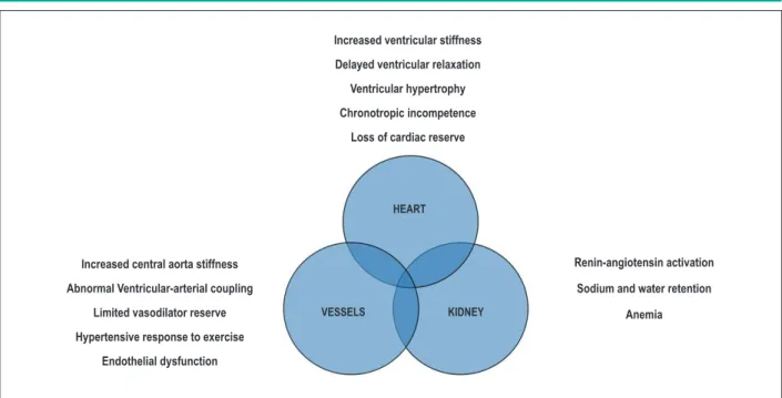
Several studies have recently demonstrated that the pathophysiology of HFpEF involves other mechanisms, including “cardiac” and “extracardiac” factors (Figure 2)27,28.
The explanation for the symptomatic difference among patients with asymptomatic diastolic dysfunction and HFpEF may be due to the simultaneous existence of these additional pathophysiological abnormalities only in patients with HFpEF. Among “extracardiac” abnormalities found in HFpEF, particular emphasis has been placed on the abnormalities found in the arterial vessels, including increased arterial stiffness, changes in ventricular-arterial coupling29,30, endothelial dysfunction and reduced vasodilator reserve 31.
There are other extracardiac factors potentially involved in HFpEF. It was found that in these patients, increased ventricular filling pressure is also due to an increased effective circulating volume due to increased sodium and water retention in the kidneys32. It should be stressed that in HFpEF, due to the simultaneous increase of ventricular and arterial stiffness, patients are very sensitive to small changes in the “central” volume16.
Recently, new “cardiac” factors have been found to contribute to HFpEF pathophysiology, such as chronotropic incompetence31 and changes in ventricular stretching, radial deformation and twisting, evaluated by speckle tracking analysis33.
Finally, HFpEF patophysiology is usually accessed at rest. However, several studies have shown that additional alterations occur during exercise in HFpEF patients31,34-36.
Is systolic function completely normal in HFpEF?
By definition, HFpEF patients have a normal ejection fraction. Nevertheless, because ejection fraction is an imprecise parameter for the evaluation of minor alterations in systolic function, it has been demonstrated that patients with HFpEF also have changes in systolic function assessed by Tissue Figure 1 -Left ventricular loops and pressure-volume (P-V) ratios in systolic and diastolic dysfunction. Panels A, B and C show dashed loops and P-V ratios of a normal
heart. Line 1 corresponds to the end diastolic pressure-volume relation, line 2 to P-V loop and line 3 to the end systolic pressure volume relation. In the presence of systolic dysfunction (panel B, full line) there is a decreased in ejection fraction (translated by the smaller width of the P-V loop) and a reduced myocardial contractility, expressed by the lower slope of the end systolic pressure volume relation (arrow). As opposed to that, in diastolic dysfunction (Panel C), the end diastolic pressure-volume relation is shifted upwards and to the left (gray line). That makes a certain amount of ventricular illing to be only achieved at the expense of much higher illing pressures than those observed for the same volume in a normal individual (see points A and B of panel C). (Adapted from Rev Port Cardiol, 2009. 28: p. 63-82).
A. NORMAL B. SYSTOLIC DYSFUNCTION C. DIASTOLIC DYSFUNCTION
LV
P
R
E
S
S
U
R
E
LV VOLUME
LV
P
R
E
S
S
U
R
E
LV
P
R
E
S
S
U
R
E
Doppler analysis37,38. Also, a recent study has shown that in HFpEF patients important alterations in systolic function also occur especially in response to exercise33.
Misconception 5: there are no objective criteria for the diagnosis of HFpEF
Part of the controversy and misconceptions concerning HFpEF resulted from the lack of consensus about its diagnostic criteria.
Such limitation was overcome in 2007 after the publication of a consensus document of the European Society of Cardiology with updated diagnostic criteria for HFpEF24. According to this document, three prerequisites should be fulfilled simultaneously to diagnose HFpEF: 1) symptoms and signs of HF; 2) EF> 50% in a non-dilated LV (defined as LV with a end diastolic volume < 97 ml/m2); 3) evidence of high LV filling pressures. The demonstration of high LV filling pressures can be made by invasive hemodynamic evaluation (which is the gold-standard method, but is difficult to apply in clinical practice) or by combining several echocardiographic parameters together with natriuretic peptides quantification. By echocardiography several diastolic parameters can be obtained that allow LV filling pressures estimation39. The most widely used parameter, and also the easiest to analyse, is the E/e’ ratio, which is obtained from the ratio between the peak transmitral flow velocity (E wave) and the mitral annulus velocity, determined from Tissue Doppler analysis (the e’ wave) (Fig. 3). When the E/e’ ratio at the level of the septal wall is > 15, LV filling pressures are certainly increased, whereas a E/e’ ratio < 8 represents normal LV filling pressures24. However when the E/e’ ratio is between 8 and 15, it is necessary to combine this value with other diastolic function echocardiographic parameters, as discussed later.
The new diagnostic algorithm of HFpEF, despite a few limitations28 allowed standardizing the diagnosis of HFpEF.
Misconception 6: diastolic function evaluation by echocardiography is inaccurate and has no inluence on clinical management strategies
The assessment of LV diastolic function should be an integral part of routine echocardiographic evaluation40, especially in patients with dyspnoea and/or heart failure, due to its diagnostic24 and prognostic significance41.
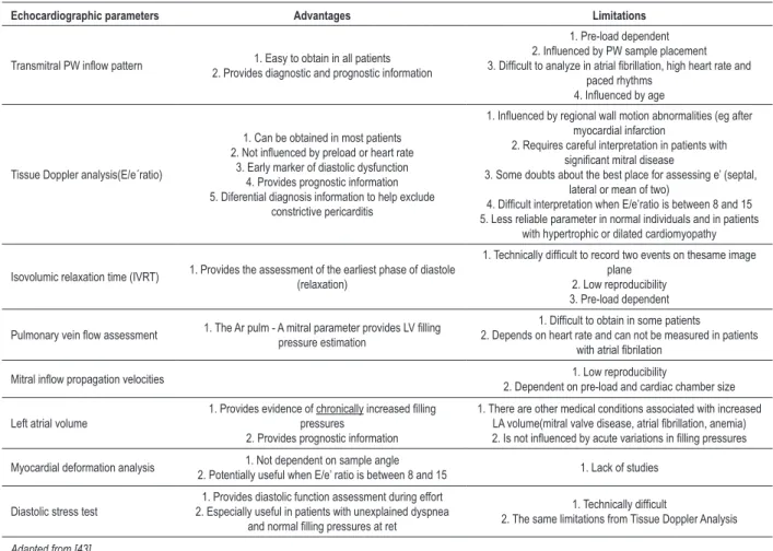
Initially, the diastolic function by echocardiography was mainly assessed through pulsed Doppler analysis of transmitral flow pattern. When this parameter is used alone, it is little specific, and has several limitations. This fact has led to the emergence of the (misconceived) idea that the echocardiographic assessment of diastolic function is little specific and little useful in clinical practice. Today, there are several echocardiographic parameters in the assessment of diastolic function, whose applications, advantages and limitations have been the target of a consensus document of the European and American societies of Echocardiography39, which will be briefly addressed in this study (Table 2).
Pulsed Doppler transmitral inlow pattern
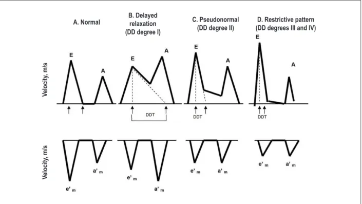
The analysis of transmitral flow by pulsed Doppler is easy to obtain in almost all patients (Fig. 4, A). By analyzing transmitral filling pattern, it is possible to define four degrees of diastolic dysfunction (Fig. 3).
Nevertheless, this parameter has several limitations (Table 2)39 because when used alone, it is not possible to distinguish a normal pattern from a pseudonormal, which indicates a grade II diastolic dysfunction (Figure 3). Despite its limitations, when combined with other diastolic dysfunction parameters, the evaluation of the E/A ratio can be useful in clinical practice to support the diagnosis of HFpEF24. and give prognostic information, when a restrictive pattern is present41.
Increased ventricular stiffness
Delayed ventricular relaxation
Ventricular hypertrophy
Chronotropic incompetence
Loss of cardiac reserve
Increased central aorta stiffness
Abnormal Ventricular-arterial coupling
Limited vasodilator reserve
Hypertensive response to exercise
Endothelial dysfunction
Renin-angiotensin activation
Sodium and water retention
Anemia
KIDNEY VESSELS
HEART
Isovolumic relaxation time (IVRT)
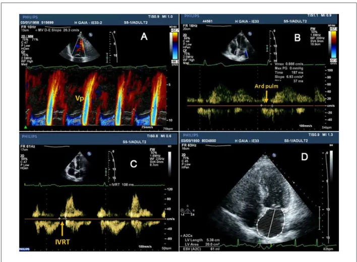
This parameter, which assesses primarily the ventricular relaxation, measures the time interval between aortic valve closure and mitral valve opening (Fig. 5, panel C). The normal value of IVRT is 70-90 msec, a value that increases with delayed relaxation, but shortens when filling pressures are markedly increased39.
Mitral low propagation velocity (Vp)
The mitral flow propagation velocity (Vp) is evaluated according to Figure 5, panel A. When ventricular relaxation is delayed, the Vp slope is reduced.
Pulmonary vein low velocity assessment
The pulmonary vein flow assessment can provide several measurements for diastolic function evaluation. However, the most reliable parameter is the Ar pulm - Ad mitral, which is the time difference between the duration of reversed pulmonary vein flow during atrial systole (Ar pulm) and the duration of the mitral A wave flow (Figure 5, B); when the Ar pulm - Ad mitral difference is > 30 msec, LV filling pressures are increased24.
Tissue Doppler assessment at the mitral annulus and E/e’ ratio
The most widely used echocardiographic parameter for diastolic function evaluation is the E/e’(see figure 4)24,40, which is the ratio of the E wave velocity from transmitral flow divided by the e’ wave velocity obtained by Doppler tissue at the mitral annulus level. By applying the pulsed tissue Doppler
at the septal or lateral side of the mitral annulus, it is possible to evaluate the velocity of the mitral annulus displacement and calculate the velocity of the systolic wave (S wave), of the early diastolic wave (E’, e’ or Ea) and of the late diastolic wave (A’, a’ or Am).
Several studies have shown that E/e’ ratio correlates closely with LV filling pressures, independently from ejection fraction values. When E/e’ ratio at the septal side of the mitral annulus is > 15, LV filling pressures are increased, whereas an E/e’ value < 8 indicates normal filling pressures. However, when the e’ is evaluated at the lateral side of the mitral annulus, and not at the septal wall, a cut-off of E/e’ > 12 (instead of 15) should be used, because displacement velocities are greater at the lateral side42.
Left atrial volume
Increased left atrial volume (LA) (Fig. 5, D) is a morphological marker of chronically increased diastolic filling pressures43 and is an important mortality predictor44. LA volume can also be increased in atrial fibrillation or significant mitral valve disease, therefore it is important to combine this parameter with the patient’s clinical condition and with other echocardiographic markers of diastolic dysfunction39.
Myocardial strain analysis
Myocardial strain can now be evaluated by echocardiography using speckle tracking, which can provide essential information regarding diastolic function39, and may be a more reliable marker of diastolic dysfunction than the E/e’ ratio45.
V
elocity
, m/s
B. Delayed relaxation (DD degree I)
C. Pseudonormal (DD degree II)
D. Restrictive pattern (DD degrees III and IV) A. Normal
Figure 3 -Evaluation of different degrees of diastolic dysfunction using data obtained from the transmitral low pattern (top) and analysis of tissue Doppler at the mitral
annulus level (bottom). Legend: DD - diastolic dysfunction; DDT - diastolic deceleration time; E - transmitral low velocity during early ventricular illing; A - transmitral low velocity during atrial contraction; e’- Tissue Doppler velocity at the mitral annulus level during early ventricular illing.
V
elocity
Table 2 – Advantages and limitations of various echocardiographic parameters of diastolic function assessment
Echocardiographic parameters Advantages Limitations
Transmitral PW inlow pattern 2. Provides diagnostic and prognostic information1. Easy to obtain in all patients
1. Pre-load dependent 2. Inluenced by PW sample placement 3. Dificult to analyze in atrial ibrillation, high heart rate and
paced rhythms 4. Inluenced by age
Tissue Doppler analysis(E/e´ratio)
1. Can be obtained in most patients 2. Not inluenced by preload or heart rate
3. Early marker of diastolic dysfunction 4. Provides prognostic information 5. Diferential diagnosis information to help exclude
constrictive pericarditis
1. Inluenced by regional wall motion abnormalities (eg after myocardial infarction
2. Requires careful interpretation in patients with signiicant mitral disease
3. Some doubts about the best place for assessing e’ (septal, lateral or mean of two)
4. Dificult interpretation when E/e’ratio is between 8 and 15 5. Less reliable parameter in normal individuals and in patients
with hypertrophic or dilated cardiomyopathy
Isovolumic relaxation time (IVRT) 1. Provides the assessment of the earliest phase of diastole
(relaxation)
1. Technically dificult to record two events on thesame image plane
2. Low reproducibility 3. Pre-load dependent
Pulmonary vein low assessment 1. The Ar pulm - A mitral parameter provides LV illing pressure estimation
1. Dificult to obtain in some patients
2. Depends on heart rate and can not be measured in patients with atrial ibrilation
Mitral inlow propagation velocities 2. Dependent on pre-load and cardiac chamber size1. Low reproducibility
Left atrial volume
1. Provides evidence of chronically increased illing
pressures 2. Provides prognostic information
1. There are other medical conditions associated with increased LA volume(mitral valve disease, atrial ibrillation, anemia) 2. Is not inluenced by acute variations in illing pressures
Myocardial deformation analysis 1. Not dependent on sample angle
2. Potentially useful when E/e’ ratio is between 8 and 15 1. Lack of studies
Diastolic stress test
1. Provides diastolic function assessment during effort 2. Especially useful in patients with unexplained dyspnea
and normal illing pressures at ret
1. Technically dificult
2. The same limitations from Tissue Doppler Analysis
Adapted from [43].
Figure 4 -E/e’ ratio evaluation. The left panel shows transmitral inlow Doppler pattern, with the E wave velocity (E), the A wave velocity (A), the E wave deceleration
Figure 5 -Demonstration of different echocardiographic parameters used for diastolic dysfunction analysis. Panel A shows how to measure the mitral low propagation
velocity (Vp); panel B shows the pulmonary vein low velocity assessment, from which it is possible to determine the time duration of reversed pulmonary vein low during atrial systole (Ar pulm), that is used to calculate the Ar pulm - Ad mitral: the time difference between the Ar pulm and the duration of mitral A wave low (see left panel, Figure 4); panel C shows how to calculate the isovolumic relaxation time (IVRT); and inally, panel D shows left atrial volume measurement, using Simpson’s method.
Diastolic stress test
A great number of patients with diastolic dysfunction only develop symptoms during exercise. Therefore, it is important to evaluate LV filling pressures in response to exercise, by conducting a diastolic stress test.
This test can evaluate the E/e’ ratio variation in response to exercise. While in individuals with normal relaxation, both E and e’ velocities increase proportionally (keeping a normal E/e’ ratio), in patients with diastolic dysfunction there is a progressive increase of the E/e’ ratio with exercise46.
In conclusion, although some limitations still exist47, diastolic function can be reliably assessed by echocardiography, using an integrated step-by-step approach, starting with E/e’ ratio evaluation. Moreover, diastolic dysfunction evaluation provides essential information for diagnosis, prognosis and management of patients with HF, particularly those with HFpEF42.
Misconception 7: there are effective strategies to treat HF with preserved EF
Probably the biggest misconception in HFpEF management is to think that there are effective therapeutic strategies for
HFpEF, or to believe that such treatment may be similar to that used in systolic HF.
Firstly, despite its clinical and epidemiological significance, HFpEF treatment remains largely empirical and not evidence based. Unlike in SHF, few randomized clinical trials have been conducted in these patients.
Secondly, the few clinical trials conducted in HFpEF patients, only evaluated the effectiveness of renin-angiotensin system inhibitors. In all such studies the results were disappointing, since there was no survival benefit by using such agents. Hence, the use of other therapeutic agents in HFpEF can only be recommended theoretically or based on data obtained from observational studies.
Finally, in recent decades, the prognosis of HFpEF has remained unchanged over time, contrasting with the survival benefit observed in SHF patients6. This observation also demonstrates that HFpEF management strategies are still not appropriate.
Use of the renin-angiotensin system modulators
reduction or survival benefit, as demonstrated using perindopril (PEP-CHF trial)48, irbesartan (I-PRESERVE)49 or candesartan (CHARM-Preserved)50.
Role of beta blockers in HFpEF
In theory, beta blockers (BB) have various potential benefits in HFpEF treatment: i) by reducing the heart rate they increase the duration of diastole and hence ventricular filling time; ii) they decrease myocardial oxygen requirements; iii) they lower blood pressure; and iv) they may induce regression of LVH. On the other hand, these beneficial effects may be partially mitigated since BB delay ventricular relaxation and reduce contractility16.
Although there are no clinical trials assessing BB efficacy in HFpEF, it is expected that these agents can be potentially beneficial, especially those with a vasodilator effect (e.g. carvedilol and nebivolol), because they can also reduce arterial stiffness.
Data from observational studies indicate that beta-blockers in HFPEF may reduce mortality51. Recently, a subanalysis derived from the SENIORS trial showed that in the subgroup of patients with EF > 35%, the benefits of this BB were similar, which suggests that the effectiveness of BB is not depend on ejection fraction52. With so many uncertainties, there is an urging need for a clinical trial to test the use of BB in HFpEF.
Aldosterone antagonists in HFpEF
The use of antagonists aldosterone in HFpEF can be beneficial, at least from a theoretical standpoint. Aldosterone acts both on the myocardium and vessels, promoting myocyte hypertrophy, fibrosis and collagen deposition, all of which may contribute to increased myocardial and arterial stiffness, contributing HFpEF progression53. A small clinical trial demonstrated that spironolactone improved echocardiographic parameters of diastolic dysfunction54. A randomized clinical trial - the TOPCAT study - is currently in progress aimed at assessing the role of aldosterone antagonists in HFPEF patients.
Other therapeutic strategies
Given so many uncertainties, only some general principles are recommended for HFpEF treatment1: i) aggressive blood pressure control, to prevent the onset of HFpEF, to reduce the number of HF hospitalizations, to induce left ventricular hypertrophy regression and to improve ventricular-arterial coupling; ii) reduction of ventricular filling pressures, by restricting salt intake and administration of diuretics, which is particularly important since HFpEF patients are highly sensitive to changes in central volume and pre-load; iii) maintaining sinus rhythm, to preserve atrial contraction; iv) heart rate control, preventing tachycardia, which shortens diastole duration; and v) treatment of underlying comorbidities, using an integrated and multidisciplinary approach.
Potential new therapeutic targets in HFpEF
The future treatment for HFpEF is dependent on a better understanding of its pathophysiology and on multiple interventions on the various underlying physiopathological mechanisms. Due to the heterogeneous mechanisms that cause HFpEF, its treatment will always be multifactorial and individualized to each patient.
Assuming that changes in relaxation and increased stiffness are the main pathophysiological alterations in HFpEF, it is necessary to develop new therapeutic strategies that specifically target these alterations. Alagebrium, or ALT-177, is a new drug that breaks the crosslinks that form between advanced glycosylation endproducts, thereby improving diastolic function (by reducing ventricular stiffness), vascular function (by improving arterial distensibility), and ventricular-arterial coupling. Small clinical trials have shown promising results in HFpEF55.
Given the importance of fibrosis in increasing ventricular stiffness, several studies are analyzing (with promising results) the antifibrotic effects of several growth factors, cytokines and signaling molecules56.
In recent years, our research group has also contributed to clarifying the determinants of left ventricular passive properties, demonstrating that ventricular stiffness is not just a passive property, but that it can be actively modulated (and reduced) using neuro-hormonal manipulation (e.g. renin-angiotensin system and endothelin, among others), opening new therapeutic targets for ventricular stiffness reduction57-60.
In HFpEF, ventricular relaxation should also be improved. As previously mentioned, relaxation is dependent on the uptake of calcium back into the sarcoplasmic reticulum by the action of SERCA2A, which in turn is regulated by phospholamban61. Animal studies have shown that genetic transfer of SERCA2A or modified phospholamban improves ventricular diastolic function62, 63.
Given the beneficial effects of nitric oxide (NO) on endothelial, vascular and myocardial functions, type 5 phosphodiesterase inhibitors (e.g. sildenafil) may have a role in HF treatment, including in HFpEF64. A clinical trial is currently in progress to assess this possibility.
Potential Conflict of Interest
No potential conflict of interest relevant to this article was reported.
Sources of Funding
There were no external funding sources for this study.
Study Association
References
1. ESC Guidelines for the diagnosis and treatment of acute and chronic heart failure 2008: the Task Force for the Diagnosis and Treatment of Acute and Chronic Heart Failure 2008 of the European Society of Cardiology.Eur Heart J. 2008; 29 (19): 2388-442.
2. Tribouilloy C, Resinaru D, Mahjoub H, Souliére V, Levy F, Peltier M, et al. Prognosis of heart failure with preserved ejection fraction:a 5 year prospective population-based study. Eur Heart J. 2008; 29 (3): 339-4.
3. Bhatia RS, Tu J, Lee DS, Austin PC, Fang J, Haouzi A, et al. Outcome of heart failure with preserved ejection fraction in a population-based study. N Engl J Med. 2006; 355 (3): 260-9.
4. Fonarow GC, Stough WG, Abraham WT, Albert NM, Gheorghiade M, Greenberg BH, et al. Characteristics, treatments, and outcomes of patients with preserved systolic function hospitalized for heart failure: a report from the OPTIMIZE-HF Registry. J Am Coll Cardiol. 2007; 50 (8): 768-77.
5. Zile MR, Brutsaert DL. New concepts in diastolic dysfunction and diastolic heart failure. Part I. Diagnosis, prognosis, and measurements of diastolic function. Circulation. 2002; 105 (11): 1387-93.
6. Owan TE, Hodge D, Herges RM, Jacobsen SJ, Roger VL, Redfield MM. Trends in prevalence and outcome of heart failure with preserved ejection fraction. N Engl J Med. 2006; 355 (3): 251-9.
7. Yancy CW, Lopatin M, Stevenson LW, De Marco T, Fonarow GC. Clinical presentation, management, and in-hospital outcomes of patients admitted with acute decompensated heart failure with preserved systolic function: a report from the ADHERE Database. J Am Coll Cardiol. 2006; 47 (1): 76-84.
8. Moutinho MA, Colucci FA, Alcoforado V, Tavares LR, Rachid MB, Rosa ML, et al. Heart failure with preserved ejection fraction and systolic dysfunction in the community. Arq Bras Cardiol. 2008; 90 (2): 132-7.
9. Brutsaert DL, De Keulenaer. Diastolic heart failure: a myth. Curr Opin Cardiol. 2006; 21 (3): 240-8.
10. De Keulenaer GW, Brutsaert D. The heart failure spectrum: time for a phenotype-oriented approach. Circulation. 2009; 119 (24): 3044-6.
11. Zille MR. Heart failure with preserved ejection fraction: is this DHF? J Am Coll Cardiol. 2003; 41 (9): 1519-22.
12. Lee DS, Gona P, Vasan RS, Larson MB, Benjamin EJ, Wang TJ, et al. Relation of disease pathogenesis and risk factors to heart failure with preserved or reduced ejection fraction: insights from the Framingham heart study of the NHLB Institute. Circulation. 2009; 119 (24): 3070-7.
13. Chen HH, Lainchbury J, Senni M, Bailey KR, Redfield MM. Diastolic heart failure in the community: clinical profile, natural history, therapy, and impact of proposed diagnostic criteria. J Card Fail. 2002; 8 (5): 279-87.
14. Zile MR, Baicu C, Bonnema DD, Diastolic heart failure: definitions and terminology. Prog Cardiovasc Dis. 2005; 47 (5): 307-13.
15. Leite-Moreira AF. Current perspectives in diastolic dysfunction and diastolic heart failure. Heart. 2006; 92 (5): 712-8.
16. Fontes-Carvalho R, Leite-Moreira AF. The pathophysiology of heart failure with preserved ejection fraction and its therapeutic implications. Rev Port Cardiol. 2009; 28 (1): 63-82.
17. van Heerebeek L, Borbely A, Niessen HW, Bronzwaer JG, van Der Velden J, Stienen GJ, et al. Myocardial structure and function differ in systolic and diastolic heart failure. Circulation. 2006; 113 (16): 1966-73.
18. Borbély A, Falcao-Pires I, van Heerebeek L, Hamdani N, Edes I, Gavina C, et al. Hypophosphorylation of the Stiff N2B titin isoform raises cardiomyocyte resting tension in failing human myocardium. Circ Res. 2009; 104 (6): 780-6.
19. Ahmed SH, Clark L, Pennington WR, Webb CS, Bonnema DD, Leonardi AH, et al. Matrix metalloproteinases/tissue inhibitors of metalloproteinases: relationship between changes in proteolytic determinants of matrix composition and structural, functional, and clinical manifestations of hypertensive heart disease.Circulation. 2006; 113 (17): 2089-96.
20. Spinale FG, Coker M., Heung LJ, Bond BR, Gunasinghe HR, Etoht, et al. A matrix metalloproteinase induction/activation system exists in the human left ventricular myocardium and is upregulated in heart failure. Circulation. 2000; 102 (16): 1944-9.
21. Periasamy M, Jansen P. Molecular basis of diastolic dysfunction. Heart Fail Clin. 2008; 4 (1): 13-21.
22. Hunt SA, Abraham W, Chin MH, Feldman AM, Francis S, Ganiats TG, et al. 2009 Focused update incorporated into the ACC/AHA 2005 Guidelines for the Diagnosis and Management of Heart Failure in Adults. A Report of the ACC Foundation/AHA Task Force on Practice Guidelines Developed in Collaboration With the International Society for Heart and Lung Transplantation.J Am Coll Cardiol, 2009; 53 (15): e1-e90.
23. Zile MR, Baicu C, Gaasch WH. Diastolic heart failure: abnormalities in active relaxation and passive stiffness of the left ventricle. N Engl J Med. 2004; 19 (19): 1953-9.
24. Paulus W, Tchoppe C, Sanderson J, Rusconi C, Flachskampf FE, Rademakers FE, et al. How to diagnose diastolic heart failure: a consensus statement on the diagnosis of heart failure with normal left ventricular ejection fraction by the Heart Failure and Echocardiography Associations of the European Society of Cardiology. Eur Heart J. 2007; 28 (20): 2539-50.
25. Redfield MM, Jacobsen S, Burnett JC Jr, Mahoney DW, Bailey KR, Rodeheffer RJ. Burden of systolic and diastolic ventricular dysfunction in the community: appreciating the scope of the heart failure epidemic. JAMA. 2003; 289 (2): 194-202.
26. Kuznetsova T, Herbotz L, Lopez B, Jin Y, Richart T, Thys L, et al. Prevalence of left ventricular diastolic dysfunction in a general population. Circ Heart Fail. 2009; 2 (2): 105-12.
27. Maurer MS, King D, El-Khoury R, Packer M, Burkhoff D. Left heart failure with a normal ejection fraction: identification of different pathophysiologic mechanisms. J Card Fail. 2005; 11 (3): 177-87.
28. Maeder M, Kaye D, Heart failure with normal left ventricular ejection fraction. J Am Coll Cardiol. 2009; 53 (11): 905-18.
29. Kawaguchi M, Hay I, Fetics B, Kass DA. Combined ventricular systolic and arterial stiffening in patients with heart failure and preserved ejection fraction: implications for systolic and diastolic reserve limitations. Circulation. 2003; 107 (5): 714-20.
30. Borlaug BA, Kass DA. Ventricular-vascular interaction in heart failure. Heart Fail Clin. 2008; 4 (1): 23-36.
31. Borlaug BA, Melenovski V, Russell SD, Kessler K, Pacak K, Becker LC, et al. Impaired chronotropic and vasodilator reserves limit exercise capacity in patients with heart failure and a preserved ejection fraction. Circulation. 2006; 114 (20): 2138-47.
32. Maurer MS, Burkhoff D, Fried LP, Gottdiener J, King DL, Kitzman DW. Ventricular structure and function in hypertensive participants with heart failure and a normal ejection fraction: the Cardiovascular Health Study. J Am Coll Cardiol. 2007; 49 (9): 972-81.
33. Wang J, Khoury D, Yue Y, Torre-Amione G, Nagueh SF. Preserved left ventricular twist and circumferential deformation, but depressed longitudinal and radial deformation in patients with diastolic heart failure. Eur Heart J. 2008; 29 (10): 1283-9.
34. Tan YT, Wenzelburger F, Lee E, Heatlie G, Leyva F, Patel K, et al. The pathophysiology of heart failure with normal ejection fraction: exercise echocardiography reveals complex abnormalities of both systolic and diastolic ventricular function involving torsion, untwist, and longitudinal motion. J Am Coll Cardiol. 2009; 54 (10): 36-46.
35. Westermann D, Kasner M, Steendijk P, Spillmann F, Riead A, Weitmann K, et al. Role of left ventricular stiffness in heart failure with normal ejection fraction. Circulation. 2008; 117 (16): 2051-60.
36. Kitzman DW, Higginbotham M, Cobb FR, Sheikh KH, Sullivan MJ. Exercise intolerance in patients with heart failure and preserved left ventricular systolic function: failure of the Frank-Starling mechanism. J Am Coll Cardiol. 1991; 17 (5): 1065-72.
37. Bruch C, Gradaus R, Gunia S, Breithardt G, Wichter T. Doppler tissue analysis of mitral annular velocities: evidence for systolic abnormalities in patients with diastolic heart failure. J Am Soc Echocardiogr. 2003; 16 (10): 1031-6.
39. Nagueh SF, Appleton C, Gillebert TC, Marino PN, Oh JK, Smiseth AO, et al. Recommendations for the evaluation of left ventricular diastolic function by echocardiography. Eur J Echocardiogr. 2009; 10 (2): 165-93.
40. Mesquita E, Jorge A. Heart failure with normal ejection fraction: new diagnostic criteria and pathophysiological advances. Arq Bras Cardiol. 2009; 93 (2): 180-7.
41. Somaratne JB, Whalley G, Gamble GD, Doughty R. Restrictive filling pattern is a powerful predictor of heart failure events post acute myocardial infarction and in established heart failure: a literature-based meta-analysis. J Card Fail. 2007; 13 (5): 346-52.
42. Little WC, Oh J. Echocardiographic evaluation of diastolic function can be used to guide clinical care. Circulation. 2009; 120 (9): 802-9.
43. Lester SJ, Tajik A, Nishimura RA, Oh JK, Khanderia BK, Seward JB. Unlocking the misteries of diastolic function. J Am Coll Cardiol. 2008; 51 (7): 679-89.
44. Douglas P. The left atrium: a biomarker of chronic diastolic dysfunction and cardiovascular disease risk. J Am Coll Cardiol. 2003; 42 (7): 1206-7.
45. Dokainish H, Sengupta R, Pillai M, Bobek J, Lakkis N. Usefulness of new diastolic strain and strain rate indexes for the estimation of left ventricular filling pressure. Am J Cardiol. 2008; 101 (10): 1504-9.
46. Ha JW, Oh JK, Pellikka PA, Ommen SR, Stussy VL, Bailey KR, et al. Diastolic stress echocardiography: a novel noninvasive diagnostic test for diastolic dysfunction using supine bicycle exercise Doppler echocardiography. J Am Soc Echocardiogr. 2005; 18 (1): 63-8.
47. Tschöpe C, Paulus W. Is echocardiographic evaluation of diastolic function useful in determining clinical care? Doppler echocardiography yields dubious estimates of left ventricular diastolic pressures. Circulation. 2009; 120 (9): 810-20.
48. Cleland JG, Tendera M, Adamus J, Freemantle N, Polonski L, Taylor I. PEP-CHF Investigators. The perindopril in elderly people with chronic heart failure (PEP-CHF) study. Eur Heart J. 2006; 27 (19): 2338-45.
49. Massie BM, Carson P, McMurray JJ, Komajda M, McKelvie R, Zile MR. I-PRESERVE Investigators. Irbesartan in patients with heart failure and preserved ejection fraction. N Engl J Med. 2008; 359 (23): 2456-67.
50. Yusuf S, Pfeffer M, Swedberg K, Granger CB, Held P, McMurray JJ, et al. Effects of candesartan in patients with chronic heart failure and preserved left-ventricular ejection fraction: the CHARM-Preserved Trial. Lancet. 2003; 362 (9386): 777-81.
51. Dobre D, van Veldhuisen D, DeJongste MJ, Lucas C, Cleuren G, Sanderman R, et al. Prescription of beta-blockers in patients with advanced heart failure and preserved left ventricular ejection fraction: clinical implications and survival. Eur J Heart Fail. 2007; 9 (3): 280-6.
52. van Veldhuisen DJ, Cohen-Solal A, Böhm M, Anker SD, Babalis D, Roughton M, et al. SENIORS Investigators. Beta-blockade with nebivolol in elderly heart failure patients with impaired and preserved left ventricular ejection fraction: data from SENIORS. J Am Coll Cardiol. 2009; 53 (23): 2150-8.
53. Weber KT. Aldosterone in congestive heart failure. N Engl J Med. 2001; 345 (23): 1689-97.
54. Mottram PM, Haluska B, Leano R, Cowley D, Stowasser M, Marwick TH. Effect of aldosterone antagonism on myocardial dysfunction in hypertensive patients with diastolic heart failure. Circulation. 2004; 110 (5): 558-65.
55. Little WC, Zile M, Kitzman DW, Hundley WG, O´Brien TX, Degroof RC. The effect of alagebrium chloride (ALT-711), a novel glucose cross-link breaker, in the treatment of elderly patients with diastolic heart failure. J Card Fail. 2005; 11 (3): 191-5.
56. Kaye DM, Krum H. Drug discovery for heart failure: a new era or the end of the pipeline? Nat Rev Drug Discov. 2007; 6 (2): 127-39.
57. Leite-Moreira AF, Castro-Chaves P, Pimentel-Nunes P, Lima-Carneiro A, Guerra MS, Soares JB, et al. Angiotensin II acutely decreases myocardial stiffness: a novel AT1, PKC and Na+/H+ exchanger-mediated effect. Br J Pharmacol. 2006; 147 (6): 690-7.
58. Leite-Moreira AF, Bras-Silva C, Pedrosa CA, Rocha-Sousa AA. ET-1 increases distensibility of acutely loaded myocardium: a novel ETA and Na+/H+ exchanger mediated effect. Am J Physiol Heart Circ Physiol. 2003; 284 (4): 1332-9.
59. Castro-Chaves P, Fontes-Carvalho R, Pintalhao M, Pimentel-Nunes P, Leite-Moreira AF. Angiotensin II-induced increase in myocardial distensibility and its modulation by the endocardial endothelium in the rabbit heart. Exp Physiol. 2009; 94 (6): 665-74.
60. Ladeiras-Lopes R, Ferreira-Martins J, Leite-Moreira AF. Acute neurohumoral modulation of diastolic function. Peptides. 2009; 30 (2): 419-25.
61. Periasamy M, Jansen PM. Molecular basis of diastolic dysfunction. Heart. Fail Clin. 2008; 4 (1): 13-21.
62. Gupta D, Palma J, Molina E, Gaughan JP, Long W, Houser S, et al. Improved exercise capacity and reduced systemic inflammation after adenoviral-mediated SERCA-2a gene transfer. J Surg Res. 2008; 145 (2): 257-65.
63. Schmidt U, del Monte F, Miyamoto MI, Matsui T, Gwathmey JK, Rosenzweig A, et al. Restoration of diastolic function in senescent rat hearts through adenoviral gene transfer of sarcoplasmic reticulum Ca(2þ)-ATPase. Circulation. 2000; 101 (7): 790-6.