831
IMAGES IN NEUROLOGY
Trigeminal perineural spread of renal
cell carcinoma
Propagação perineural trigeminal do carcinoma de células renais
Alejandro Hornik
1, Jordan Rosenblum
2, José Biller
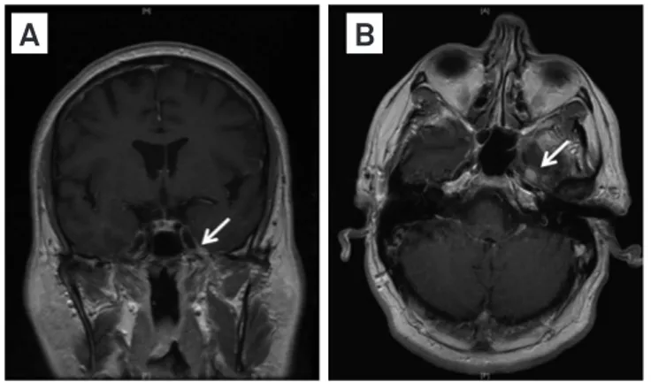
3A 55-year-old man had a ive-day history of “pins and
needles” sensation on the left chin. Examination showed
decreased pinprick sensation on the territory of the left
mandibular branch of the trigeminal nerve. Brain
magnet-ic resonance imaging (MRI) with gadolinium showed
en-hancement involving the left mandibular branch (Fig 1).
Computed tomography (CT) of the chest, abdomen, and
pelvis showed a left kidney mass (Fig 2) diagnosed as renal
carcinoma following nephrectomy.
The “numb-chin” syndrome heralds or accompanies
systemic malignancies
1. Trigeminal perineural spread
has been well-documented in head and neck neoplasms
2,
however, to our knowledge, it has not been reported in
renal neoplasms.
1Department of Neurology, Chief Resident, Stritch School of Medicine, Loyola University Medical Center, Chicago, USA; 2Department of Radiology, Director Neuro-Radiology, Stritch School of Medicine, Loyola University Medical Center, Chicago, USA; 3Department of Neurology, Chairman, Stritch School of Medicine, Loyola University Medical Center, Chicago, USA.
Correspondence: José Biller; Department of Neurology, Loyola University Chicago, Stritch School of Medicine; 2160 S. 1st Avenue / Bldg. 105, Room 2700; Maywood, IL 60153 - E-mail: jbiller@lumc.edu
Conflict of interest: There is no conflict of interest to declare.
Received 01 March 2012; Received in final form 30 April 2012; Accepted 07 May 2012
References
1. Lossos A, Siegal T. Numb chin syndrome in cancer patients: etiology, response to treatment, and prognostic significance. Neurology 1992;42:1181.
2. Warden KF, Parmar H, Trobe JD. Perineural spread of cancer along the three trigeminal divisions. J Neuro-Ophthalmology 2009;29: 300-307.