BRAIN SPECT IN DEMENTIA
A CLINICAL-SCINTIGRAPHIC CORRELATION
CARLOS A. BUCHPIGUEL**, SANDRA C. MATHIAS*, LILIAM Y. ITAYA**, NÉLIO G.BARROS**, LUIS AP. PORTELA*, JOSE M.M.FREITAS*, PAULO CARAMELLI*, PAULO E. CARRILHO*,
LUIZ A. BACHESCHI*. FAUSTO H. HIRONAKA * *, RICARDO NITRINI*
ABSTRACT - The aim of this study was to compare the accuracy of computed tomography (CT) and single photon emission computerized tomography (SPECT) in the diagnosis of dementia. Fifty-two patients with clinical diagnosis of dementia and 11 controls were studied. The scans were interpreted by one experienced neuroradiologist and one nuclear radiologist, both blinded to the clinical data. In the diagnosis of dementia, CT and SPECT showed equal sensitivity (82.7%) and statistically similar specificity (63.8 and 81.8%, respectively). The specificity of SPECT in diagnosing Alzheimer's disease (100%) was statistically superior to CT (69%). However, both methods showed similar sensitivity in detecting Alzheimer's disease. In conclusion, SPECT and CT showed similar accuracy in the diagnosis of dementia. The quite high specificity of SPECT in Alzheimer's disease may be useful for confirming that diagnosis, particularly for patients with presenile onset of the disease.
KEY WORDS: SPECT, computed tomography, dementia, Alzheimer's disease, HMPAO-Tc99m.
SPECT cerebral na demência: uma correlação clínico-cintilográfica
RESUMO - O objetivo deste estudo foi comparar a acurácia da tomografia computadorizada (TC) e da tomografia computadorizada por emissão de fóton único (SPECT) no diagnóstico de demência. Cinquenta e dois pacientes com diagnóstico clínico de demência e 11 controles foram estudados. Os exames foram interpretados por um neuroradiologista e um radiologista nuclear, ambos cegos quanto aos dados clínicos. No diagnóstico de demência, a TC e a SPECT mostraram sensibilidades iguais (82,7%) e estatisticamente especificidades semelhantes (63,8 e 81,8%, respectivamente). A especificidade da SPECT no diagnóstico da doença de Alzheimer (100%) foi significativamente superior à da TC (69%). Contudo, ambos os métodos mostraram sensibilidades semelhantes na detecção de doença de Alzheimer. Em conclusão, TC e SPECT mostraram acurácia similar no diagnóstico de demência. A alta especificidade observada no diagnóstico de doença de Alzheimer pode ser útil na confirmação do diagnóstico clínico, especialmente na forma pré-senil da doença.
PALAVRAS-CHAVE: SPECT, tomografia computadorizada, demência, doença de Alzheimer, HMPAO-Tc99m.
The clinical confirmation of dementia consists of two main distinct phases. The first is designed for confirming the cognitive decline and the second for determining the involved etiology. However, the lack of a gold-standard test is the main obstacle for establishing the diagnosis of dementia.
Anatomical imaging modalities such as X-ray computed tomography (CT) and magnetic resonance imaging (MRI) are useful in the work up of patients with dementia. They can detect space-occupying lesions, neoplasm, stroke, hydrocephalus and frontal lobe atrophy that might be found in association with trauma or Pick's disease1 8
. However, neither of those techniques is valuable nor specific for the diagnosis of Alzheimer's disease.
Centro de Medicina Nuclear - Department of Radiology (**) and Department of Neurology (*) of São Paulo University School of Medicine. Aceite: 4-março-1996.
The use of functional images has been shown to be valuable in the differential diagnosis of certain types of dementia. Regional alteration in cerebral blood flow and glucose metabolism has been seen in demented patients using single photon emission computerized tomography (SPECT) and positron emission tomography ( P E T )1 0 , 1 7
-2 0 , 2 5
. Quite specific functional patterns have been described for multi-infarct dementia, frontal dementia and dementia of Alzheimer's type4
.
The purpose of this study was to prospectively evaluate the accuracy of brain SPECT and C T in the differential diagnosis of dementia.
MATERIAL AND METHODS
Patients
Fifty-two patients with clinical evidence of cognitive impairment were studied. All patients had enough clinical follow-up to establish the definite diagnosis of dementia. The inclusion criterion was the following: 1) diagnosis of dementia according to DSM-III-R criteria1
,2) a score lower than 24 in the mini-mental state examination (MMSE)1 1
, and 3) patients not younger than 20 years old. Specifically for the diagnosis of Alzheimer's disease, the National Institute of Neurological and Communicative Disorders and Stroke-Alzheimer's Disease and Related Disorders Association (NINCDS-ADRDA) criteria were used2 1
. According to the age of onset of the clinical picture, Alzheimer's disease patients were separated in presenile (onset < 65 years-old) or senile (onset > 65 years-old) groups. In case of vascular dementia, it was applied the criteria proposed by the National Institute of Neurological Disorders and Stroke and by Association Internationale pour la Recherche et I'Enseignement en Neurosciences (NINDS-AIRENj23
. Hachinski1 4
and Cummings & Benson scores6
, MMSE and Blessed dementia scale3
were also obtained for patients with Alzheimer's disease and vascular dementia. Eleven age-matched normal volunteers were also studied (ages ranging from 58 to 84 years; mean: 70.9 year), and all gave verbal consent to be included in this trial.
Imaging techniques
CT was done in all patients and normal controls. The scans were interpreted by one experienced neuroradiologist, blinded to the clinical data.
SPECT was obtained in all patients and normal controls. A single-head rotating scintillation camera (ZLC 75-Orbiter, SIEMENS-USA) equipped with a low energy high resolution collimator was used. The scans were obtained 15 to 30 minutes after an intravenous injection of 740 MBq of HMPAO-Tc99m (CERETEC, Amerham). The acquisition protocol comprised 64 frames with 30 seconds of duration each, in a circular orbit of 360 degrees and a 64x64 matrix. A special head holder was used to avoid undesired motion during the study. The images were back-projected and filtered with a Butterworth filter, using a 0.7 nyquist frequency and order number of 10. No prefiltering or attenuation correction was applied. The images were reconstructed according to the orbito-meatal line. All images were interpreted by one nuclear medicine physician who was also completed blinded to the clinical and radiographic data.
The images were interpreted using a visual criterion based on changes in color scale. The color scale represents a percentile of change in cerebral blood flow. Colors lower in scale than a pre-defined threshold were considered abnormal. In borderline cases, a semiquantitative approach was used, drawing regions of interest (ROIs) in the suspected cortical areas. A lateralization index (ratio between left and right cortical areas) higher than 10% was considered abnormal.
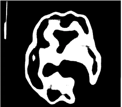
Eight perfusion patterns were defined as following (Figs 1-5): A, normal; B, bilateral posterior temporal-parietal perfusion defect; C, pattern B plus additional changes; D, unilateral temporal and/ or parietal perfusion defect; E, frontal perfusion defect; F, single or multiple large cortical perfusion defects; G, multiple small cortical perfusion defects; H, diffuse cortical perfusion defect.
Statistical analysis
The x2
Fig 5. Brain SPECT with a single large cortical perfusion defect in the posterior left parietal lobe. Example of pattern F.
RESULTS
From the 52 patients, 34 had Alzheimer disease, 8 vascular dementia, 2 Parkinson's disease, 2 Lewy inclusion body dementia, 1 frontal dementia, 1 normal-pressure hydrocephalus, 1 paretic neurosyphilis and 3 undetermined etiology
Comparison ofCT and SPECT in the diagnosis of dementia
The result of both methods in the diagnosis of dementia is shown in Table 1. Equal sensitivity was observed for C T and SPECT (82.7%), but the specificity was higher with SPECT in comparison to C T (81.8% and 63.6%, respectively; p>0.2). Two controls showed mild functional abnormalities on SPECT, one with mild frontal perfusion defect (pattern E) and the other with mild right temporal-parietal defect (pattern D). The other 9 controls showed normal perfusion pattern (pattern A). C T was falsely positive in 4 normal subjects (3 indicative of Alzheimer's disease and 1 hydrocephalus).
C T and SPECT in the diagnosis of Alzheimer's disease
Using only the patterns B and C as indicative of Alzheimer's disease, both methods showed low sensitivity as illustrated in Table 2. However, again SPECT showed higher specificity (100%) while compared to C T (69%) (p<0.01). If the pattern D is included as indication of Alzheimer's disease, the sensitivity would change to 64.7% (p>0.2) and the specificity to 93.1 % (p<0.05). Table 3 shows the probability of Alzheimer's disease based on the different scintigraphic patterns and according to Bayes' theorem. Patterns B and C showed to be highly indicative of Alzheimer's disease (100% of probability).
the B and C patterns showed more severe cognitive deficits than patients with patterns A and D (p<0.05). The time of disease did not show any correlation with these 4 scintigraphic patterns. However, the presenile group showed higher prevalence of patterns B and C and consequently lower M M S E scores than the senile group. Again, the time of disease was statistically similar in the two groups (Table 4).
Eleven patients showed asymmetric perfusion in posterior temporal and/or parietal lobes. No correlation could be observed between this pattern and the occurrence of any neurological or neuropsychological signs of asymmetric brain involvement.
C T and SPECT in the diagnosis of vascular dementia
Both methods showed similar sensitivity (62% and 50%) and specificity (94.5% and 85.4%) (Table 5). Even though the specificity for C T was slightly higher, the difference was not statistically significant (p>0.2). The patterns F and G were used as indication of vascular disease.
CT and SPECT in the other subtypes of dementia
T h e case with frontal dementia was correctly diagnosed by SPECT (pattern E) and CT. Two cases presented with meningoencephalitis and showed diffuse cortical perfusion defect (pattern H). SPECT did not show any particular specific pattern in the two patients with Parkinson's disease (one normal and the other with pattern D) and in the patients with Lewy body inclusion disease.
DISCUSSION
Controversies exist concerning the clinical importance of detecting functional impairments of the brain. Since the early 70's , the use of Xe-133 and multiprobe scintillation systems have permitted to measure the regional cerebral blood flow in terms of ml/min/lOOg of t i s s u e1 6 , 2 2 , 2 4
. Even tough this technique gained good acceptance by neurologists and neurophysiologists in recent years, a poor spatial resolution and high cost limited its clinical use. The most complete imaging technique for studying the brain function is the PET. F-18 fluor-2-deoxyglucose (FDG) is the commonest radiopharmaceutical used in the clinical setting. Various groups have used such technology in patients with dementia. All have shown metabolic deficits in posterior temporoparietal, and occasionally in frontal l o b e s2 , 7 , 1 2
. Friedland et al.1 3
showed good correlation between reduced FDG uptake and histological evidence of cell loss in Alzheimer's disease. Despite the very good diagnostic accuracy of PET, its clinical use is limited due mainly to high cost and low availability.
The technological evolution and the development of new radiopharmaceuticals opened new perspectives for the SPECT functional imaging. Cohen et al.5
showed similar accuracy of SPECT and PET in the differential diagnosis of dementia, being the former a much more affordable test. Using high sensitivity Xe-133 SPECT, Bonte et al.4
been reported. Devous & Bonte9
showed high sensitivity (83%) but limited specificity (60%) using HMPAO and a dedicated three-headed system in 29 patients with Alzheimer's disease. However, one of the most important drawbacks of SPECT is the lack in quantitation and the limited spatial resolution in comparison to PET. There are also a few truly prospective studies comparing the diagnostic efficacy in a selected population. Other aspect that has to be mentioned is the relative lack of standardization among the different centers. It is really difficult to compare results obtained using a single-headed camera with those obtained with a multi-headed system. Holman1 5
was the first to propose a standard visual classification for interpreting functional images of the brain. In our opinion, it is important to define guidelines for interpreting brain SPECT studies. Lack of reproducibility among different laboratories may be related not only to the type of population but also to technical factors. We chose a slight modified visual classification proposed by Holman, but using a single-head SPECT camera. The limited resolution of our camera could explain the low sensitivity (47%) of SPECT in detecting Alzheimer's disease in contrast to a higher sensitivity obtained with a dedicated system (65%). Similarly to Holman's data, the posterior bilateral temporoparietal defect had a very high predictive value for Alzheimer's disease. The other patterns, including the normal, had a very low predictive value according to Bayes' theorem. Even though other authors have reported the occurrence of bilateral temporoparietal defect in Parkinson's disease1 9
and in vascular dementia8 , none of our cases with Parkinson's or vascular disease showed such functional patterns.
As far as we are concern, we are the first group to report a correlation between specific functional patterns and early or late-onset of Alzheimer's disease. Even though no correlation could be observed between the different patterns and the time of evolution of disease, the presenile group showed more frequently patterns B and C and more severe cognitive impairment, in contrast to senile group that showed more varied results and milder cognitive impairment. This could suggest two different clinical forms of that disease, although more prospective studies need to be done in order to confirm such findings.
In the detection of vascular dementia, our results are quite disappointing comparing to CT. This could be explained by the poor spatial resolution of our single-headed SPECT system, but also for the heterogeneous and limited number of patients with such disease included in this trial. We did not classify the patients with vascular disease in the three possible categories: thromboembolic disease (large vessel disease), small vessel disease with multi-lacunar infarcts and the Binswanger's disease.
Our preliminary results suggest that SPECT may be an important diagnostic tool for confirming the diagnosis of Alzheimer's disease, particularly in the presenile group of patients. Even using a single head SPECT camera, very high probability of Alzheimer's disease can be observed with the functional patterns B and C. In our experience, the role of SPECT in confirming multi-infarct dementia is more limited and did not show any advantages in comparison to CT.
REFERENCES
1. American Psychiatric Association. Diagnostic and statistical manual of mental disorders. Ed3 rev. Washington,DC: American Psychiatric Association, 1987.
2. Benson DF, Kuhl DE, Hawkins RA, Phelps M E , Cummings JL, Tsai SY. T h e fluorodeoxyglucose F-18 scan in A l z h e i m e r ' s and multi-infarct dementia. Arch Neurol 1983;40:711-714.
3. Blessed G, Tomlinson BE, Roth M. T h e association between quantitative measures of dementia and of senile change in the cerebral grey matter of elderly subjects. Br J Psychiatry 1968;114:797-811.
4. Bonte FJ, Horn J, Tintner R, Weiner MF. Single photon tomography in Alzheimer's disease and the dementias. Semin Nucl Med 1990;4:342-352.
5. Cohen M B , Metter FJ, Grahan LS, Wasterlain C, Spolter L, Lake RR, Rose G, Yumada L, Chang C C . Differential diagnosis of dementia with " p u r e " 1-123 iodoamphetamine and a clinical camera. J Nucl Med 1983;24:P106.
6. Cummings JL, Benson F. Dementia of the Alzheimer type: an inventory of diagnostic clinical features. J Am Geriatr Soc 1986;34:12-19. 7. Dauru R, Grady C, Haxby J, Saudaram M, Cutler M, Heston L, Moore A, Schlageter N, Larson S, Rapoport SI. Positron
emission tomography in Alzheimer's disease. Neurology 1986;36:879-887.
8. Deutsch G, Tweedy JR. Cerebral blood flow in severity-matched Alzheimer and multi-infarct patients. Neurology 1987,37:431-438. 9. Devous M D Sr, Bonte FJ. Initial evaluation of cerebral blood flow imaging with a high-resolution, high-sensitivity
10. Fazekus F, Alavi A, Chawluk J B , Zimmerman RA, Hackney D, Bilaniuk L, Rosen M, Alves W M , Hurtig HI, Jamieson DG, Kushner MJ, Reivjch M. Comparison of CT, MR, and PET in Alzheimer's dementia and normal aging. J Nucl Med 1989;30:1607-1615.
11. Folstein SE, Folstein SE, McHugh PR. "Mini-mental state": a practical method for grading the cognitive state of patients for the clinician. J Psychiatr Res 1975; 12:189-198.
12. Foster NL, Chase TN, Fedio P, Patronas NJ, Brooks RA, Di Chiro G. Alzheimer's disease: focal cortical changes shown by positron emission tomography. Neurology 1983;33:961-965.
13. Friedland RP, Brun A, Budinger TF. Pathological and positron emission tomographic correlations in Alzheimer's disease [ letter]. Lancet 1985,1:228.
14. Hachinski VC, Linette D, Phil M, Zilhka E, Boulay GH, McAllister VL, Marshall J, Russel RWR, Simon L. Crerebral blood flow in dementia. Arch Neurol 1975;32:632-637.
15. Holman BL, Johnson KA, Gerada B, Carvalho PA, Satlin A. The scintigraphic appearance of Alzheimer's disease: a prospective study using technetium-99m-HMPAO SPECT. J Nucl Med 1992;33:181 -185.
16. Ingvar DH, Risberg J. Increase of regional cerebral blood flow during mental effort in normals and in patients with focal brain disorders. Exp Brain Res 1967;3:195-211.
17. Jagust W J , Budinger TF, Reed BR. The diagnosis of dementia with single photon emission computed tomography. Arch Neurol 1987;44:258-262.
18. Jagust W J , Eberling JL. MRI, CT, SPECT, PET: their use in diagnosing dementia. Geriatrics 1991 ;46:28-35.
19. Kuwabara Y, Ichiya Y, Otsuka M, Tuhara T, Fukumura T, Gunasekera R, Masuda K.. Differential diagnosis of bilateral parietal abnormalities in 1-123 IMP S P E C T imaging. Clin Nucl Med 1990;10:893-899.
20. M c G e e r PL, Kamo H, Harrop R, McGeer EG, Martin WRW, Pate BD, Li D K B . Comparison of PET, MRI and C T with pathology in a proven case of Alzheimer's disease. Neurology 1986;36:1569-1574.
2 1 . M c K h a n n G , Drachman D, Folstein M, Katzman R, Price D, Stadlan EM. Clinical diagnosis of Alzheimer's disease: report of the NINCDS-ADRDA Work Group under the auspices of Department of Health and Human Services Task Force on Alzheimer's disease. Neurology 1984;34:939-944.
22. Obrist W D , Chivian E, Cronquist S, Ingvar DH. Regional cerebral blood flow in senile and presenile dementia. Neurology 1970;20:315-322.
2 3 . Roman G C , Tatemichi TK, Erkinjuntti T, Cummings JL, Masdeu JC, Garcia JH, Amaducci L, Orgogozo J M , Brun A, Hofman A, Moody D M , Obrien M D , Yamagushi J, Grafman J, Drayer BP, Bennet DA, Fisher M, Ogata J, Kokmen E, Bermejo F, Woff PA, Gorelick P B , Bick KL, Pejeau AK, Bell MA, De Carli C, Culebras A, Korczyn A D , Bogousslavsky J, Hartamnn A, Sheinberg P. Vascular dementia: diagnostic criteria for research studies: report of the N I N D S - A I R E N International Workshop. Neurology 1 9 9 3 , 4 3 : 250-260.
24. Simard D, Olensen O B , Paulson O B , Lassen NA, Skinhoj E.. Regional cerebral blood flow and its regulation in dementia. Brain 1971;94:273-288.