Coronary Artery Disease in Patients with Rheumatic and
Non-Rheumatic Valvular Heart Disease Treated at a Public Hospital in
Rio de Janeiro
Dany David Kruczan, Nelson Albuquerque de Souza e Silva, Basílio de Bragança Pereira, Vítor André Romão, Wilson
Braz Correa Filho, Fidel Ernesto Castro Morales
Instituto Estadual de Cardiologia do Rio de Janeiro Aloysio de Castro/IECAC, Universidade Federal do Rio de Janeiro - UFRJ, Rio de Janeiro, RJ - Brazil
Summary
Objective: to estimate the prevalence of coronary artery disease (CAD) in valvular heart disease of rheumatic (RVHD) and non-rheumatic (NVHD) etiology, assessing possible predictive factors for the presence of CAD.
Methods: This is a cross-sectional study of a series of cases obtained from a pre-defined population, wherein 1,412
patients referred for heart surgery of any etiology were evaluated. Of these, 294 primary heart disease patients aged ≥40
submitted to cinecoronary arteriography (CA) were identified and studied.
Results: patients with RVHD presented lower prevalence of CAD (4%) when compared to NVHD (33.61%), p<0.0001. The logistic regression analysis showed that age, typical angina-like chest pain (TACP), systemic arterial hypertension (SAH), diabetes and dyslipidemia were significantly related to CAD, and that the rheumatic etiology was not a disease determinant. Smoking and gender were clinically important in CAD, although not statistically significant. In the whole group, the Log-linear
analysis showed that, regardless of the etiology, gender, age ≥55, SAH, TACP, diabetes and dyslipidemia were all related
directly to CAD, with the latter three being the most important variables for the disease.
Conclusion: the prevalence of CAD among RVHD patients is low, whereas it is high among NVHD patients; the rheumatic etiology does not seem to have any beneficial effects on the prevalence of CAD; gender, age, SAH, TACP, dyslipidemia and diabetes were identified as being strongly associated with the presence of CAD. It is possible to define the criteria that indicate the need for pre-surgical CA in heart valve replacements, so that the standard indication after the age of 40
years can be avoided. (Arq Bras Cardiol 2008; 90(3):197-203)
Key words: Coronary arteriosclerosis / epidemiology; rheumatic heart disease / diagnosis; heart valve diseases / diagnosis; coronary angiography.
Mailing address: Dany David Kruczan •
Av. Nsa.Sra.Copacabana, 195/1106 - Copacabana - 22020-002, Rio de Janeiro, RJ - Brazil
E-mail: [email protected], [email protected]
Manuscript received May 13, 2007; manuscript revised received on August 1, 2007; accepted on October 8, 2007.
Introduction
The coronary angiography has been indicated as a routine procedure in the preoperative assessment of patients with valvular heart disease that meet the following criteria:
males aged ≥ 35 years, pre-menopausal females aged ≥35
years with cardiovascular risk factors and post-menopausal females1,2. The procedure is justified by the diagnosis of coronary obstructions, especially the asymptomatic ones and the significant prevalence of coronary artery disease (CAD) in these patients. The diagnosis of associated coronary obstruction would indicate myocardial revascularization during the valve replacement, regardless of the clinical presentation and the presence or absence of symptoms. In our country, the coronary angiography has been indicated almost exclusively for patients
≥ 40 years. However, in clinical practice, we have observed
a low prevalence of CAD among patients with valvulopathies, especially those with rheumatic etiology. It is noteworthy that although the non-rheumatic etiology of the valvulopathies is often found in our population, the rheumatic etiology still considerably outnumbers the first3,4.
The present study was developed considering these observations and in view of the restrictions regarding the rationale adopted for the very often indiscriminate indications of a costly, invasive and non-morbidity-free method. Our objectives were: to assess the prevalence of CAD in patients with orovalvular injuries
with rheumatic and non-rheumatic etiology aged ≥ 40 years,
referred to evaluation of surgical indication for valve replacement in a public hospital specialized in cardiology in the city of Rio de Janeiro and identify possible predictive factors of the presence of CAD in these patients, aiming at assessing the influence of the rheumatic etiology on the disease.
use of anti-hypertensive medication, CAD, family history and diagnoses related to this disease, diabetes mellitus
(considered when fasting glycemia was ≥ 126mg/dl and dyslipidemia (considered when total cholesterol was ≥ 200mg/dl and/or triglycerides ≥150 mg/dl.
At the statistical analysis the Chi-square test was used for the comparison of proportions and the Student’s t test was used for the analysis of the continuous variables, with p value being
considered significant when ≤0.05. The logistic regression
model was used to estimate the influence of the selected variables on the outcome, i.e., the presence of coronary artery disease and the Log-linear model was used to analyze the interdependence relations of the first, second and third-degrees among the variables. The R System statistical program, version 2.3, was used for the statistical analyses. A database (Access) was created with the patients’ clinical information and results of the complementary tests.
The study was approved by the Ethics Committee in Research of the Instituto Estadual de Cardiologia Aloysio de Castro, Rio de Janeiro, on 10/05/2005.
Results
1. Analysis of the groups with rheumatic and non-rheumatic valvular cardiopathy.
Table 1 compares the variables of the groups with rheumatic and non-rheumatic valvular cardiopathy. The ones that presented statistical difference were: sex (predominantly female in the rheumatic group and predominantly male in the rheumatic group); age, older among the non-rheumatic patients; typically anginal chest pain (more frequent in the non-rheumatic group); absence of chest pain (higher frequency in the rheumatic group); SAH, diabetes mellitus, dyslipidemia and presence of coronary artery disease (more frequent in the non-rheumatic group). On the other hand, there was no significant difference between the two groups regarding the variables: BMI (normal, overweight and obese); atypical chest pain; dyspnea; family history of coronary artery disease; being a smoker and being an ex-smoker. Thus, these two groups presented heterogeneous clinical characteristics.
2. Correlation of coronary artery disease with the other variables.
To assess whether the valvulopathy etiology had any influence determining the presence of CAD, a logistic regression model analysis was carried out in which the disease was considered the dependent variable and the others were considered determinants. The variables included in this analysis were: sex, age, BMI (overweight and obesity), typical chest pain, SAH, diabetes mellitus, dyslipidemia, smoking and having a rheumatic etiology. Considering the median of
54 years, the cutoff for age was <55 and ≥55 years. The
rheumatic etiology showed it was not a determinant variable in CAD, which allowed us to proceed with the analysis involving the 294 patients with valvulopathies, regardless of the etiology (Table 2). Considering its clinical importance in coronary artery disease, the variables smoking and sex were maintained in the model, although they did not present statistical significance. hospital in the city of Rio de Janeiro. During this period, a
broad experience was acquired on the treatment of patients
with orovalvular injuries, especially adults aged ≥40 years, of
low socioeconomic level, many of them with a history of acute episodes of rheumatic fever, referred to evaluation of surgical indication for valve replacement. All patients underwent coronary angiography.
Methods
In a transversal case series study carried out in a pre-defined population between the years 1999 and 2005, a total of 1,412 patients were evaluated, after being referred with heart surgery indication due to any etiology. Of these, 294 patients with primary heart valve lesions were identified and selected for the study, with 175 presenting a rheumatic
etiology and 119 a non-rheumatic etiology, aged ≥40 years,
previously evaluated through coronary angiography. All patients met the clinical criteria for the diagnosis of stenosis, heart failure or double lesion of the aortic and/or mitral valves, supported by anamnesis and clinical assessment5. The echocardiogram was used to define the criteria of rheumatic and non-rheumatic etiology6.
To define the rheumatic valvular cardiopathy, the following criteria were considered: 1) in the mitral valve: thickening of the free border of the leaflets with or without commissure fusion and of the subvalvular apparatus with reduced mobility and posterior leaflet fixation, and 2) in the aortic valve: cusp thickening, from the border to the base. For the non-rheumatic valvular cardiopathy, the following degenerative disease criteria were considered: 1) in the mitral valve: leaflet thickening with preserved mobility in the free border; 2) in the aortic valve: fibrocalcific degeneration, characterized by calcification that started on the cusp bases and went towards their borders, and 3) in the bicuspid aortic valve: visualization of only two leaflets with cusp thickening.
The CAD was assessed by angiographic criteria of obstructive
lesions ≥50% for the left coronary trunk (LCT) and ≥70% in
the following arteries: anterior descending (AD), right coronary artery (RCA) and circumflex artery (CxA). For the marginal (MG) and diagonal (DG) arteries, or even an intermediate branch of anatomic importance (diagonalis – DI) the criteria
considered were lesions ≥70% and arteries with a diameter
> 3mm. Although only the severe lesions, of anatomically surgical nature, were considered in the present study, the mild and moderate lesions were classified for recording purposes, also by angiographic criteria. Mild lesions were classified as the obstructions <50% of the vessel lumen and moderate lesions were classified as those presenting obstruction between 50% and <70% of the vessel lumen7.
The studied variables were: age, sex, body mass index (BMI - patients with BMI < 25kg/m² were considered normal; those with BMI between 25 and 30kg/m² were overweight and those with BMI >30kg/m² were considered obese), cheat pain (typical, atypical and no pain), dyspnea, smoking and tobacco smoke load (number of years during which the patient smoked 1 pack a day), systemic arterial hypertension (SAH), considered systolic arterial pressure
Those variables that significantly correlated with the disease at the final model analysis were: age, typical chest pain, SAH, diabetes mellitus and dyslipidemia, as shown in (a)
(a) Final expression of the logistic regression model:
Logit (CAD) = -5.07 + 0.78 sex + 0.04 age + 2.05 typical chest pain + 0.91 SAH+2.04 diabetes +1.64 Dyslipidemia + 0.57 smoking; where:
Logit (CAD) = Ln{ Pr(CAD = 1) / Pr (CAD = 0) }
3. Interdependence Relations between coronary artery disease and the other variables
To support the results obtained at the logistic regression, the Log linear model was also used, which assessed the interdependence relations among the variables. It is a model of multivariate analysis, characterized by treating all variables with the same level of importance and not as outcome and explicative variables. The variables that showed to be significant in the model, when isolated, were: coronary artery disease, sex, age, typical chest pain, SAH, diabetes mellitus and dyslipidemia. At the interdependence relation (interaction) of the second-degree, an association was observed between coronary artery disease and
male sex, age ≥ 55 years, typical chest pain, SAH, diabetes and dyslipidemia as well as an association between age ≥ 55 years and typical chest pain; age ≥ 55 years and SAH; typical chest pain
and diabetes; SAH and diabetes; diabetes and dyslipidemia. At the interdependence relation of the third-degree, an association of coronary artery disease with typical chest pain and diabetes was observed, as well as an association between SAH and diabetes and of diabetes with dyslipidemia. In summary, the variables most strongly associated to coronary artery disease were typical chest pain, diabetes mellitus and dyslipidemia (Fig. 1).
4. Clinical characteristics of the population with and without coronary artery disease
Table 1 - Characteristics of the rheumatic and non-rheumatic groups.
Variables Rheumatic
(n=175) %
Non-rheumatic
(n=119) % P value
Sex male 54 30,86 85 71.43 <0.0001
female 121 69.14 34 28.57 <0.0001
Age (mean) 50.87 63.66
BMI
normal 147 84.00 89 74.79 0.072
overweight 22 12.57 23 19.33 0.157
obesity 06 3.43 7 5.88 0.470
Chest pain
typical pain 12 6.86 57 47.90 <0.0001
atypical pain 34 19.43 22 18.49 0.959
No pain 129 73.71 40 33.61 <0.0001
dyspnea 169 96.57 102 85.71 0.140
SAH 52 29.71 70 58.82 <0.0001
diabetes 07 4.00 17 14.29 0.003
dyslipidemia 14 8.00 21 17.65 0.020
CAD 07 4.00 40 33.61 <0.0001
family history 76 43.43 66 55.46 0.056
smoker and ex-smoker 87 49.71 58 48.73 0.789
n - number of patients; BMI - body mass index; SAH - systemic arterial hypertension; CAD - coronary artery disease.
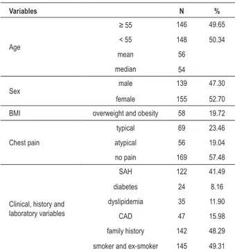
Table 2 - Demographic and clinical characteristics of the total population.
Variables N %
Age
≥ 55 146 49.65
< 55 148 50.34
mean 56
median 54
Sex male 139 47.30
female 155 52.70
BMI overweight and obesity 58 19.72
Chest pain
typical 69 23.46
atypical 56 19.04
no pain 169 57.48
Clinical, history and laboratory variables
SAH 122 41.49
diabetes 24 8.16
dyslipidemia 35 11.90
CAD 47 15.98
family history 142 48.29 smoker and ex-smoker 145 49.31
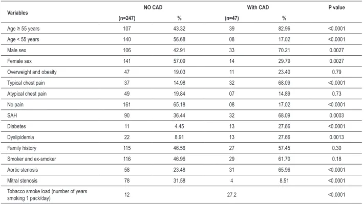
Table 3 shows the clinical characteristics of the population according to the presence and absence of CAD. The findings are summarized as follows:
At the age range ≥55 years, o percentage of the disease
prevalence was higher than in the age range <55 years. The frequency of the disease was higher in the male than in
the female sex. In the group with presence of the disease, significantly higher percentages were observed in the following variables: typical chest pain, SAH, diabetes, dyslipidemia, aortic stenosis and tobacco smoke load. In the group with absence of the disease, significantly higher percentages were observed for the variables: absence of pain and mitral stenosis. There was no significant difference between the groups regarding the following variables: overweight and obesity, family history, atypical pain, smoker and ex-smoker (although with a higher percentage in the group with coronary artery disease than in the group without the disease).
Discussion
The present study analyzes the prevalence of CAD of
rheumatic and non- rheumatic etiology in patients ≥ 40 years.
In its severe form, the disease was considered not only when there was a trunk lesion as well as three-vessel lesions that affected simultaneously the main coronary arteries, but also the one-vessel and two-vessel lesions and the severe lesions that affected secondary branches of anatomical importance. Our criteria seem to be quite comprehensive, considering that most of the revised studies consider a trunk lesion to be
severe when it is ≥50%, and the coronary artery lesions (CD,
CX and DA) when they are >70%8,9. For some authors10, the ideal condition for the surgical indication of myocardial revascularization is that in which the patients exhibit proximal lesions of at least 70% in the main coronary arteries with good distal portions.
Age range ≥ 55 SAH
CP
SX
CAD
DYSL DM
Fig. 1 -Interdependence of risk factors from CAD in the age range ≥ 55 years.
CAD – Coronary Artery Disease; CP – chest pain; DM – Diabetes mellitus; DYSL – Dyslipidemia; SAH - systemic arterial hypertension.
Table 3 - Clinical characteristics of the population with CAD and without CAD
Variables NO CAD With CAD P value
(n=247) % (n=47) %
Age ≥ 55 years 107 43.32 39 82.96 <0.0001
Age < 55 years 140 56.68 08 17.02 <0.0001
Male sex 106 42.91 33 70.21 0.0027
Female sex 141 57.09 14 29.79 0.0027
Overweight and obesity 47 19.03 11 23.40 0.79
Typical chest pain 37 14.98 32 68.09 <0.0001
Atypical chest pain 49 19.84 07 14.89 0.73
No pain 161 65.18 08 17.02 <0.0001
SAH 90 36.44 32 68.09 0.0003
Diabetes 11 4.45 13 27.66 <0.0001
Dyslipidemia 22 8.91 13 27.66 0.0013
Family history 115 46.56 27 57.45 0.30
Smoker and ex-smoker 116 46.96 29 61.70 0.18
Aortic stenosis 58 23.48 31 65.96 <0.0001
Mitral stenosis 78 31.58 4 8.51 <0.0001
Tobacco smoke load (number of years
smoking 1 pack/day) 12 27.2 <0.0001
Our findings show that it is possible to clinically identify orovalvular patients with a lower probability of presenting CAD and based on that, we suggest that the coronary angiography should not be indicated indiscriminately for all patients, but for those presenting clear clinical evidence and predictive factors of the disease.
Regarding the etiology, we observed a low prevalence of severe CAD among the rheumatic patients11; it was initially thought that this etiology could confer some degree of protection against the development of coronary atherosclerosis, as observed by other investigators12. This was based on the fact that, in general, these patients receive the prophylaxis for rheumatic fever with the use of intramuscular antibiotics every 21 days for many years and this could have a protective antibacterial and antiinflammatory effect on the genesis of coronary atherosclerosis. However, our findings do not corroborate the idea. A review of the clinical history showed that most of the patients had never received prophylaxis for rheumatic fever, which seems to make sense, as if this had really been adequately performed, certainly their cardiac valves would not have reached such state of damage as commonly found in these patients.
The data showed an alternative explanation: that the low prevalence could be due to the demographic and clinical characteristics of this population13,14. In a group with predominantly young individuals of the female sex, and thus, with fewer comorbidities, it is reasonable to assume that the prevalence of the disease is lower than in another group, with predominantly older individuals of the male sex.
The prevalence of coronary artery disease was much higher in the non-rheumatic group, which was characterized by the predominance of the male sex and older age15,16, in opposition to what was observed in the rheumatic group. Other clinical characteristics significantly differed between these two groups: typically anginal chest pain and risk factors such as systemic arterial hypertension, diabetes and dyslipidemia17,18, more often found in the non-rheumatic group, with aortic stenosis due to fibrocalcific degeneration the most frequent diagnosis in this group19,20 and mitral stenosis the most frequent one in the rheumatic group13.
The demographic and clinical aspects that characterized the patients from the CAD group were (as shown in Table 3):
age ≥55 years, male sex, typical chest pain, SAH, diabetes,
dyslipidemia, tobacco smoke load and the presence of two or more risk factors, with aortic stenosis being the diagnosis most often associated to the disease. Mitral stenosis was the least frequently associated diagnosis to CAD, similar to what has been described in other studies9,21,22.
Some aspects observed in our database data are noteworthy. Few patients with CAD were in the 40 to 60-year age range, but they presented clinical signs that suggested the presence of the disease. Thus, few of them would have been missed at a clinical stratification. Below the age range of 60 years, the prevalence of the disease was only 1.7%, i.e., it is almost non-existent within the range where most of the patients with rheumatic valvulopathy are concentrated, leading us to question the validity of indiscriminately perform coronary angiographies in this population. Regardless of the etiology,
the prevalence of the disease was 6.95% among the patients aged < 60 years, increasing to 31.8% among those aged
≥60 years, which is expected, as the comorbidities increase
with age. In a comparative analysis between the absence of risk factors with the presence of 1, 2, 3, 4 and 5 risk factors, respectively, we observed that patients who presented 2 or more risk factors were predominantly concentrated in the group with coronary artery disease, whereas in the group without CAD, higher percentages of patients with only one or no risk factors were observed.
Therefore, we considered that the hemodynamic study could be dispensed with in patients without typical angina and no associated risk factors. Regarding the coronary lesions, some considerations should also be made. According to our data, of 187 patients younger than 60 years, only 1 (0.53%) presented a trunk lesion and only 2 (1.06%) presented severe three-vessel lesions, that is, only three anatomical and truly surgical patients were identified below this age range. They presented typical angina and the association of 2 or more risk factors, and therefore, surely they would not be missed by a good clinical stratification.
In literature, the indication of routine preoperative catheterism has a level of evidence C4, which means that this evidence is based solely on the experts’ opinion, on case studies or on the measure taken as a precaution, with no other well-established basis. For instance, patients with severe aortic failure who do not present symptoms of myocardial ischemia or risk factors, which are known to increase the prevalence of coronary artery disease, could safely dispense with the preoperative coronary angiography, according to some researchers23. Others24 correlate the low prevalence of the disease and the valvulopathy, pointing out to the fact that the coronary angiography is unnecessary in these patients, except in the presence of risk factors or clinical findings such as angina and previous myocardial infarction.
Conclusions
In our opinion, the routine indication of preoperative coronary angiography based solely on the age criterion must be reconsidered. Although the diagnostic coronary angiography is a low-mortality method, it has an incidence of complications that vary from less than 1% to close to 5%. However, when such complications occur, they can have a quite significant adverse effect. Among other complications, it is important to mention the occurrence of brain injuries due to gaseous and solid microembolisms that can result in the cognitive impairment of patients, particularly the predisposed ones25.
the two groups30. The authors did not consider an advantage to submit patients who were candidates to major valvular surgery, with coronary artery disease, to cardiac surgery and even to preoperative angioplasty. In our opinion, the same consideration applies to the investigation of coronary artery disease in patients with valvulopathies with low probability to present the disease, especially considering that the hemodynamic study brings an additional risk, many times with a higher likelihood of complications than those caused by the disease itself. However, we consider it reasonable to indicate the myocardial revascularization at the moment of the valve replacement, when this indication is made for the patients who present clinical evidence of ischemia, translated as the presence of chest angina associated to the predictive factors of coronary artery disease. Another aspect that we consider important is that the indication of revascularization associated to valve replacement is very often of “prophylactic” nature, considering that, in general, the coronary artery disease is randomly explored and casually found. In many cases, the surgical indication is made because a certain lesion was disclosed at the coronary angiography. Once the indication is
made due to the presence of the lesion, the surgical procedure is seen as an opportunity to revascularize the patient.
Our approach in the present study is based on the fact that there is no solid evidence that such management benefits the patients. In contrast, what is observed is that the valve replacement associated to the myocardial revascularization procedure significantly increases short- and mid-term mortality.
Potential Conflict of Interest
No potential conflict of interest relevant to this article was reported.
Sources of Funding
There were no external funding sources for this study.
Study Association
This article is part of the thesis of doctoral submitted by Dany David Kruczan, from Universidade Federal do Rio de Janeiro.
References
1. Pomerantzeff PM, Barbosa GV, Sousa Fº BS, Brandão CMA, Ribeiro EJ, Costa FDA, et al. Diretrizes para a conduta dos pacientes com doenças das valvas cardíacas. In: Portal Cardiol [online]; 2003. [citado 2006 ago 20]. Disponível em: http://publicacoes.cardiol.br/consenso/2003/site/036.asp.
2. American College of Cardiology. American Heart Association. Task Force on Practice Guidelines (Writing Committee to revise the 1988 guidelines for the management of Patients with valvular heart disease). Society of Cardiovascular Anesthesiologists. Bonow RO, Carabello BA, Chattergee K, de Leon AC Jr, Faxon DP, Freed MD, et al. ACC/AHA 2006 guidelines for the management of patients with valvular heart disease: a report of the American College of Cariology/AHA Task Force on Practice guidelines. J Am Coll Cardiol. 2006; 48 (3): e1-148.
3. Argüelles E, Fiszman P, Fakoury L. Febre reumática e doenças valvulares do coração. Rio de Janeiro: Intermédica, 1984.
4. Terreri MT, Len C, Hilário MOE, Goldenberg MB. Utilização de recursos e custos de pacientes com febre reumática. Rev Bras Reumatol. 2002; 42 (4): 211-7.
5. Azevedo AC, Sekeff J. Cardiologia clínica. São Paulo: Sarvier, 1994.
6. Suaide Silva CE, Ferreira LDC, Monaco CG, Gil MA, Peixoto LB, Leal SMB, et al. O ecocardiograma no apoio à decisão clínica. 3a. ed. São Paulo: Revinter, 2003.
7. King III. SB, Douglas JS Jr. Atlas of heart diseases. New York: McGraw Hill; 1985.
8. Marchand E, Pichard, A, Casanegra P. Association of coronary artery disease and valvular heart disease in Chile. Clin Cardiol. 1983; (6): 352- 6.
9. Savova A, Stoitchev S, Ovanesyan H, Finkov B, Belov J. Coronary artery disease in patients with valvular heart disease. Z. Cardiol. 1986; 75 (Suppl 2): 73-5.
10. Sousa JEMR, Batlouni M, Jatene AD. Insuficiência coronária. São Paulo: Sarvier, 1984.
11. Rangel A, Hernández J, Iris JM, Badui E. Indicacion de la coronariografia en
las valvulopatias cardiacas. Arch Inst Cardiol Méx. 1996; 66: 60-9.
12. Jose VJ, Gupta SN, Joseph G, Chandy ST, George NJ. Prevalence of coronary artery disease in patients with rheumatic heart disease in the current era. Indian Heart J. 2004; 56 (2): 129-31.
13. Chu PH, Chiang CW, Hsu LA, Lin KH, Cheng NJ. Low prevalence of coronary arterial disease in Chinese adult with mitral stenosis. Chang Gung Med J. 2001; 24 (2): 97-102.
14. Guray Y, Guray U, Yilmaz BM, Mecit B, Kisacik H, Korkmaz S. Prevalence of angiographically significant coronary artery disease in patients with rheumatic mitral stenosis. Acta Cardiol. 2004; 59 (3): 305-9.
15. Alexopoulos D, Kolovou G, Kyriakidis M, Antonopoulos A, Adamopavlos S, Sleight P. Angina and coronary artery disease in patients with aortic valvular disease. Angiology. 1993; 44 (9): 707-11.
16. Bozbas H, Yildirir A, Küçük MA, Ozqül A, Atar I, Sezgin A, et al. Prevalence of coronary artery disease in patients undergoing valvular surgery due to rheumatic involvement. The Anadolu Kardiyol Derq. 2004; 4: 223-6.
17. Ramsdale DR, Faragher EB, Bennett DH, Bray CL, Ward C, Beton DC. Preoperative prediction of significant coronary artery disease in patients with valvular heart disease. Br Med J. 1982; 284: 223-6.
18. Gomez Doblas JJ, Jimenez Navarro M, Rodriguez Bailón I, Alonso Briales JH, Hernandez Garcia JM, Montiel Trujillo A. Coronariografia preoperatoria en pacientes valvulares: análisis de probabilidad de lesión coronária. Rev Esp Cardiol. 1998; 51 (9): 756-61.
19. Mandal AB, Gray IR. Significance of angina pectoris in aortic valve stenosis. Br Heart J. 1976; 38: 811-5.
20. Rapp AH, Hillis LD, Lange RA, Cigarroa JE. Prevalence of coronary artery disease in patients with aortic stenosis with and without angina pectoris. Am J Cardiol. 2001; 87 (10): 1216-7.
22. Raungratanaamporn O, Srivanasont N, Chaithiraphan S, Bhuripanyo K. Prevalence of coronary artery disease in patients with valvular heart disease. J Med Assoc Thai. 1995; 78 (1): 1-4.
23. Timmermans P, Willems JL, Piessens J, De Geest H. Angina pectoris and coronary artery disease in severe aortic regurgitation. Am J Cardiol. 1988; 61 (10): 826-9.
24. Meruane SJ, Kauffmann QR, Florenzano FF. Associacion de enfermedad coronaria y valvulopatias: implicaciones para la indicacion de coronariografia. Rev Med Chil. 1989; 117 (6): 641-6.
25. Lund C, Nes RB, Ugelstad TP, Due-Tennesen P, Andersen R, Hol PK, et al. Cerebral emboli during left heart catheterization may cause acute brain injury. Eur Heart J. 2005; 26 (13): 1269-75.
26. Clark RE. Outcome as a function of annual coronary artery bypass graft volume. Ann Thorac Surg. 1996; 61: 21-6.
27. Tu JV, Sykora K, Naylor CD. Assessing the outcomes of coronary artery bypass graft surgery: how many risk factors are enough. J Am Coll Cardiol. 1997; 30: 1317-23.
28. Godoy PH, Klein CH, Souza e Silva NA, Oliveira GMM, Fonseca TMP. Letalidade na cirurgia de revascularização do miocárdio no Estado do Rio de Janeiro - SIH/SUS - no Período 1999-2003. Rev SOCERJ. 2005; 18: 123-30.
29. Herlitz J, Brandrup-Wognsen G, Caidahl K, Haglid M, Karlsson T, Albertsson P, et al. Mortality and morbidity among patients who undergo combined valve and coronary artery bypass surgery: early and late results. Eur J Cardiothorac Surg. 1997; 12: 836-46.