Case Report
Introduction
Stress-induced ventricular dysfunction (also known as Takotsubo cardiomyopathy) was originally described in Japan in patients presenting a condition similar to an acute myocardial infarction (AMI), although with normal coronary arteries. Later, a number of reports were presented from Caucasian populations, with strong predominance among females, and fast recovery. Triggering factors were associated to physical or emotional stress and a number of non-cardiac conditions1-6. This report describes a patient whose condition was compatible with Takotsubo cardiomyopathy, in whom no triggering factor found.
Case Report
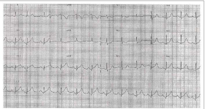
A seventy-four-year old female patient with a history of systemic hypertension and chronic peripheral vascular failure had the onset of condition with oppressive precordial pain irradiating to left upper limb and sweating while showering or bathing. The patient denied any stress condition before the pain. The patient was admitted at the emergency service 3 hours after the onset of symptoms, with systemic hypertension (BP 80/40 mmHg) and 12-lead ECG showing mild elevation of segment ST in DII, DIII, aVF, V5 and V6, and mild unlevelling of segment ST in V1, V2, and V3 (Figure 1).
An emergency coronary angiography demonstrated coronary arteries with no significant obstructive lesions and parietal irregularities in anterior descending artery. Ventriculography showed abnormalities in parietal motility, with akinesia in mid and apical segments, and baseline hyperkinesia, resulting in ventricular aneurysmal dilation, which suggested the takotsubo cardiomyopathy condition (Figure 2). The transthoracic ECG at rest also showed akinesia of mid-apical segments and hyperkinesia of left ventricle baseline segments (Figure 3). Enzyme curve showed cardiac troponin I at 9.29 and 6.39 ng/ml 14 hours and 38 hours after precordial pain, respectively. Normal value for cardiac troponin at our institution is <2.0 ng/ml. Corresponding values for creatine-kinase mB fraction (mass) were 21.2 and 11.3 ng/ml (normal value <4.0 ng/ml).
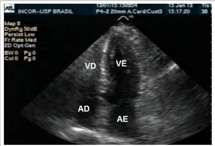
The patient was clinically stable, with no relapse of symptoms, and was discharged. A new echocardiography two weeks after the onset of symptoms showed full recovery of systolic function and left ventricle segmental motility (Figure 4).
Discussion
A number of reports have described patients with reversible left ventricular dysfunction and symptoms that are similar to those in the acute myocardial infarction condition - typically after emotional stress or concurrent to a number of pathologies that include pneumothorax, subarachnoid hemorrhage, and pheochromocytoma, among others with no coronary arteries stenosis1-8. No evident emotional stressor was identified in this particular patient, but the acute coronary syndrome clinical condition, coronariography pattern, ventriculography of segmental myocardial dysfunction with aneurysmal dilation, and ventricular dysfunction reversibility as presented are recurrent to takotsubo cardiomyopathy diagnosis.
In literature, takotsubo cardiomyopathy (from the Japanese, meaning an octopus trap, represented by a jar with a flattened bottom and narrow neck) or stress cardiomyopathy accounts for approximately 1% of acute myocardial infarctions, affecting mostly women, predominantly those over 60 years of age4. Early symptoms are similar to those reported in acute myocardial infarction condition, and typically associated to a triggering emotional or physical stressor. Coronary arteries are compatible with ventricular dysfunction pattern and similar to a takotsubo on ventriculography. The systolic dysfunction is fully reversible within a few weeks. Once the acute phase is over, the condition usually has good prognosis. Despite some fatal reports, such as ventricular rupture, the good prognosis for the condition is confirmed by rapid decrease of atrial natriuretic peptide levels when compared to those in acute
Key words
Stress, psychological; ventricular dysfunction; catecholamines; cardiomyopathies.
This is the report of a 74-year-old female patient with a history of systemic hypertension and peripheral vascular disease who presented acute coronary syndrome symptoms. Coronary angiography showed coronary arteries with no significant obstructions. Ventriculography and echocardiography showed akinesia in mid and apical segments; and hyperkinesia of left ventricle basal segments. Two weeks after the onset of symptoms, a new echocardiogram demonstrated normal global and regional systolic function. The uncommon, reversible pattern for systolic dysfunction and segmental compromising that gives left ventricle a takostubo-like shape is known today as stress cardiomyopathy.
Takotsubo Cardiomyopathy Causing Transitory Ventricular
Dysfunction
Angele A. Alves, Ingrid Kowatsch, Jeane Mike Tsutsui, José C. Nicolau, Marta F. Lima, Wilson Mathias Junior
Instituto do Coração (InCor)- HCFMUSP – São Paulo, SP - Brazil
Mailing address: Jeane Mike Tsutsui •
Av. Dr. Enéas de Carvalho Aguiar, 44 - 05403-000, São Paulo, SP - Brazil E-mail: [email protected] / [email protected]
Manuscript received March 6, 2007; revised manuscript received May 9, 2007; accepted May 9, 2007.
Case Report
Alves et al Cardiomyopathy of Takotsubo
Arq Bras Cardiol 2008; 90(3) : e16-e19 seen in acute myocardial infarction10. Although studies have shown an association between sympathetic activation and stress cardiomyopathy, they are no evidence of a causal relationship. High catecholamine levels may occur due to a secondary response, or be a major cause in this pathology. Further studies are necessary for better understanding of the pathogenesis of this syndrome. However, it should be kept in mind as a differential diagnosis for thoracic discomfort at the emergency room, and a cause of sudden cardiac death in those with known cardiac disease.
myocardial infarction, in correlation with fast improvement of ventricular systolic function9. Strong prevalence is reported among females, and management consists primarily in support and symptomatic therapy.
Although stress cardiomyopathy mechanisms have not been clearly demonstrated, coronary vasospasm or myocarditis are not unlikely causes for the pathology. Patients with the syndrome show evidence of exacerbated sympathetic activation, with catecholamines levels higher than those
Fig. 1 - C12-lead ECG on patient admittance at the emergency room.
Fig. 2 - Ventriculography - diastole and systole to show takotsubo shape as a result of left ventricle mid-apical segments akinesia and baseline segments hyperkinesia.
Left Ventricle
Diastole
Left Ventricle
Systole
Case Report
Alves et al
Cardiomyopathy of Takotsubo
Arq Bras Cardiol 2008; 90(3) : e16-e19
Fig. 4 - Echocardiogram performed two weeks after the onset of symptoms to show the recovery of left ventricle systolic function, with no change in segmental motility. AD - right atrium, AE - left atrium, VD - right ventricle, VE - left ventricle.
Fig. 3 - Echocardiogram performed on admission day shows left ventricle diastole and systole with apical aneurysmal dilation – typical in Takotsubo cardiomyopathy. EA - left atrium, VE - left ventricle.
Systole
Diastole
Case Report
References
1. Akashi YJ, Sakakibara M, Miyake F. Reversible left ventricular dysfunction “takotsubo” cardiomyopathy associated with pneumothorax. Heart. 2002; 87: E1.
2. Rossor AM, Pearce SH, Adams PC. Left ventricular apical ballooning (takotsubo cardiomyopathy) in thyrotoxicosis. Thyroid. 2007; 17 (2): 181-2.
3. Abe Y, Kondo M, Matsuoka R, Araki M, Dohyama K, Tanio H. Assessment of clinical features in transient left ventricular apical ballooning. J Am Coll Cardiol. 2003; 41: 737-42.
4. Akashi YJ, Nakazawa K, Sakakibara M, Miyake F, Koike J, Sasaka K. The clinical features of takotsubo cardiomyopathy. QJM. 2003; 96: 563-73.
5. Girod JP, Messerli AW, Frank Z, Tang WH, Brener SJ. Takotsubo-like transient left ventricular dysfunction. Circulation. 2003; 107: e120-e121.
6. Pollick C, Cujec B, Parker S, Tator C. Left ventricular wall motion abnormalities in subarachnoid hemorrhage: an echocardiographic study. J Am Coll Cardiol. 1988; 12: 600-5.
7. Yamanaka O, Yasumasa F, Nakamura T, Okno A, Endo Y, Yoshimi K, et al. Myocardial stunning-like phenomenon during a crisis of pheochromocytoma. Jpn Circ J. 1994; 58: 737-42.
8. Akashi YJ, Tejima T, Sakurada H, Matsuda H, Suzuki K, Kawasaki K, et al. Left ventricular rupture associated with takotsubo cardiomyopathy. Mayo Clin Proc. 2004; 79: 821-4.
9. Shah DP, Sugeng L, Goonewardena SN, Coon P, Lang RM. Takotsubo cardiomyopathy. Circulation. 2006; 113: e762.
10. Wittstein IS, Thiemann DR, Lima JA, Baughman KL, Schulman SP, Gerstenblith G, et al. Neurohumoral features of myocardial stunning due to sudden emotional stress. N Engl J Med. 2005; 352: 539-48.
Arq Bras Cardiol 2008; 90(3) : e16-e19
Alves et al Cardiomyopathy of Takotsubo