Arq Bras Cardiol volume 73, (nº6), 1999
Canesin et al Endomyocardial fibrosis with massive calcification of the left ventricle
5 0 3
Hospital Universitário da Universidade Estadual de Londrina and Instituto do Coração do Hospital das Clínicas - FMUSP
Mailing address: Manoel Fernandes Canesin Rua Dr. Elias Cesar, 155/701 -86015-640 - Londrina, PR, Brazil.
Submitted: 04/01/99 Accepted: 06/16/99
Manoel Fernandes Canesin, Renato Faria da Gama, Débora Lee Smith, Flávio Jun Kazuma, Arlei Takiuchi, Antonio Carlos Pereira Barretto
Londrina, PR – São Paulo, SP - Brazil
Endomyocardial Fibrosis Associated with Massive
Calcification of the Left Ventricle
Case Report
This is the report of a rare case of endomyocardial fi-brosis associated with massive calcification of the left ven-tricle in a male patient with dyspnea on great exertion, which began 5 years earlier and rapidly evolved. Due to lack of information and the absence of clinical signs that could characterize impairment of other organs, the case was initially managed as a disease with a pulmonary ori-gin. With the evolution of the disease and in the presence of radiological images of heterogeneous opacification in the projection of the left ventricle, the diagnostic hypothesis of endomyocardial disease was established. This hypothesis was later confirmed on chest computed tomography. The patient died on the 16th day of the hospital stay, probably because of lack of myocardial reserve, with clinical findin-gs of refractory heart failure, possibly aggravated by pul-monary infection. This shows that a rare disease such as endomyocardial fibrosis associated with massive calcifi-cation of the left ventricle may be suspected on a simple chest X-ray and confirmed by computed tomography.
Endomyocardial fibrosis is a rare disease, but in Brazil and other tropical and subtropical countries its prevalence has been increasing1. It is characterized by fibrosis of the endocardium and myocardium, and of the inlet and apex of either the right or left or both ventricles. Most of the time, its clinical evolution is progressive with findings of refractory heart failure.
We report a case of difficult diagnosis due to its dis-creet initial clinical manifestation, and also rare due to its association with massive endomyocardial calcification. Initially, the diagnostic hypothesis of primary pulmonary di-sease was formulated, and the investigation followed that direction. Later, based on radiological and tomographic ima-ges of calcification in the cardiac area, the hypothesis of endomyocardial fibrosis was formulated.
Report of the case
The patient is a 23-year-old white male, 1.73 m tall and weighing 60 kg, who sought medical treatment complaining of progressive dyspnea. The patient reported that at the age of 18 years, dyspnea on great exertion began and e-volved with paroxysmal periods of worsening and improve-ment. One month prior to hospital presentation, the dysp-nea evolved to dyspdysp-nea on medium and mild exertion. He also reported having repetitive episodes of pulmonary infection since childhood. He denied orthopnea, paroxys-mal nocturnal dyspnea, and chest pain. He experienced sporadic diarrheas prior to hospital admission. He denied fever. The patient reported the presence of a painless pre-cordial prominence since the age of 16 years, and also a fa-milial antecedent of coronary heart disease. He denied smo-king or having other associated diseases.
On physical examination, the patient was in regular condition, with peripheral cyanosis and tachypnea. The precordial prominence referred to in the history was evident, with no local inflammatory signs or evidence of bone deformity. Blood pressure was 130/80 mmHg and the heart rate was 90 bpm. Cardiac rhythm was regular with normal cardiac sounds and no murmurs. The lungs had disseminated crepitant rales and sibili bilaterally. The remaining findings of the physical examination were within the normal range.
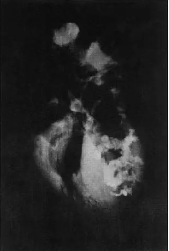
Spirometry revealed a severe restrictive disorder. During the first days at the hospital, the patient developed fever with a dry cough and worsening of the dyspnea. Treatment with bronchodilators and antibiotics was started. Blood and urine tests were within the normal range. The following tests were negative: anti-HIV, tubercle bacilli, and fungus in the sputum. Rest electrocardiogram showed left bundle-branch block, with deviation of the electrical axis to the left, and right ventricle overload. On chest X-ray, lungs with moderate congestion were evidenced, as was an enlargement of the cardiac silhouette (+/4+), with an image of heterogeneous opacification in the projection of the left ventricle (Fig. 1). Computed tomography of the chest showed condensation of some foci located in the pulmonary parenchyma, enlargement in cardiac volume, and intra-cardiac images of massive calcification (Fig. 2).
transthora-5 0 4
Canesin et al
Endomyocardial fibrosis with massive calcification of the left ventricle
Arq Bras Cardiol volume 73, (nº 6), 1999
cic echocardiogram was inconclusive due to lack of a thoracic window for performing the test. The transesopha-geal echocardiogram was also inconclusive because of the patient’s intolerance due to dyspnea and nausea at the time of the test. However, mild thickening of the septum was evi-denced, with mild hypokinesia of the left ventricle. Left ventricle ejection fraction could not be quantified. Tricus-pid regurgitation and pulmonary arterial hypertension with an increase in the right ventricle volume were observed.
The patient’s condition significantly worsened. He was transferred to the intensive care unit and died on the 16th day of the hospital stay.
On autopsy, the gross examination showed fibrosis and massive calcification of the left ventricle, particularly at the apex, spreading to the left ventricle outflow tract (Fig. 3). Microscopy confirmed the presence of significant fibrosis covering a large area of the endocardium (Fig. 4). The
radio-Fig. 2 – Chest computed tomography showing intracardiac image of massive calcification.
Arq Bras Cardiol volume 73, (nº6), 1999
Canesin et al Endomyocardial fibrosis with massive calcification of the left ventricle
5 0 5
Fig. 5 – Chest computed tomography showing intracardiac image of massive calcification.
Fig. 4 – Histopathology of the endomyocardium with significant fibrosis and calcification.
logical image obtained after the autopsy (Fig. 5) showed in greater detail the intense calcification of the heart.
Discussion
Conditions leading to impairment of left ventricle filling are various, endomyocardial fibrosis being one of them1. Endomyocardial fibrosis is an infrequent disease, more prevalent in tropical countries. Its clinical findings
depend on the cardiac chambers affected and on their degree of involvement. Precordial pain suggests a predominant involvement of the left ventricle; ascites, pericardial effusion and a low prothrombin time suggest a predominant impairment of the right ventricle. Dyspnea and edema are common findings in the involvement of both ventricles, and it is difficult to differentiate which ventricle is more affected based on these data2. Invol-vement of both ventricles, right ventricle fibrosis, and tricuspid or mitral regurgitation are associated with a higher mortality3. Endocardial calcification is described in some case series but is not a frequent finding4-6.
Endomyocardial fibrosis is a disease of unknown etiology, first described in Brazil in 19547. In 1984, Silver et al1 described the first case of massive endocardial calcification of the left ventricle, suggesting it was a different entity causing restrictive cardiomyopathy. This suggestion was refuted by Lengyel et al8, who suggested that the endocar-dial calcification was a clue for the diagnosis of endomyocar-dial fibrosis.
In our patient’s clinical history, complaints of dyspnea and wheezing predominated, with previous repetitive respiratory infections. In addition, fever during hospital stay and absence of cardiac murmur or gallop rhythm suggested that the disease was primarily of pulmonary origin. A simple chest X-ray was performed showing a heterogeneous opacification in the left ventricle projection, with a mild increase in the cardiac silhouette and pulmonary congestion. Later, a computed tomography was performed, showing an image of significant calcification in the left ventricle.
After these results, we began to consider the possibi-lity of primary heart disease and started treatment with a diuretic, which resulted in a significant improvement in symptoms. An echocardiogram was also performed, with little help for the diagnosis, showing only pulmonary hyper-tension and tricuspid regurgitation.
In a retrospective analysis, the first evidence of heart disease was provided by the chest X-ray, because more specific tests, such as transthoracic and transesophageal echocardiograms were technically impaired. The left ventri-cle calcification present in the simple X-ray and later confir-med on the chest computed tomography was the great sign pointing to the diagnosis of endomyocardial fibrosis with massive calcification of the left ventricle. The rapid evoluti-on to death was attributed to a pulmevoluti-onary infectievoluti-on, which exhausted the patient’s small myocardial reserve. Endo-myocardial fibrosis could have been diagnosed by ventricu-lography, whose image is considered the gold standard for the diagnosis of that disease. The patient, however, died before undergoing that test. Therefore, the findings of the chest computed tomography made the diagnosis possible. Later, the anatomicopathological study confirmed the diag-nosis of endomyocardial fibrosis.
5 0 6
Canesin et al
Endomyocardial fibrosis with massive calcification of the left ventricle
Arq Bras Cardiol volume 73, (nº 6), 1999
References
1. Silver MA, Bonow RO, Deglin SM, et al. Acquired left ventricular endocardial constriction for massive mural calcific deposits: a newly recognized cause of in-pairment to left ventricular filling. Am J Cardiol 1984; 53: 1468-70. 2. Barretto ACP, Mady C, Arteaga E, et al. Quadro clínico da endomiocardiofibrose.
Correlação com a intensidade da fibrose. Arq Bras Cardiol 1988; 51: 401-5. 3. Barretto ACP, Luz PL, Oliveira AS, et al. Determinants of survival in
endomyo-cardial fibrosis. Circulation 1989; 80: (suppl. I): I-177 - I-182.
4. Cockshott, WP, Saric S, Ikeme AC. Radiological findings in endomyocardial fi-brosis. Circulation 1967; 35: 913-22.
5. Fernandes F, Mady C, Vianna CB, et al. Aspectos radiológicos da endomiocardio-fibrose. Arq Bras Cardiol 1996; 67: 103-5.
6. Morrone LFP, Moreira ELC, Lopez HM. Endomiocardiofibrose com calcificação endocárdica maciça biventricular. Arq Bras Cardiol 1996; 67: 103-5.
7. Ball JD, Williams AW, Davies JNP. Endomyocardial fibrosis. Lancet 1954; 1: 1049.
8. Lengyel M, Árvay A, Palik I, et al. Massive endocardial calcification associated endomyocardial fibrosis. Am J Cardiol 1985; 56: 815-6.
fibrosis with calcification. In addition, the density of the image of the massive calcium deposit, even denser than the patient’s own body of vertebra, indicates the diagnosis. Its finding, even though uncommon, helps in the clinical charc-terization of the disease, allowing the identification of the most affected chamber2. Massive calcification, as in this case, is even rarer.
It is worth emphasizing that even with the massive calcification here presented, the patient remained with only a few symptoms for a long period of time, worsening more intensely as death approached. Another peculiarity of the case is the anatomicopathological finding of marked fibrosis with
calcification spreading to the outflow tract of the left ventricle, which is also rare in patients with endomyocardial fibrosis.

