Instituto de Cardiologia do Rio Grande do Sul/Fundação Universitária de Cardiologia
Mailing address: Paulo R. Prates – IC/FUC – Av. Princesa Isabel, 395 - 90620-001 Porto Alegre, RS, Brazil - E-mail: [email protected]
English version by Stela Maris C. e Gandour
Objective - To assess the surgical results of endoven-tricular patch plasty repair in akinetic and dyskinetic left ventricular areas.
Methods - We studied 52 patients who had undergone endoventricular patch plasty repair associated with myo-cardial revascularization. The preoperative functional class distribution was as follows: class I in 1 (1.9%) pati-ent; class II in 2 (3.8%) patients; class III in 23 (44.2%) patients; and class IV in 26 (50%) patients.
Results - The immediate mortality rate was 7.6% (4 patients). The clinical outcome of 44 patients followed up within a mean postoperative time of 29±25 months was as follows: class I in 33 (75%) patients; class II in 7 (15.9%) patients; class III in 2 (4.5%) patients; and class IV in 2 (4.5%) patients. Comparison between pre- and postopera-tive catheterization in 21 patients showed that the ejection fraction increased from 46.3% to 51.3% (p=0. 17); the left ventricular systolic volume decreased from 76.4 mL to 57.5 mL, (p=0.078); and the left ventricular diastolic volume decreased from 141.2 mL to 105.8 mL (p=0.0 73). These findings showed the tendency toward improvement, but with nonsignificant results.
Conclusion - The technique proved to be effective, to have a low mortality rate, to cause significant clinical im-provement, an increase in ejection fraction, and a reduc-tion in left ventricular volumes.
Key words: ventricular aneurysm, ventricular dysfunction, cardiac surgery, hemodynamic assessment
Arq Bras Cardiol, volume 79 (nº 2), 112-6, 2002
Paulo R. Prates, Abud Homsi Neto, Lucas Maynard Lovato, Guaracy F. Teixeira Fº, João R. M. Sant’Anna, Luiz M. Yordi, Renato A. K. Kalil, Ivo A. Nesralla
Porto Alegre, RS - Brazil
Late Results of Endoventricular Patch Plasty Repair in
Akinetic and Dyskinetic Areas After Acute Myocardial
Infarction
An ischemic event may impair the interventricular sep-tum with both akinetic and dyskinetic alterations. As that is not a resectable tissue, special techniques have been deve-loped for its surgical treatment. In 1985, Cooley et al 1
repor-ted on the endoaneurysmorrhaphy technique for correcting left ventricular aneurysms, according to which aneurysms were more appropriately treated using a Dacron flap. Since 1991, we have been using this technique with preserved bovine pericardium, and our first results were published in 1991 and 1993 2,3.
The incidence of left ventricular aneurysms after acu-te myocardial infarction depends on the diagnostic methods used. Studies based on clinical criteria and autopsy findings indicate an incidence ranging from 10% to 15%. These data were obtained prior to thrombolytic therapy and angioplas-ty, and the impact of these types of treatment in the inciden-ce of aneurysms remains unknown 4.
Transmural myocardial infarction may cause early or late alterations in the left ventricular wall 5. Dyskinesia
re-presents the paradoxical movement of the infarction scar 6
and is considered the true aneurysm. Akinesia represents the situation in which the scar remains motionless.
Both akinetic and dyskinetic segments may lead to chronic heart failure and a reduction in left ventricular ejec-tion fracejec-tion throughout the years. Enlargement of the left ventricular cavity results in an increase in ventricular wall tension, causing an increase in the work of the residual he-althy heart muscle. This results in ischemic distress, which, in the future, may lead to a reduction in systolic function 7,8.
In true aneurysms, surgery has always been indicated in the presence of difficult to control heart failure, ventricular arrhythmias, angina refractory to clinical treatment, throm-boembolism, or when the patients are referred for revascula-rization surgery 3-9. Current studies have shown that early
Both akinetic and dyskinetic alterations are treated with the use of endoaneurysmorrhaphy, and, in the cases of akinesia, endoventricular patch plasty repair is the most ap-propriate denomination for the technique. The literature 11-13
shows that this is the most physiological manner of repai-ring the ventricular geometry, reconstructing the apex, and eliminating distension.
The objective of this study was to analyze early and late clinical results and also to hemodynamically assess en-doaneurysmorrhaphy or endoventricular patch plasty re-pair in patients with left ventricular dysfunction and akinetic or dyskinetic areas. Clinical and hemodynamic data of the patients who died early and of those who survived were compared and revealed factors that influenced mortality.
Methods
From January 1991 to December 2000, 108 patients underwent endoaneurysmorrhaphy according to Cooley’s technique 1, using a flap of preserved bovine pericardium
(fig. 1). The first 52 patients were studied.
Endoaneurysmorrhaphy surgery begins with a ventri-culotomy at the left ventricular apex. Thrombi, when present in the cavity, are removed. The left ventricle is carefully ins-pected, and the precise transition zone between the fibrous and healthy myocardial tissue is identified.
A flap of bovine pericardium is molded according to the reconstitution of the left ventricular apex and sutured with Prolene 3.0 suture thread in the transition line between the fibrous and the healthy endocardial tissue, following the suture line adjacent to the interventricular septum, remo-deling the apex.
Distortions present in the left ventricular apex and in its septum are excluded with this process, aiming at
redu-cing the left ventricular cavity, with a consequent reduction in ventricular wall pressure and reorganization of the heal-thy myocardial area. This leads to a reduction in cardiac work and oxygen consumption.
Then the ventriculotomy is sutured with Prolene 2.0 suture thread and myocardial revascularization is performed with mammary artery or saphenous vein bypass grafts, ac-cording to the need assessed during the catheterization pri-or to surgery.
In regard to the characteristics of our sample, we obser-ved that the mean age of the patients was 58 years, the youngest was 35 and the oldest 76. Of these 52 patients, 34 (65.4%) were males and 18 (34.6%) were females. The mean age for both sexes was 58 years. All patients had experienced a previous acute myocardial infarction, which resulted in left ventricular akinetic or dyskinetic alterations, or both. The preoperative functional class according to New York Heart Association (NYHA) criteria was as follows: class I in 1 (1.9%) patient; class II in 2 (3.8%) patients; class III in 23 (44.2%) patients; and class IV in 26 (50%) patients.
In regard to cardiopulmonary events prior to surgery, we observed 2 (3.8%) patients with chronic obstructive pul-monary disease, 24 (45.6%) patients with systemic hyper-tension, 21 (40.4%) patients with previous heart failure, and 1 (1.9%) patient with reverted cardiopulmonary arrest.
In our study, 17 (32.7%) patients had had an acute myo-cardial infarction (AMI) in the preceding 6 months, 6 (11.5%) patients had had an AMI between 6 months and 1 year before, 8 (15.4%) had had an AMI between 1 and 2 ars before, and 21 (40.4%) had had an AMI more than 2 ye-ars before. The number of preoperative AMI varied as fol-lows: 42 (80.8%) patients had had 1 previous AMI; 8 (15.4%) patients had had 2 AMIs; 1 (1.9%) patient had had 3 AMIs; and 1 (1.9%) patient had had 4 AMIs.
Preoperative electrocardiography revealed 1 (1.9%) patient with sinus bradycardia, 1 (1.9%) patient with atrial fi-brillation, and 1 (1.9%) patient with sustained ventricular ta-chycardia. The remaining patients had sinus rhythm. Atrio-ventricular conduction was normal in 49 patients; 2 (3.8%) patients had first-degree atrioventricular block, and 1 (1.9%) had complete left bundle-branch block. Of the 52 patients, 1 (1.9%) had atrial septal defect.
Data from the preoperative catheterization were also analyzed. Some patients underwent catheterization in other services, which did not provide us with total access to the results. We were able to obtain the ejection fraction of 47 (89.3%) patients and the left ventricular dimensions of 43 (81.7%) patients. The catheterization showed 22 (42.3%) patients with left ventricular akinesia and 30 (57.7%) with dyskinesia. The mean ejection fraction was 45.6±14%, the left ventricular final diastolic volume was 139±84 mL, and the left ventricular final systolic volume was 73.7±50 mL.
In regard to the expansion of coronary artery disease, 6 (11.5%) patients had lesions in 1 vessel, 17 (32.7%) had sions in 2 vessels, 26 (50%) in 3 vessels, and 3 (5.8%) had le-sions in 4 vessels.
The mean duration of extracorporeal circulation was
a b
c
d e
87.5 minutes, the shortest being 47 minutes and the longest 147 minutes. The mean duration of ischemia was 60 tes, the shortest being 30 minutes and the longest 105 minu-tes. Of this series, 50 (96.2%) patients underwent myocardial revascularization concomitant with surgery, and 2 (3.8%) patients did not undergo myocardial revascularization. Of those undergoing that procedure (fig. 2), 11 (20.9%) patients received a mammary artery graft to the anterior descending artery, 29 (55.1%) patients received a saphenous vein by-pass graft to the anterior descending artery, 22 (41.8%) recei-ved a saphenous vein bypass graft to the right coronary ar-tery, 32 (60.8%) received a saphenous vein bypass graft to the marginal branch, and 10 (19%) received a saphenous vein bypass graft to the diagonal branch. These patients were followed up with clinical examinations, analysis of the medical records, and telephone interviews.
Results
The characteristics of our sample are similar to those of other studies, with a prevalence of male patients and a mean age over 50 years.
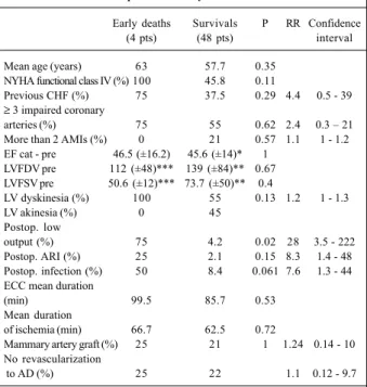
After surgery, we had 4 (7.6%) early deaths in the first postoperative month, 1 (1.9%) death due to low cardiac output, 1 (1.9%) due to respiratory insufficiency, 1 (1.9%) due to arrhythmia, and 1 (1.9%) due to septicemia (tab. I).
The complications in the early postoperative period were as follows: 1 (1.9%) patient had alterations in conduc-tion of the first- and second-degree atrioventricular block type; 4 (7.6%) had atrial fibrillation; 2 (3.8%) had ventricular tachycardia; 5 (9.6%) patients had low cardiac output, 2 due to bleeding and requiring reoperation; and 3 patients had acute left ventricular dysfunction. Of these 3 patients, 2 re-quired the Bio-Pump to leave extracorporeal circulation, and 1 required intraaortic balloon. In the postoperative period, 2 patients had acute respiratory insufficiency.
Postoperative electrocardiography was not performed in 1 (1.9%) patient, and in 41 (77.9%) patients it was identical
to that of the preoperative period. One (1.9%) patient had AV conduction disorders, 1 (1.9%) had ventricular extrasys-tole, 4 (7.7%) had acute atrial fibrillation, 2 (3.8%) had supra-ventricular tachycardia, and 2 (3.8%) had supra-ventricular tachy-cardia. In regard to alterations in the ST segment, 1 (1.9%) patient had alterations in the anterior wall, and the other (1.9%) patient had alterations in the inferior wall.
Of our patients, 1 (1.9%) had sepsis and another (1.9%) had mediastinal infection.
Forty-seven (89.3%) patients were discharged in good condition, and 1 (1.9%) persisted with arrhythmia difficult to control.
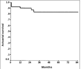
From July to August 1998, we screened 44 patients through clinical-medical visits or telephone interviews. The mean time between surgery and the interview was 29 months, the shortest being 1 month and the longest 92 months.
Six patients died in a later phase as follows: 2 (3.8%) patients died in the 12th month; 1 (1.9%) died in the 26th month;
1 (1.9%) patient in the 29th month; and 2 (3.8%) patients after
the last clinical review, 1 (1.9%) patient in the 86th month, and 1
(1.9%) patient in the 87th postoperative month (fig. 3).
The functional class of the patients reviewed was as fol-lows: 33 (75%) patients were in class I; 7 (15.9%) patients were in class II; 2 (4.5%) patients were in class III; and 2 (4.5%) patients were in class IV. Three patients died in the 12th, 26th, and 86th month
without their clinical follow-up. Reports from their relatives indicated that they were asymptomatic until their death. One patient died in the 29th month with important cardiac symptoms.
Twenty patients agreed to undergo postoperative
ca-Table I - Comparison of early deaths and survivals
Early deaths Survivals P RR Confidence (4 pts) (48 pts) interval
Mean age (years) 63 57.7 0.35 NYHA functional class IV (%) 100 45.8 0.11
Previous CHF (%) 75 37.5 0.29 4.4 0.5 - 39
≥ 3 impaired coronary
arteries (%) 75 55 0.62 2.4 0.3 – 21 More than 2 AMIs (%) 0 21 0.57 1.1 1 - 1.2 EF cat - pre 46.5 (±16.2) 45.6 (±14)* 1
LVFDV pre 112 (±48)*** 139 (±84)** 0.67 LVFSV pre 50.6 (±12)*** 73.7 (±50)** 0.4
LV dyskinesia (%) 100 55 0.13 1.2 1 - 1.3
LV akinesia (%) 0 45
Postop. low
output (%) 75 4.2 0.02 28 3.5 - 222
Postop. ARI (%) 25 2.1 0.15 8.3 1.4 - 48 Postop. infection (%) 50 8.4 0.061 7.6 1.3 - 44 ECC mean duration
(min) 99.5 85.7 0.53
Mean duration
of ischemia (min) 66.7 62.5 0.72
Mammary artery graft (%) 25 21 1 1.24 0.14 - 10 No revascularization
to AD (%) 25 22 1.1 0.12 - 9.7
RR - relative risk; CHF - congestive heart failure; AMI - acute myocardial infarction; EF - left ventricular ejection fraction; LVFDV - left ventricular final diastolic vo-lume; LVFSV - left ventricular final systolic vovo-lume; ECC - extracorporeal circulation; ARI - acute respiratory insufficiency; LV - left ventricle; AD - anterior descending artery; * 47 patients; ** 43 patients; *** 3 patients.
19%
60.8% 41.8%
55.1%
20.9%
94.3%
DG bypass graft MG bypass graft
RC bypass graft AD bypass graft
bypass graft mammary artery graft
0% 50% 100%
theterization, and 1 patient had an examination performed in another service comprising only the ejection fraction. The mean time between surgery and the examination was 33.8±24 months. The mean ejection fraction of 21 patients was 51.5± 16.7 mL, and the left ventricular final diastolic and systolic volumes of 20 patients were 105±46 mL and 57.5±31 mL, respectively. In regard to the grafts, their patencies were as follows: of the mammary artery grafts to the anterior descending artery, 3 (50%) were patent; of the saphenous vein bypass grafts to the anterior descending artery, 6 (60%) were patent; of the saphenous vein bypass grafts to the right coronary artery, 8 (88.9%) were patent; of the saphenous vein bypass grafts to the diagonal branch, 3 (60%) were patent, and of the saphenous vein bypass grafts to the marginal branch, 8 (66.7%) were patent.
Discussion
Acute myocardial infarction in the left ventricular ante-rior wall leads to important alterations in left ventricular sha-pe and volume, which do not significantly improve even af-ter successful reperfusion. A loss in the anaf-terior wall and septal contractility occurs, leading to dilation of the nonim-paired muscular mass as an adaptive mechanism to maintain systolic volume. This remodeling causes progressive left ventricular dysfunction and subsequent congestive heart failure. This fact, even in the absence of significant symp-toms, focuses attention on surgical repair as a possible treatment for patients with this type of lesion 10.
Surgical treatment of left ventricular aneurysms is usu-ally indicated in patients with dyskinetic areas. However, the repair technique comprising resection of those areas and reapproximation of the margins reported in 1985 1 does
not restore left ventricular morphology and impairs hemo-dynamic function. According to Jatene 7, the patient who
survives a left ventricular aneurysm should improve with its repair. This concept was the motivation for the constant
search for ways to refine the technique for correction of left ventricular aneurysms. In 1984, Jatene made an important contribution to the technique of correction of that patho-logy, emphasizing the relevance of maintaining left ven-tricular geometry. Dor et al 5 and Cooley et al 1 introduced
new concepts with the techniques of endoventricular patch plasty repair and endoaneurysmorrhaphy. The technique reported by Cooley et al 1 and modified with the use of
preserved bovine pericardium restores morphology and improves hemodynamic function both in true aneurysms, dyskinetic areas, and in patients with dysfunction due to Months
Actuarial survival
Fig. 3 – Actuarial curve of survival (Kaplan-Meyer) in 84 months.
1.0
.9
.8
.7
.6
.5
.4
.3
.2
.1
0.0
0 12 24 36 48 60 72 84
Fig. 4 - Alteration of pre-and postoperative hemodynamic values. LV - left ventricular.
LV ejection fraction
LV diastolic volume
LV systolic volume
PRE - POST
-PRE - POST
-PRE - POST
-141.2±88
105.8±46
76.4±48.2
1. Cooley DA, Collins HA, Morris Jr GC, Chapman DW. Ventricular aneurysms af-ter myocardial infarction: surgical excision with use of temporary cardiopulmo-nary bypass. JAMA 1985; 167: 557-60.
2. Prates PR, Vitola D, Santana JR, et al. Técnica de Cooley modificada para correção ci-rúrgica do aneurisma do ventrículo esquerdo. Arq Bras Cardiol 1991; 56: 219-2. 3. Prates PR, Vitola D, Sant’Anna JRM, et al. Surgical repair of ventricular aneurysms.
Texas Heart Inst J 1993; 20: 19-22.
4. Gay WA. Ventricular Aneurism - In: Sabiston D. Textbook of Surgery. 15th
edi-tion. Durham: W.B. Saunders Co.; 1997; XIV: 1965-8.
5. Dor V, Sabatier M, Di Donato M, Montiglio F, Toso A, Maioli A. Efficacy of endo-ventricular patch plasty in large pos infartion akinetic scar and severe left ventri-cular disfunction comparison with a series of large disknetic scars. J Thorac Car-diovasc Surg 1998; 116: 50-7.
6. Dor V. Advanced therapy in cardiac surgery. In: Decker BC. Endoventricular Patch Plasty Repair for Pos Infarction Severe Left Ventrcular Disfunction. Lon-don: Hamilton, 1999; 38: 354-61.
7. Jatene A. Left ventricular aneurismectomy: resection or reconstruction. J Thorac Cardiovasc Surg 1985; 89: 321-31.
8. Cooley DA. Ventricular aneurisms and akinesis. Clev Clin Q 1978; 45: 130-2. 9. Cohen M, Packer M, Gorlin R. Indications of left ventricular aneurismectomy.
Current Topics 1983; 67: 717-22.
10. Athanasuleas CL, Stanley AWH, Backberg GD, Dor V, DiDonato M, Blacksto-ne EH and the RESTORE Group. Surgical anterior ventricular endocardial res-taration (SAVER) in the dilated remodeled ventricle after anterior myocardial infarction. J Am Coll Cardiol 2001; 37: 1199-209.
11. Santos GG, Haddad V, Avelar SF, et al. Tratamento cirúrgico dos aneurismas do ventrículo esquerdo com reconstrução geométrica: aspectos cirúrgicos e resul-tados imediatos. Rev Bras Cir Cardiovasc 1991; 6: 116-23.
12. Cooley DA. Ventricular endoaneurysmorraphia results of an improved method of repair. Texas Heart Inst J 1989; 16: 72-5.
13. Hutching GM, Brawley RK. The influence of cardiac geometry on the result of ventricular aneurysms repair. Am J Pathol 1980; 99-221.
References
14. Rivera R, Delcan JL. Factors influencing better results in operation for post in-farction ventricular aneurism. Ann Thorac Surg 1979; 27: 445-50.
15. Di Donato M, Sabatier M, Dor V, Toso A, Maioli M, Fantini F. Akinetic versus dys-kinetic postinfarction scar: relation to surgical outcome in patients undergoing endoventricular circular patch plasty repair. J Am Coll Cardiol 1997; 29: 1569-75. 16. Nesralla IA, Sant’Anna, JR, Kalil RA, et al. Resultados tardios com a ressecção de
aneurismas ventriculares. Arq Bras Cardiol 1979; 32: 9-13.
17. Coogerman M, Stimon EB, Griepp RB, Shunway NE. Survival and function after left ventricular aneurismectomy. J Thorac Cardiovasc Surg 1995; 69: 321-8. 18. Tudge TL, Olsmer JL, Miells NL. Surgical experience with post infarction
ventri-cular aneurism. South Med J 1979; 70: 29-32.
19. Akins CW. Resection of left ventricular aneurysm during hipotermic fibrilato arrest without aortic oclusion. J Thorac Cardiovasc Surg 1986; 91: 610-8. 20. Brawley RK, Magovern CJ, Gott VL, Donahoo JS, Gardner TJ, Watkins L. Left
ventricular aneurysmectomy factors influence post operative results. J Thorac Cardiovasc Surg 1983; 85: 712-7.
21. Burton NA, Stinson EB, Oyer RE, Shunway NE. Left ventricular aneurysm: preoperative risk factors and long term postoperative results. J Thorac Cardiovasc Surg 1979; 77: 65-75.
22. Gosgrove DM, Loop ED, Irarrazaval MS, Groves LK, Taylor PC, Golding LA. Determinants of long term survival after ventricular aneurysmectomy. Ann Thorac Surg 1978; 26: 317-63.
23. Crosby IK, Wellons HA, Martin RP, Schuch D, Muller WH. Employability: a new indication for aneurysmectomy and coronary revascularization. Circulation 1980; 62: 79-83.
24. Harken HA, Horowtz LN, Josephson ME. Comparison of standart aneurysmec-tomy with directed endocardial resection for the treatment of recurent sustained ventricular tachycardia. J Thorac Cardiovasc Surg 1980; 80: 527-34. 25. Moran JM, Scandon PJ, Nemickas R, Pifarre R. Surgical treatment of post
infarc-tion ventricular aneurysm. Ann Thorac Surg 1976; 21: 107-12.
26. Lefemine AA, Govindarajam R, Ramaswamy K, Black H, Maldoff I, Sanella N. Left ventricular resection for aneurysm and akinesia due to coronary artery disease: fifty consecutive patients. Ann Thorac Surg 1977; 23: 461-6.
akinetic areas, as reported by Dor et al 5. This more
physio-logical technique alters factors that influence the Laplace law, reduces the left ventricular cavity, restores wall pressure to normality, prevents septal paradoxical mo-vements, and brings about an improvement in the hemodynamic and clinical conditions of the patients. Studies have already been published confirming these findings 4,5,14-16.
The technique reported by Cooley consists of ope-ning the aneurysm area and suturing a Dacron graft between the normal endocardial area and the fibrous area, which is excluded without resecting normal myocardial structures. This surgery allows revascularization of the impaired area. In a quite different way from that recommended by Co-oley, we used a bovine pericardial graft to exclude the akine-tic or dyskineakine-tic areas after acute myocardial infarction. This way, we were able to exclude distortions produced in the septum without resecting the viable myocardial wall, allowing the associated revascularization of the anterior descending artery when necessary. The bovine pericardium, which is a more malleable tissue than Dacron is, acts like a
cover for the impaired wall, not allowing its dyskinetic mo-vement, therefore increasing left ventricular stroke volume. We attributed the high index of occlusion of the grafts to the anterior descending artery to the fact that we implan-ted mammary artery or saphenous vein bypass grafts whe-never that artery was patent, independent of its distal bed. Most of the time, the anterior descending artery was located in the fibrous portion of the left ventricular anterior wall, and we intended to use the patent anterior descending artery, even in fibrous areas, as a tube to irrigate the septum throu-gh its septal branches.
In conclusion, the repair of left ventricular aneurysms and akinetic areas with the endoventricular patch plasty and endoaneurysmorrhaphy techniques using bovine pericar-dium showed an increase in left ventricular ejection fraction and a reduction in left ventricular volumes. These values were not statistically significant perhaps because of the low num-ber of patients with pre- and postoperative catheterization (fig. 4). The early mortality rate was low, 7.6%, which is ac-ceptable according to the international literature 17-26. A