Topical continuous use of
Lippia sidoides
Cham. essential oil induces
cutaneous in
fl
ammatory response, but does not delay wound
healing process
Maria Liduína Maia de Oliveira
a, Belise Maria Oliveira Bezerra
a, Luana Oliveira Leite
a,
Virgínia Cláudia Carneiro Girão
b, Diana Célia Sousa Nunes-Pinheiro
a,naPrograma de Pós-Graduação em Ciências Veterinárias, Faculdade de Veterinária, Universidade Estadual do Ceará, Campus do Itaperi, CEP 60740-903 Fortaleza, Ceará, Brazil
bDepartamento de Morfologia, Faculdade de Medicina, Universidade Federal do Ceará, Campus do Porangabuçu, CEP 60430-170 Fortaleza, Ceará, Brazil
a r t i c l e
i n f o
Article history:
Received 14 October 2013 Received in revised form 5 February 2014 Accepted 15 February 2014 Available online 25 February 2014
Keywords: Lippia sidoides
Verbenaceae Essential oil Inflammation Wound healing Skin
a b s t r a c t
Ethnopharmacological relevance: The essential oil ofLippia sidoides(EOLS) has been used in Brazilian folk medicine as a topical antiseptic agent in skin for treatment of wounds and superficial infections of the body. The aim of this study was to investigate the effects of EOLS on intact and damaged skin, including its action on expression of mediators, COX-2 and VEGF, involved in healing full-thickness cutaneous lesions in vivo.
Material and methods: EOLS was analyzed chemically and used at different concentrations to dose-response experiments in skin mice. Skin irritation tests by one-dosage and multiple-dosages and irritation to damaged skin were assessed by macroscopy, morphometry and histological and immuno-histochemical analyses. To evaluate the effects of EOLS on wound healing, excision wounds were surgically created on the dorsum of rats, and the ointments at 6% and 12% were applied daily to the wound area. Cutaneous lesions were assessed by planimetric (wound contraction) and macroscopic parameters.
Results: Skin irritation tests showed that topical application of EOLS promoted cutaneous inflammation in varying degrees, which was demonstrated by increase of skin thickness and formation of cutaneous edema and erythema. Topical administration of EOLS in high concentrations presented an irritant response to skin, but this irritation is lighter when low concentrations this oil were used. Histological evaluation supported the outcome of these models, which revealed accentuated presence of infl amma-tory cells infiltration. In wound healing process, the lesions treated with EOLS showed intense edema and exsudation up to day 5, but there were not significant differences in the wound contraction on days 14 and 21. No immunohistochemical staining was verified to COX-2 and VEGF mediators in skin treated with EOLS 12%.
Conclusion: The continuous application of EOLS in adequate concentrations on cutaneous wounds increases inflammatory response without delay the lesions closure. The association of these results with antimicrobial action previously related to EOLS allows its indication as an alternative therapeutic modality for topical treatment of infected cutaneous wound. Nevertheless, further studies need to be performed to determine the mechanism of action and support its application in clinical practice.
&2014 Elsevier Ireland Ltd. All rights reserved.
1. Introduction
As the primary interface between the body and the
environ-ment, the skin provides a first line of defense against infection,
trauma, or injury. Upon skin injury, a series of events take place aiming at the reconstruction of the wounded and cutaneous
homeostasis maintenance (Bangert et al., 2011). Wound repair is
a natural process of regenerating tissue with multiple pathways, which are immediately activated after an injurious stimulus and
can be divided into three overlapping phases: inflammation,
proliferation or granulation tissue formation, and tissue
remodel-ing (Velnar et al., 2009). During the inflammatory phase, leukocyte
cells play a key role in protecting the tissue against infections through phagocytosis, the antibacterial effects of oxygen radicals,
and the activation of complement (Rock et al., 2010). This response
is executed and regulated by an equally complex signaling network
Contents lists available atScienceDirect
journal homepage:www.elsevier.com/locate/jep
Journal of Ethnopharmacology
http://dx.doi.org/10.1016/j.jep.2014.02.030
0378-8741&2014 Elsevier Ireland Ltd. All rights reserved.
n
involving numerous enzymes, growth factors, cytokines and che-mokines. Of particular importance is the cyclooxygenase-2 (COX-2),
which is involved in the production of inflammatory mediators such
as prostaglandins (Rajakariar et al., 2006), and vascular endothelial
growth factor (VEGF), which regulated the angiogenesis during the
wound repair (Bao et al., 2009). In general, the inflammatory
response is a beneficial event that leads to removal of the offending
factor, repair the damage, and then the recruited cells need to be
removed themselves with resolution of inflammation (Widgerow,
2011).
In inflammatory and wound healing processes, several
medic-inal plants and their diverse biological compounds such as terpenes, phenols, lignols and essential oils (EO) have been traditionally used to inhibit or accelerate these events, respectively (Monteiro et al., 2007; Cavalcanti et al., 2012; Riella et al., 2012; Veras et al., 2013).Lippia sidoidesCham. (Verbenaceae) is a native aromatic bush from semiarid areas of the northeast Brazil,
popu-larly known as “alecrim-pimenta” (Matos, 2007). Essential oil
obtained from its leaves presented high concentration of thymol and exhibits multiple biological activities, including antimicrobial (Bertini et al., 2005; Fontenelle et al., 2007; Veras et al., 2012),
antioxidant, gastroprotective and topical anti-inflammatory
(Monteiro et al., 2007; Veras et al., 2013), and oral antiseptic (Girão et al., 2003; Botelho et al., 2009). In northeast Brazil,
pharmaceutical formulations from the essential oil of Lippia
sidoides(EOLS) have been available by programs of herbal medi-cine in primary health care to treat cutaneous wounds and
super-ficial infections in people served at regional hospitals (Matos,
2002), which make the skin a common target to pharmacologic
actions of this plant.
Despite topical acute anti-inflammatory (Monteiro et al., 2007;
Veras et al., 2013) and chronic inflammatory effects recently
attributed to EOLS (Veras et al., 2013), its safe and continuous
use on skin in different physiologic conditions, including the use in open cutaneous wound was not reported. Thus, the aim of this work was to evaluate the effects of topical application of EOLS on intact and damaged skin, including its action on the expression of mediators, COX-2 and VEGF, involved in the wound healing process in experimental models.
2. Material and methods
2.1. Plant material and chemical analysis
EOLS was purchased commercially from Technological Devel-opment Center (PADETEC) of the Federal University of Ceará. The chemical composition of EOLS was determined by gas chromato-graphy coupled to mass spectrometry, using a Shimadzu 5050 GCMS-QP instrument under the following conditions-column: W
Scientific DB-5MS fused silica capillary column (50 m0.25 mm);
carrier gas: He (1 mL/min); injector temperature: 2501C; detector
temperature: 2001C; column temperature: 35–1801C at 41C/min
and then 2501C/15 min; mass spectrum: electronic impact 70 eV.
The identification of the constituents was performed by a
computer-based library search, retention indices and visual
inter-pretation of the mass spectra (Alencar et al., 1984; Adams, 1989).
2.2. Experimental animals
Male Swiss albino mice (25–30 g) and male Wistar rats (150– 180 g) were used in the study. Animals were individually housed in polypropylene cages under standard experimental conditions of
humidity (40–45%), temperature (23–251C), 12 h light/dark cycle
and fed on normal pellet diet and water ad libitum. All experi-mental protocols were approved by Ethics Committee for Use of
Animals of the State University of Ceará (protocol no 10340180-6/ 2010).
2.3. Skin irritation tests
Skin irritation tests were performed as previously described
procedure (Jia et al., 2008) with some modifications. Swiss mice
were shaved on the dorsal surface of the body, and then were left under close observation for 24 h in order to ascertain no abnormal skin responses. The shaved animals were randomly allocated into
fifteen groups (n¼6/group), that received EOLS (10
μ
L) at 100% (innatureEOLS), 50%, 25% and 12% (v/v) in mineral oil (control group),
applied topically in skin nude area of about 1 cm2, as following:
2.3.1. One-dosage irritation to healthy skin
Mice were treated with EOLS at different concentrations in a single dose.
2.3.2. Multiple-dosages irritation to healthy skin
Mice were repeatedly treated with EOLS at different concen-trations, once per day, for consecutive 7 days.
2.3.3. Irritation to damaged skin
Mice skin abrasion was made using a scalpel blade until the
presence of the noticeable tissuefluid, but not blood. The animals
were treated with EOLS in a dose (10
μ
L) fractionated three times aday for one day.
Dermal reactions to the skin challenge, including edema and erythema, were evaluated by macroscopic examination daily for 7 consecutive days. Scoring method for skin irritation was
per-formed as follows: 1–absent; 2–light; 3–moderate; 4–intense.
Skin thicknesses were measured before and after application of treatments, every 24 h, using a micrometer (MMD IP54). On day 7, the animals were euthanized and skin samples were removed for histological and immunohistochemical analyses.
2.4. Histological analysis
Skin sample werefixed in 10% neutral buffered formalin and
were embedded in paraffin wax by usual histological processing.
Five-micrometer sections were cut and stained with hematoxylin-eosin. A representative area was selected for descriptive light
microscopic analysis (Nikon, Tokyo, Japan) at 400x magnification.
2.5. Immunohistochemical analysis
Immunohistochemical investigations were performed on paraffi
n-embedded sections from skin tissue on day 7 for COX-2 and
VEGF. Sections of 5
μ
m were mounted on positive-charged glassslides and submitted to retrieval antigen process with Dako EnVision TM FLEX Target Retrieval Solutions High pH (Code
DM828) or Low pH (Code DM829) for 20 min at 971C using the
Dako pre-treatment (PT) link module (Dako, Glostrup, Denmark). Endogenous peroxidase activity was inhibited by Peroxidase Block (Dako) for 5 min, and slides were subjected either to mouse monoclonal anti-human COX-2 (clone CX-294; Dako) diluted 1:100; and mouse monoclonal anti-human VEGF (clone VG1; Dako) diluted 1:100 and incubated for 1 h at room temperature. The slides were then washed three times in phosphate-buffered
saline (PBS, pH¼7.2) and then incubated with EnVision polymer
reagent (EnVision TMþDual Link System/HRP; Dako) for 30 min at
room temperature andfinally diaminobenzidine (DAB; Dako) for
endometrium were used as positive control for COX-2 and VEGF, respectively, according to the manufacturer's instructions (Dako Corporation, Carpinteria, CA, USA). The intensity of the staining was analyzed by light microscopy (Nikon, Tokyo, Japan) at 400x
magnification.
2.6. Wound healing evaluation
Excision wound model was used to evaluate the effects of EOLS on wound healing. Wistar rats were anesthetized with an intra-peritoneal injection of xylazine (5 mg/kg) and ketamine (80 mg/
kg) (Sadigh-Eteghad et al., 2013). The dorsal surface of each animal
was shaved and area was disinfected with povidone-iodine.
Then, an area approximately 4 cm2 was delimited on the dorsal
medial line, and one full-thickness wound was created with sterile
surgical blade and scissors as previously described (Magalhães
et al., 2008).
The animals were randomly divided into five groups (n¼
6/group). Test group was treated with EOLS ointment 6% and 12% (v/w). Control groups received ointment vehicle (vaseline and
lanolin–1:2) or 0.9% saline. Reference group was treated with 5%
clostebol acetate and neomycin sulfate cream. The treatments were applied topically once a day, starting from the wound induction until complete healing in enough quantity to cover all wounds. The wounds were left undressed to the open
environ-ment and observed daily (Oliveira et al., 2010). Inflammatory
parameters, edema and exudation, were monitored by
macro-scopic examination and graded on a four-point scale: 1–absent; 2
–light; 3–moderate; 4–intense.
Planimetrical analysis was performed on days 0, 3, 7, 14 and 21 on anesthetized animals. The contraction rate was assessed by tracing the raw wound on each evaluation day using transparency paper and a permanent marker. The wound area and one piece of
millimeter paper with known area (1 cm2) were digitalized using a
scanner (Hewlett–Packard, Palo Alto, CA, USA). The measuring wound area was obtained with images analyses as previously
described (Oliveira et al., 2010). Thus, the unhealed wound area
and the percentage of wound contraction were calculated as reduction of initial wound size and used for statistical analysis.
2.7. Statistical analysis
Statistical analysis was performed using GraphPad Prism 5.0
software (San Diego, CA, USA). The comparison between groups was carried out by ANOVA followed by Student-Newmann-Keuls
test. The analysis of the inflammatory parameters was performed
by Kruskal–Wallis test, followed by Dunn test. Results were
expressed as mean7standard deviation (SD) and values of
po0.05 were considered as statistically significant
3. Results
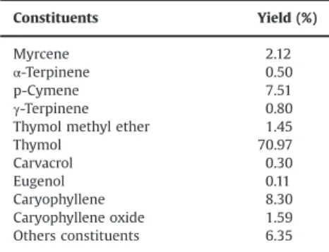
The chemical analysis of EOLS is displayed inTable 1. The main
constituent was thymol, but other minor constituents were also
identified.
The results of skin irritation tests showed that topical
applica-tion of EOLS promoted cutaneous inflammation in varying
degrees. In one-dosage irritation to healthy skin model, EOLS
100% significantly increased skin thickness (po0.05) throughout
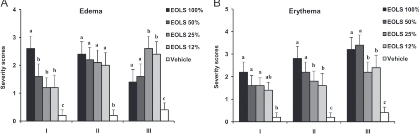
the study when compared to the control group (Fig. 1). Cutaneous
edema and erythema were more intense in mice treated with EOLS 100% in comparison to the others treatment groups on day 3 (Fig. 4). On day 7, it was observed epidermal discontinuity and
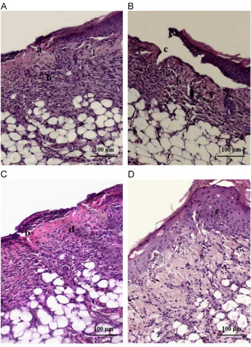
moderate polymorphonuclear cells infiltration (Fig. 5A).
In multiple-dosages irritation to healthy skin model, EOLS 100%
treated group revealed intense inflammatory parameters in
cuta-neous tissue on day 3 (Fig. 4). On day 5, this group showed a
Table 1
Percentage composition of EOLS obtained by gas chromatography/mass spectrometry.
Constituents Yield (%)
Myrcene 2.12
α-Terpinene 0.50
p-Cymene 7.51
γ-Terpinene 0.80
Thymol methyl ether 1.45
Thymol 70.97
Carvacrol 0.30
Eugenol 0.11
Caryophyllene 8.30 Caryophyllene oxide 1.59 Others constituents 6.35
400 800 1200 1600 2000 2400
0 1 3 5 7
Skin thickness (
m)
Time (days)
EOLS 100% EOLS 50% EOLS 25%
EOLS 12% Vehicle
a a
ab
b
b
c a
b
c c
b a
b b
b
b a
b b
b
Fig. 1. Effect of topical treatment with EOLS (100%, 50%, 25% and 12%) on one-dosage irritation to healthy skin model at different time points. Measurement skin thickness was performed at the times 0, 1, 3, 5, and 7 days after the single exposure to EOLS. When used in natura, EOLS significantly increased skin thickness throughout the study when compared to the control group (po0.05). Different
small letters on the same time point indicate statistical significance among groups (po0.05). Each value represents the means7S.D. (n¼6/group).
300 1000 1700 2400 3100
0 1 3 5 7
Skin
thickness
(
m)
Time (days)
EOLS 100% EOLS 50% EOLS 25%
EOLS 12% Vehicle
b a
bc
c
bc b
a
b a
b
c a
b a
a b
a
c a
b
Fig. 2.Effect of topical treatment with EOLS (100, 50, 25 and 12%) on multiple-dosage irritation to healthy skin model at different time points. Measurement skin thickness was performed at the times 0, 1, 3, 5, and 7 days after the continuous exposure to EOLS. When usedin natura, EOLS showed a significant decrease in skin thickness when compared to EOLS 12% treated group (po0.05) and control group
(po0.05). Different small letters on the same time point indicate statistical
significant decrease in skin thickness when compared to EOLS 12%
treated group (po0.05) and control group (po0.05). EOLS 50 and
25% induced higher skin thickness in relation to EOLS 100 and 12%
(po0.05) on day 7 (Fig. 2). In histological evaluation, the main
result verified was the presence of ulcer and moderate infl
amma-tory cells infiltration in EOLS 100% treated group (Fig. 5B).
In irritation to damaged skin model, the cutaneous thickness
was significantly higher in EOLS 25 and 12% treated groups in
relation to EOLS 100 and 50% treated group (po0.05) and control
group (po0.05) on day 3 (Fig. 3). On the other hand, skin
erythema and edema were more intense in EOLS 50% and 25% treated groups, respectively, as compared to control group
(po0.05) on day 3 (Fig. 4). This result was associated to the skin
peeling, dehydration and tissue shrinkage, which were more intense from day 3 post-treatment with EOLS 100% and increased
progressively during the study. On day 7, it was verified intense
inflammatory cells infiltration andfibroblast proliferation in EOLS
100% treated group (Fig. 5C), while the group that received EOLS
50% presented intense epidermal thickening with keratinocytes
proliferation (Fig. 5D). This can have contributed to the significant
increase in skin thickness observed in EOLS 50% treated group
(po0.05) when compared to the other groups at the end of
experiment (Fig. 3).
When the wounds were induced experimentally, the lesions appeared clean and free of exudate throughout the study in all groups, except in groups treated with EOLS at 6 and 12%. In these groups, it was observed intense edema and exudation on wounds up to day 5. Despite the presence of edema in all groups on day 3,
except in saline 0.9% control group, this inflammatory parameter
was more intense and noticeable in animals treated with EOLS
(po0.05) (Fig. 6); however, EOLS ointments promoted healing.
Table 2shows the reduction in wound area in the different groups over the 21-day study period. The wound areas decreased in all
groups compared with the initial wound size, but no significant
differences were verified in the wound contraction on days 14 and
21 (p40.05).
Additionally, immunohistochemical analysis in skin treated with EOLS 12% showed that the expression of COX-2 and VEGF
mediators in both inflammatory and epidermal cells were absent
on day 7. Differences in the staining intensity of COX-2 and VEGF
were not verified to EOLS 12% treatment in any skin irritation tests.
4. Discussion
In folk medicine of northeast Brazil, EOLS has been used as
antiseptic agent for local use in skin (Matos, 2002). This oil
presents thymol and others phenolic compounds, which are effective biological molecules when used in appropriate manner. Although the bioactive constituents of EOLS substantiate their use
as the wound healing agent (Cavalcanti et al., 2012; Riella et al.,
2012), they are nonselective in their action and can cause damage
to host cells. Consequently, inadequate use might cause skin
damage and interfere with or prevent healing (Chang et al.,
2000). In the present study, we investigated the effects of
continuous topical administration of EOLS on intact and damaged skin, in order to evaluate safety of EOLS on cutaneous tissues.
Data present here indicate that EOLS in high concentrations,
mainly when it was usedin nature, showed irritant effects on mice
skin, which were demonstrated by increase cutaneous thick-ness, edema and erythema formation, loss of skin hydration and elasticity. On the other hand, in low concentrations (12%), irritant effects of EOLS on the mice skin, after seven days of treatment, were limited to discrete erythema and edema, and the skin response was estimated as light irritation. Although not indicated using pure essential oils on the skin, it must be considered that
there may be improper use (Vigan, 2010). However, essential oils
600 1500 2400 3300 4200
0 1 3 5 7
Skin
thickness
(
m)
Time (days)
EOLS 100% EOLS 50% EOLS 25%
EOLS 12% Vehicle
ab a ab ab b a a b b b a a b a a b a c b b
Fig. 3.Effect of topical treatment with EOLS (100%, 50%, 25% and 12%) on irritation to damaged skin model at different time points. Measurement skin thickness was performed at the times 0, 1, 3, 5, and 7 days after the skin abrasion and exposure to EOLS. On day 3, the cutaneous thickness was significantly higher in EOLS 25 and 12% treated groups in relation to EOLS 100 and 50% treated group (po0.05) and
control group (po0.05). When used in natura, EOLS promoted skin peeling, dehydration and tissue shrinkage, which increased progressively during the study. Different small letters on the same time point indicate statistical significance among groups (po0.05). Each value represents the means7S.D. (n¼6/group).
0 1 2 3 4
I II III
S
everi
ty
scores
Edema EOLS 100%
EOLS 50% EOLS 25% EOLS 12% Vehicle 0 1 2 3 4 5
I II III
S
everi
ty
scores
Erythema EOLS 100%
EOLS 50% EOLS 25% EOLS 12% Vehicle a b
b b a a a a a b b a c c b a a a a ab a a b a b b b c c b
Fig. 5.Skin sections in the group treated with EOLS 100% (A, B and C) and EOLS 50% (D) on day 7 in different skin irritation models: (A) one-dosage irritation to healthy skin; (B) multiple-dosage irritation to healthy skin; (C) and (D) irritation to damaged skin. (a) epidermal discontinuity; (b) inflammatory cells infiltration; (c) ulcer; (d)fibroblast proliferation; (e) epidermal thickening with keratinocytes proliferation. Haematoxylin and eosin staining. Original magnification: 200x. Scale bar: 100μm.
0 1 2 3 4 5
Day 1 Day 3
S
e
veri
ty
scores
Edema EOLS 6%
EOLS 12%
Vehicle
Reference
Saline 0.9%
0 1 2 3 4 5
Day1 Day 5
S
e
veri
ty
scores
Exudation EOLS 6%
EOLS 12%
Vehicle
Reference
Saline 0.9% a
a
a
b b
a
a
ab
b
b
a a
b b
b
b b
b a
a
Fig. 6.Wound severity scores for lesions treated with EOLS (12% and 6%) ointments in excision wound model. Reference group was treated with 5% clostebol acetate and neomycin sulfate. Two variables, edema (A) and exudation (B) were assessed on days 1–3 and days 1–5, respectively, and graded on a four-point scale. A larger score represents more intense reaction of the skin wound. Significant differences in wound status were found among EOLS 12 and 6% groups and control group (po0.05) in both
edema and exudation on evaluation days. Different small letters indicate significant difference of inflammatory parameters among groups per evaluation day (po0.05).
are used in a 12% concentration to treat skin wounds, excoriations
and infections (Kerr, 2002). The skin is an immune-competent
organ capable of rapid response to chemical and physical injuries
by mounting an inflammatory response that prevents damage and
restore tissue function (Bangert et al., 2011). In this context, the
results of the current study show that an increased cellular
infiltration was observed on histological analysis in EOLS treated
skin, which may be due to increased induction of chemotactic
molecules, which might have attracted inflammatory cells towards
the wound site and intensified the inflammatory response.
Considering that relative skin irritancy in response to the topical treatment may play a role to delay or impair the wound
healing (Jia et al., 2008), an issue addressed in this study was
determining the effect of EOLS ointments on excision wound healing and evaluating their therapeutic action for topical applica-tion. Wound repair is a dynamic and complex process that requires
inflammatory reaction and formation of new tissue to heal the
lesion. In inflammatory phase, soluble mediators increase vascular
permeability, leading tofluid extravasations that result in edema,
and attract inflammatory cells, facilitating the adhesion to the
endothelium and transmigration, which result in tissue exudation (Velnar et al., 2009). A successful inflammatory response is characterized by clearance of injurious stimuli and restoration of tissue normal physiology with resolution of process. However,
when this event is not controlled, the exaggerated inflammation
is a common factor that contributes to matrix destruction, cellular
senescence, and tissue nonhealing (Rajakariar et al., 2006;
Widgerow, 2011). In our study, we found that EOLS at 6% and
12% accentuated the inflammatory response by edema and
exuda-tion formaexuda-tion, but does not delay wound closure and promoted
healing in rat skin. Despite topical anti-inflammatory activity of
EOLS in acute edema model, the repeated use of both EOLS and its
major constituent, thymol, induced pro-inflammatory effect and
cutaneous damage, suggesting that the use this oil must be limited
mainly in chronic treatment (Veras et al., 2013). On the other hand,
thymol has been related as a promising compound to be used in
treatment of inflammatory processes as well as wound healing,
once it reduced the edema and diminished the influx of leukocytes
to the injured area (Riella et al., 2012).
To understand part of the molecular mechanisms through
which topical treatment with EOLS 12% amplifies the infl
amma-tory response, we investigated the immunohistochemical staining intensity of COX-2 and VEGF. The COX-2 pathway is responsible for the production of mediators as prostaglandins, which increase microvascular permeability, promote edema and act synergisti-cally with other mediators, such as VEGF, to stimulate local neovascularization and cellular migration during the wound repair (Laulederkind et al., 2002; Rajakariar et al., 2006). Previously, it
has been suggested that single application of EOLS and thymol
exert an anti-inflammatory action by inhibition of COX enzymes
(Veras et al., 2013). However, our data showed that the
pro-inflammatory effect promoted by multiples applications of EOLS
12% in skin did not involve the COX-2 and VEGF expression. This activity could be also explained by an increased activity of 5- lipooxygenase (5-LOX), a key enzyme in the production of lipoxines, and leukotrienes, mediators with potent
chemoattrac-tant capacity, which induces the formation of ROS (Veras et al.,
2013). Therefore, further understanding of others signaling
path-ways and mediators involved in this process should be evaluated in future studies.
We and other researchers believed that the anti-inflammation
is thefirst step in the wound healing and this effect can play a
direct role in facilitating the fast healing (Jia et al., 2008; Oliveira
et al., 2010; Riella et al., 2012). However, a prolonged inflammatory
response can be beneficial when it does not cause tissues damage
and there is the need to eliminate excessive potential pathogens (Rock et al., 2010; Widgerow, 2011). In other words, the
remark-able inflammatory response and normal wound closure promoted
by EOLS in this study associated with its previous potential
antimicrobial activity (Bertini et al., 2005; Fontenelle et al., 2007;
Veras et al., 2012) can be responsible for the inhibition of
super-ficial infections of the damaged skin. Moreover, considering that
wound infection is likely the most common cause for deficient
wound healing, the efficiency of EOLS in eradiation of the potential
pathogenic microorganisms after injury can played an essential role in controlling the morbidity in patients suffering from skin wounds, such as diabetic foot ulcers in humans and traumatic ulcers in domestic animals.
5. Conclusion
In summary, we provided evidence that adequate use of EOLS has positive effects on dermal irritation response and wound healing. EOLS revealed an irritant response to skin when applied topically in high concentrations; however this irritation was light when EOLS was used in low concentrations, suggesting that the dose of EOLS should be controlled for external use. In relation to wound repair process, continuous use of EOLS in adjusted
con-centrations amplifies the inflammatory response, but does not
delay the cutaneous lesions closure. Take together, the
antimicro-bial action previously related and exacerbation of inflammatory
response during wound healing verified in our study, we suggest
that the EOLS in adequate concentrations can be used topically as an alternative therapeutic modality for treatment of infected cutaneous wound. However, further cellular and molecular Table 2
Effects of essential oil ofL. sidoides(EOLS) on wound contraction by excision wound model.
Day Unhealed wound area (mm2
) and wound contraction (%)
EOLS 6% EOLS 12% Vehicle Referencen
Saline 0.9%
0 432.95739.53a 488.38752.91a 422.08776.47a 464.08753.10a 449.15767.23a 3 503.90758.39a 539.90752.26a 456.08768.45ab 511.93742.48a 406.20769.05b 7 308.52744.40ab 386.90741.55a 237.70746.84b 310.63765.86ab 269.48760.62b
(28.5079.75)AB (20.3678.83)A (43.12710.53)B (33.02713.75)AB (39.44713.61)AB 14 72.4778.73ab 89.14726.24a 58.38716.03b 86.02719.30ab 58.52712.30b
(82.9973.81)A (81.4276.12)A (85.6275.04)A (81.4573.65)A (86.9572.09)A 21 24.6775.75a 30.26719.69a 9.84713.95a 28.33716.15a 17.1078.50a
(94.1871.97)A (93.8873.89)A (97.2974.11)A (93.8473.48)A (96.1771.82)A
Results are expressed as means7S.D. (n¼6).
Different small letters within the same line indicate significant difference of unhealed wound area among groups (po0.05). Different capital letters within the same line
indicate significant difference of wound contraction among groups (po0.05).
n
investigations to explore the mechanism of action of EOLS should be carried out before to obtain conclusions more accurate
regard-ing these heath benefits.
Acknowledgments
The authors are grateful to Fundação Cearense de Apoio ao
Desenvolvimento Científico e Tecnológico (FUNCAP) for financial support. They also extend their thanks to Maria do Socorro França Monte for help with histological processing, to Suzana Moreira de Souza for assistance with the PT link module, and to Conceição da Silva Martins for immunohistochemical techniques help.
References
Adams, R.P. (Ed.), 1989. Identification of Essential Oils by Ion Trap Mass Spectro-scopy. Academic Press, London.
Alencar, W.J., Craveiro, A.A., Matos, F.J.A., 1984. Kovats indices as preselection routine in mass spectra library search of volatiles. J. Nat. Prod. 47, 890–892.
Bangert, C., Brunner, P.M., Stingl, G., 2011. Immune functions of the skin. Clin. Dermatol. 29, 360–376.
Bao, P., Kodra, A., Tomic-Canic, M., Golinko, M.S., Ehrlich, H.P., Brem, H., 2009. The role of vascular endothelial growth factor in wound healing. J. Surg. Res. 153, 347–358.
Bertini, L.M., Pereira, A.F., Oliveira, C.L.L., Menezes, E.A., Morais, S.M., Cunha, F.A., Cavalcanti, E.S.B., 2005. Perfil de sensibilidade de bactérias frente a óleos essenciais de algumas plantas do Nordeste do Brasil. Infarma 17, 80–83.
Botelho, M.A., Santos, R.A., Martins, J.G., Carvalho, C.O., Paz, M.C., Azenha, C., Ruela, R.S., Queiroz, D.B., Ruela, W.S., Marinho, G., Ruela, F.I., 2009. Comparative effect of an essential oil mouthrinse on plaque, gingivitis and salivary Streptococcus mutanslevels: a double blind randomized study. Phytother. Res. 23, 1214–1219.
Cavalcanti, J.M., Leal-Cardoso, J.H., Diniz, L.R.L., Portella, V.G., Costa, C.O., Linard, C.F. B.M., Alves, K., Rocha, M.V.A.P., Lima, C.C., Cecatto, V.M., Coelho-de-Souza, A.N., 2012. The essential oil of Croton zehntneri and trans-anethole improves cutaneous wound healing. J. Ethnopharmacol. 144, 240–247.
Chang, Y.C., Tai, K.W., Huang, F.M., Huang, M.F., 2000. Cytotoxic and nongenotoxic effects of phenolic compounds in human pulp cell cultures. J. Endod. 26, 440–443.
Fontenelle, R.O.S., Morais, S.M., Brito, E.H.S., Kerntopf, M.R., Brilhante, R.S.N., Cordeiro, R.A., Tomé, A.R., Queiroz, M.G.R., Nascimento, N.R.F., Sidrim, J.J.C., Rocha, M.F.G., 2007. Chemical composition, toxicological aspects and antifungal activity of essential oil fromLippia sidoidesCham. J. Antimicrob. Chemother. 59, 934–940.
Girão, V.C.C., Nunes-Pinheiro, D.C.S., Morais, S.M., Sequeira, J.L., Gioso, M.A., 2003. A clinical trial of the effect of a mouth-rinse prepared withLippia sidoidesCham essential oil in dogs with mild gingival disease. Prev. Vet. Med. 59, 95–102.
Jia, Y., Zhao, G., Jia, J., 2008. Preliminary evaluation: the effects ofAloe feroxMiller and Aloe arborescens Miller on wound healing. J. Ethnopharmacol. 120, 181–189.
Kerr, J., 2002. The use of essential oils in healing wounds. Int. J. Aromather. 12, 202–206.
Laulederkind, S.J., Thompson-Jaeger, S., Goorha, S., Chen, Q., Fu, A., Rho, J.Y., Ballou, L.R., Raghow, R., 2002. Both constitutive and inducible prostaglandin H synthase affect dermal wound healing in mice. Lab. Investig. 82, 919–927.
Magalhães, M.S.F., Fechine, F.V., Macedo, R.N., Monteiro, D.L.S., Oliveira, C.C., Brito, G.A.C., Moraes, M.E.A., Moraes, M.O., 2008. Effect of a combination of medium chain triglycerides, linoleic acid, soy lecithin and vitamins A and E on wound healing in rats. Acta Cirúrgica Bras. 23, 262–269.
Matos, F.J.A., 2002. Farmácias vivas: sistemas de utilização de plantas medicinais projetado para pequenas comunidades. Editora UFC, Fortaleza, Brazil, 267p.
Matos, F.J.A., 2007. Plantas medicinais: guia de seleção e emprego de plantas usadas em fitoterapia no Nordeste do Brasil, 3ª edição Imprensa Universitária, Fortaleza, Brazil p. 394.
Monteiro, M.V.B., Leite, A.K.R.M., Bertini, L.M., Morais, S.M., Nunes-Pinheiro, D.C.S., 2007. Topical anti-inflammatory, gastroprotective and antioxidant effects of the essential oil ofLippia sidoidesCham. leaves. J. Ethnopharmacol. 111, 378–382.
Oliveira, M.L.M., Nunes-Pinheiro, D.C.S., Tomé, A.R., Mota, E.F., Lima-Verde, I.A., Pinheiro, F.G.M., Campello, C.C., Morais, S.M., 2010. In vivo topical anti-inflammatory and wound healing activities of the fixed oil of Caryocar coriaceumWittm. seeds. J. Ethnopharmacol. 129, 214–219.
Rajakariar, R., Yaqoob, M.M., Gilroy, D.W., 2006. COX-2 in inflammation and resolution. Mol. Interv. 6, 199–207.
Riella, K.R., Marinho, R.R., Santos, J.S., Pereira-Filho, R.N., Cardoso, J.C., Albuquerque-Junior, R.L.C., Thomazzi, S.M., 2012. Anti-inflammatory and cicatrizing activities of thymol, a monoterpene of the essential oil fromLippia gracilis, in rodents. J. Ethnopharmacol. 143, 656–663.
Rock, K.L., Latz, E., Ontiveros, F., Kono, H., 2010. The sterile inflammatory response. Ann. Rev. Immunol. 28, 321–342.
Sadigh-Eteghad, S., Dehnad, A., Mahmodi, J., Hoseyni, H., Khalili, I., Razmarayii, N., 2013. Healing potential of aStreptomycessp. secondary metabolite, SEM-1-111, on experimental full-thickness excision cutaneous wounds in Wistar rats. Clin. Exp. Dermatol. 38, 178–184.
Veras, H.N.H., Araruna, M.K.A., Costa, J.G.M., Coutinho, H.D.M., Kerntopf, M.R., Botelho, M.A., Menezes, I.R.A., 2013. Topical antiinflammatory activity of essential oil of Lippia sidoides Cham: possible mechanism of action. Phytother. Res. 27, 179–185.
Veras, H.N.H., Rodrigues, F.F.G., Colares, A.V., Menezes, I.R.A., Coutinho, H.D.M., Botelho, M.A., Costa, J.G.M., 2012. Synergistic antibiotic activity of volatile compounds from the essential oil ofLippia sidoidesand thymol. Fitoterapia 83, 508–512.
Velnar, T., Bailey, T., Smrkoli, V., 2009. The wound healing process: an overview of the cellular and molecular mechanisms. J. Int. Med. Res. 37, 1528–1542.
Vigan, M., 2010. Essential oils: renewal of interest and toxicity. Eur. J. Dermatol. 20, 685–692.