Multicenter and international study of MIC/MEC distributions for definition of 1
epidemiological cutoff values (ECVs) for species of Sporothrix identified by molecular 2
methods 3
4
5/22-7/16
5
6
A. Espinel-Ingroff 1, D. P. B. Abreu2, R. Almeida-Paes3, R.S.N. Brilhante4, A. Chakrabarti5, A.
7
Chowdhary6, F. Hagen7, S. Córdoba8; G. M. Gonzalez9, N. P. Govender10, J. Guarro11, E. M.
8
Johnson12; S. E. Kidd13, S. A. Pereira3, A. M. Rodrigues14, S. Rozental15, M. W. Szeszs16, R.
9
Ballesté Alaniz17, A. Bonifaz18, L. X. Bonfietti16, L. P. Borba-Santos15, J. Capilla11, AL Colombo14,
10
M. Dolande19, M. G. Isla8, M.
S. C.Melhem16, A. C. Mesa-Arango20, M. M. E. Oliveira3, M. M. 11
Panizo19, Z. Pires de Camargo14,R. M. Zancope-Oliveira3, J. F. Meis7, J. Turnidge21
12
13
1
VCU Medical Center, Richmond, VA; 2Universidade Federal Rural do Rio de Janeiro,
14
Seropédica, Brasil; 3Fundação Oswaldo Cruz-Fiocruz, Instituto Nacional de Infectologia
15
Evandro Chagas, Laboratório de Micologia, Rio de Janeiro, RJ, Brasil; 4Specialized Medical
16
Mycology Center, Federal University of Ceará, Fortaleza-CE, Brazil; 5Department of Medical
17
Microbiology, Postgraduate Institute of Medical Education & Research, Chandigarh, India;
18 6
Department of Medical Mycology, Vallabhbhai Patel Chest Institute, University of Delhi, Delhi,
19
India; 7Canisius Wilhelmina Hospital, Centre of Expertise in Mycology, Nijmegen, The
20
Netherlands; 8Departamento Micologia; Instituto Nacional de Enfermedades Infecciosas “Dr. C.
21
G. Malbrán”, Buenos Aires, Argentina; 9Universidad Autonóma de Nuevo León, Monterrey, 22
Nuevo León, México; 10National Institute for Communicable Diseases and University of the
23
Witwatersrand, Johannesburg, South Africa; 11Mycology Unit Medical School, Universitat Rovira
24
i Virgili, Reus, Spain; 12Mycology Reference Laboratory, Public Health England, Bristol, UK;
25
13National Mycology Reference Centre, SA Pathology, Adelaide, Australia;. 14Universidade
26
Federal de São Paulo, São Paulo, Brasil; 15Instituto de Biofísica,Universidade Federal do Rio
27
de Janeiro, Brasil; 16Instituto Adolfo Lutz, São Paulo, Araçatuba, Rio Claro Laboratories, Brasil;
28 17
Departamento de Laboratorio Clínico, Hospital de Clínicas Dr. M. Quintela, Facultad de
29
Medicina, Universidad de la República, Montevideo, Uruguay; 18Hospital General de Mexico,
30
Mexico City, Mexico; 19Instituto Nacional de Higiene Rafael Rangel, Caracas, Venezuela;
31
20Grupo de Investigación Dermatológica,Universidad de Antioquía, Medellín, Colombia; and
32
University of Adelaide, Adelaide, Australia.
33
on September 11, 2017 by
http://aac.asm.org/
*Corresponding author: 3804 Dover Rd., Richmond, VA 23221
34
Phone: 804-358-5895
35
Email: [email protected]
36
37
38
39
40
Abstract 41
42
Clinical and Laboratory Standards Institute (CLSI) conditions for testing the
43
susceptibilities of pathogenic Sporothrix species to antifungal agents are based on a
44
collaborative study that evaluated five clinically relevant isolates of Sporothrixschenckii sensu
45
lato and some antifungal agents. With the advent of molecular identification, there are two basic
46
needs: to confirm the suitability of these testing conditions for all agents and Sporothrix species
47
and to establish species-specific epidemiologic cutoff values (ECVs) or breakpoints (BPs) for
48
these species. We collected available CLSI MICs/MECs of amphotericin B, five triazoles,
49
terbinafine, flucytosine and caspofungin for 301 Sporothrix schenckii sensu stricto, 486 S.
50
brasiliensis, 75 S. globosa and 13 S. mexicana molecularly identified isolates. Data were
51
obtained in 17 independent laboratories (Australia, Europe, India, South Africa, South and North
52
America) using conidial inoculum suspensions and 48-72 h of incubation at 35°C. Sufficient and
53
suitable data (modal MICs within 2-fold concentrations) allowed the proposal of the following
54
ECVs for S.schenckii and S. brasiliensis, respectively: amphotericin B 4 and 4 µg/ml,
55
itraconazole 2 and 2 µg/ml; posaconazole 2and 2µg/ml; and voriconazole 64 and 32µg/ml;
56
ketoconazole and terbinafine ECVs for S. brasiliensis were 2 and 0.12 µg/ml, respectively.
57
Insufficient or unsuitable data precluded the calculation of ketoconazole and terbinafine ECVs
58
for S. schenckii as well as ECVs for S. globosa and S. mexicana or any other antifungal agent.
59
These ECVs could aid the clinician in identifying potentially resistant isolates (non-wild type)
60
less likely to respond to therapy.
61
245
62
Introduction
63
64
Sporotrichosis is considered a relatively uncommon granulomatousinfection of the
65
cutaneous and subcutaneous tissue, although dissemination to other deep-seated organs has
66
been reported (1,2). The first case of sporotrichosis was documented in the United States in the
67
on September 11, 2017 by
http://aac.asm.org/
late1800s by Benjamin Schenck (3,4). This case was followed by worldwide reports as well as
68
numerous outbreaks (e.g., in the South African mines in the 1920s and 1930s, among children
69
in relatively remote areas of Peru, the Brazilian case clusters, and in the USA (5-8). In addition,
70
several feline outbreaks caused by Sporothrix brasiliensis with transmissions from cat to human
71
to cat have been reported in Brazil (7,8). Most other outbreaks or infections have been
72
associated with traumatic inoculation of vegetative materials and/or soil. Until recently, all cases
73
were attributed to S. schenckii, according to phenotypic identification (macro and microscopic
74
studies, carbohydrate assimilations, and conversion to the yeast phase). The advent of
75
molecular methodologies and the use of internal transcribed spacer (ITS), region sequence
76
analysis of chitin-synthase, ß-tubulin and calmodulin (CAL) genes indicated that there were
77
various cryptic species nested in the medically relevant clade. The taxon was considered as the
78
Sporothrix schenckii species complex (8-12). Therefore, sporotrichosis is caused by different
79
pathogenic species, including the three clinically relevant species evaluated in the present
80
study: S. schenckii sensu stricto (referred from now only as S. schenckii), S. brasiliensis, and S.
81
globosa. We also evaluated one rare species in the environmental clade, S. mexicana (10,11).
82
83
The recommended therapeutic agents for the treatment of human sporotrichosis are
84
itraconazole, amphotericin B and its lipid formulations (invasive/disseminated disease),
85
terbinafine, and fluconazole; the saturated solution of potassium iodide has been an alternative
86
choice for lymphocutaneous/cutaneous infections (2,13-18). Ketoconazole is not used as much
87
given its low efficacy and potentially severe side effects (13,16). Among the newer triazoles, in
88
vivo and in vitro activity has been reported with posaconazole in combination with amphotericin
89
B, while voriconazole has not been considered a therapeutic choice for these infections due to
90
its high MICs (19,20).
91
92
The Clinical and Laboratory Standards Institute (CLSI) has described testing conditions
93
for the “filamentous phase of the S. schenckii species complex”, because the initial CLSI
94
collaborative evaluation predated molecular studies, which only included five isolates that were
95
documented as “S. schenckii” (21,22). Therefore, the species of Sporothrix are not mentioned in
96
the CLSI M38-A2 document (21). In addition, interpretive MIC/MEC categories, either formal
97
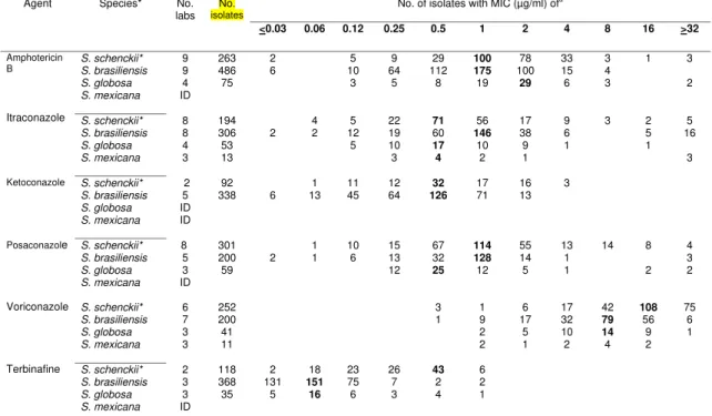
breakpoints (BPs) or epidemiological cutoff values (ECVs), have not been established for any of
98
Sporothrix species. Method-dependent and species-specific ECVs should identify the non-wild
99
type (non-WT) isolates with reduced susceptibility to the agent being evaluated due to acquired
100
mutational or other resistance mechanisms (23,24). Whilst ECVs would not predict the clinical
101
on September 11, 2017 by
http://aac.asm.org/
success to therapy, these endpoints could identify those isolates less likely to respond to the
102
specific agents. We have collected available MICs/MECs of nine antifungal agents from 17
103
laboratories for molecularly identified isolates of four Sporothrix species. These MIC/MEC
104
values represent the antifungal susceptibility of the two more prevalent species (S. schenckii
105
and S. brasiliensis) as well of those of S. globosa and S. mexicana to the different agents as
106
determined by the CLSI M38-A2 method (21). Although the in vitro data were obtained in 17
107
laboratories, the isolates originated from different geographical areas (Australia, Europe, India,
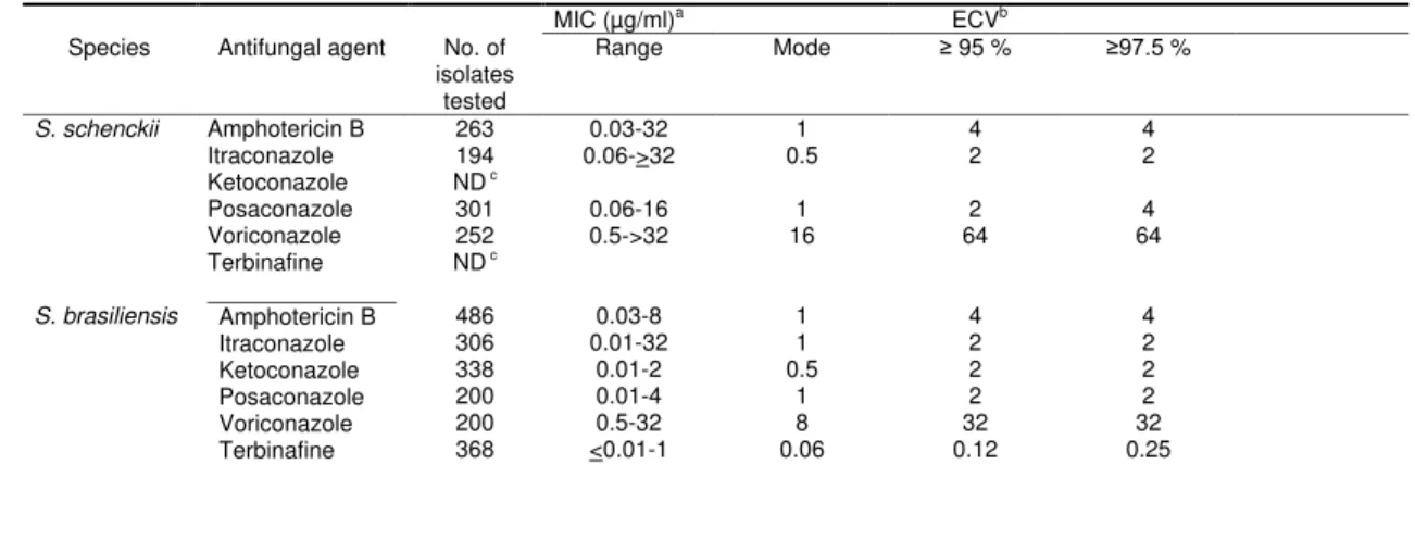
108
South Africa, and both South and North American countries).
109
110
The purpose of the present study was (i) to pool available MIC/MEC data determined by
111
the broth microdilution M38-A2 method originating from 17 independent laboratories for S.
112
schenckii, S. brasiliensis, S. globosa and S. mexicana; (ii)to define the WT susceptibility
113
MIC/MEC distributions of amphotericin B, five triazoles, terbinafine, flucytosine, and
114
caspofungin; (iii) to assess the suitability of these distributions for ECV calculation (including
115
interlaboratory modal agreement); and (iv) to propose CLSI ECVs for two of those species (S.
116
schenckii and S. brasiliensis) when the agent/species combination comprised >100 MICs that
117
originated in 3 to 9 laboratories. MICs of S. globosa and S. mexicana that originated in 3 to 4
118
laboratories were also listed when the distribution comprised at least 10 isolates from >3
119
centers; caspofungin, flucytosine and fluconazole data were summarized in the text.
120
121
Results and Discussion
122
123
CLSI BPs, which reliably predict clinical response to therapy, are not available for any
124
filamentous (mould) species including the Sporothrix species. While the establishment of BPs
125
requires, in addition to other parameters, the clinical correlation of both high and low in vitro
126
results with in vivo data, ECVs are based solely on in vitro data obtained in multiple laboratories
127
(24,25). ECVs or BPs are needed in order to identify the potential in vitro resistance to the agent
128
under evaluation. Although the scarcity of clinical data has precluded the establishment of CLSI
129
BPs for mould testing, several ECVs (e.g., for certain species of Aspergillus, Fusarium and the
130
Mucorales) are available (23,24,26,27). ECVs should distinguish the two populations (WT and
131
non-WT) that are present in the MIC/MEC distribution of a species and agent combination.
132
ECVs for S. brasiliensis and some agents were recently reported using data from a single
133
laboratory (28). However, the definition of ECVs using data from multiple laboratories allows the
134
evaluation of modal (more frequent value in each MIC/MEC distribution) compatibility among the
135
on September 11, 2017 by
http://aac.asm.org/
individual distributions included in the pool (a CLSI requirement) (24). To our knowledge, ECVs
136
have not been defined for any other Sporothrix species; therefore, we collected available MIC/
137
MEC data for S. schenckii, S. brasiliensis, S. globosa and S. mexicana from 17 laboratories
138
worldwide in order to propose ECVs for several antifungal agents.
139
140
Another requirement for the definition of ECVs is that the MIC/MEC data must be
141
accompanied by results for at least one of the quality control (QC) or reference strains (23,24).
142
Examination of the results for QC or reference isolates in our study demonstrated that
143
discrepant MICs for the QC and reference strains (21), although uncommon, were obtained in
144
some laboratories as follows: (i) lower amphotericin B, itraconazole and posaconazole MICs
145
than the expected limits for the QC Candida krusei ATCC 6258 strain from one laboratory; (ii)
146
lower amphotericin B and posaconazole MICs for the QC isolate Paecilomyces variotii ATCC
147
MYA-3630 and the reference Aspergillus flavus ATCC 204304 strains, respectively, from
148
another laboratory. As far as we know, MIC limits have not been established for terbinafine and
149
any fungal strain. However, the laboratories that provided terbinafine MICs used as their internal
150
controls some of the QC or reference isolates. Terbinafine MICs ranged from 0.25 to 1 µg/ml
151
and 0.25 to 0.5 µg/ml for both A. fumigatus ATCC MYA-3626 and A. flavus ATCC 204304,
152
respectively. Nevertheless, the MIC ranges for the C. krusei ATCC 6258 (2 to 64 µg/ml) and to
153
certain extent for C. parapsilosis ATCC 22019 (0.01 to 0.5 µg/ml) were wider than the approved
154
ranges for QC or reference isolates (21). These results indicated that both Candida QC strains
155
could be unsuitable as either QC or reference isolates for terbinafine, but future collaborative
156
studies should establish control guidelines for this agent.
157
158
Although we received MIC/MEC data from 17 laboratories for the four Sporothrix species
159
evaluated in the present study, distributions for each species/agent combination were not
160
collected from each center. In addition, the following unsuitable distributions were excluded: (i)
161
aberrant (mode at the lowest or highest concentration tested) or distributions where the mode is
162
not obvious (e.g., distributions having two or more modes), (ii) when MICs for the QC isolate(s)
163
were outside the recommended limits, or (iii) the mode of a particular distribution was more than
164
one concentration/dilution than the global mode (23,24). In addition, we only incorporated data
165
obtained by the same and unmodified M38-A2 testing parameters as per responses to the
166
survey sent to each laboratory (described below) as follows: (i) MIC distributions that were
167
obtained using conidial suspensions as the inoculum; (ii) MICs obtained after 48 to 72 h of
168
incubation at 35°C; and (iii) by the standard growth inhibition criteria for each agent. Those are
169
on September 11, 2017 by
http://aac.asm.org/
essentially the M38- A2 testing guidelines for obtaining in vitro data for a variety of
non-170
dermatophyte mould species and agents; the exception is terbinafine (only evaluated in
171
multicenter studies for dermatophytes by the CLSI reference method) (21). However, regarding
172
the Sporothrix species, the testing guidelines were based on the multicenter evaluation that
173
included five isolates of S. schenckiisensu lato and four (amphotericin B, fluconazole,
174
itraconazole and ketoconazole) of the nine agents evaluated in the present study (21,22). Since
175
collaborative studies have not been conducted with molecularly identified isolates and QC data
176
are not available for terbinafine, the present collaborative study provides important corroboration
177
about the testing conditions that could yield the most comparable values for six of the nine
178
agents (best interlaboratory modal agreement). These parameters could serve as the basis for
179
further and related studies for evaluating other agents and species, e.g., S. globosa and S.
180
mexicana.
181
182
The MIC distributions of the four Sporothrix species and six of the nine agents evaluated
183
are depicted in Table 1. The modal MICs ranged between 0.5 and 2 µg/ml for most of the
184
species and agent combinations; the exceptions were the higher voriconazole (8 to 16 µg/ml)
185
and the lower terbinafine modes for S. brasiliensis and S. globosa (0.06 µg/ml). Flucytosine,
186
fluconazole and caspofungin data were also collected for S. schenckii, S. brasiliensis and S.
187
globosa from two to five laboratories. Although most of those distributions were either abnormal
188
or unsuitable for ECV definition, both fluconazole and flucytosine modes were consistently at
189
the upper end of the distribution (>32 µg/ml) for S. brasiliensis and S. schenckii, while
190
caspofungin modes were ~1 µg/ml (data not listed in Table 1). While abundant in vitro data are
191
found in the literature in addition to those summarized in Table 1, these studies (i) predated the
192
advent of molecular identification, (ii) reported MIC/MEC data mostly for S. schenckii and S.
193
brasiliensis, and (iii) MICs were obtained for either the yeast or filamentous phase or by
194
modified versions of the CLSI reference method (e.g., supplemented RPMI broth [2%], 30°C
195
incubation, longer incubation times) (29-32). Although some MIC ranges in Table 1 were wider
196
than those in prior studies, owing perhaps to the larger number of isolates (e.g., > 200 versus <
197
100) and different testing conditions, the antifungal susceptibility trend of those species to the
198
various agents is similar. When MICs that were obtained using both the yeast and conidial
199
phases of S. schenckii were compared, the yeast phase yielded lower amphotericin B and
200
itraconazole MICs, while terbinafine MICs were similar or the same (30).There was a need to
201
ascertain which testing conditions yield the most reproducible results. Our collaborative study
202
provides such corroboration at least for the two more prevalent species and clinically relevant
203
on September 11, 2017 by
http://aac.asm.org/
therapeutic agents. In addition, our results suggest that the incubation time for S. globosa needs
204
to be longer and that further evaluation is needed for S. mexicana, among other species.
205
206
Table 2 summarizes MIC ranges, modes and more importantly our proposed ECVs for
207
the species and agents with sufficient data to fulfill the current criteria (> 100 MICs of each
208
agent and species obtained in > 3 independent laboratories) for establishing method-and
209
species-dependent ECVs by the iterative statistical method (23,24). The CLSI has selected the
210
97.5% over the 95% ECVs, both values were calculated and documented. As expected, the
211
highest ECVs were for voriconazole versus S. schenckii and S. brasiliensis (64 and 32 µg/ml,
212
respectively) and the lowest value for terbinafine and S. brasiliensis (0.12 µg/ml). Sufficient and
213
suitable terbinafine MIC data were not available to calculate the terbinafine ECV for S. schenckii
214
according to the current criteria; this species/agent combination needs to be further evaluated.
215
We are also proposing ECVs of 4 µg/ml for amphotericin B and ECVs of 2 µg/ml for three
216
triazoles and both S. schenckii and S. brasiliensis. The high ECVs for these two species (e.g.,
217
amphotericin B and voriconazole ECVs above expected and achievable serum levels) indicate
218
their resistant nature, as was the case for certain species among the Mucorales and Fusarium
219
spp. (26,27). Although the ECV is not a predictor of clinical response to therapy, the high values
220
suggest that isolates of these species could be unresponsive to therapy with these agents. On
221
the other hand, categorization of an isolate as WT does not necessarily signify that it is
222
susceptible to or treatable by the agent under evaluation.
223
224
Unfortunately, among the moulds, genetic information concerning the mechanisms of
225
resistance is mostly available for A. fumigatus and the triazoles. To our knowledge that is not
226
the case for the clinically relevant Sporothrix species. In addition, limited data have been
227
documented regarding the possible correlation between MICs for the Sporothrix infective isolate
228
and the outcome of therapy with the specific agent, including amphotericin B, itraconazole or
229
terbinafine (17,33). In one of those two studies, five patients who responded to oral itraconazole
230
(pulse, 400 mg/day one week with a three week break) for lymphangitic and fixed cutaneous
231
sporotrichosis, the itraconazole MICs for 4 of the 5 infecting S. schenckii isolates were either
232
0.25 or 0.5 µg/ml (17). Those itraconazole MICs were below our proposed ECV of 2 µg/ml for
233
this species and those strains could be considered WT strains (Table 2). In the other report,
234
seven patients with various and persistent S. brasiliensis infections (including disseminated
235
disease) were treated for > 13 weeks as follows: itraconazole 100 mg (3 patients), terbinafine
236
200 mg (3 patients) and amphotericin B, followed by 800 mg of posaconazole (1 HIV-infected
237
on September 11, 2017 by
http://aac.asm.org/
patient) (33). MICs for the serial infective isolates and the clinical response to therapy were as
238
follows: itraconazole 1 or 2 µg/ml (patients cured/infection free); terbinafine between 0.03 and
239
0.12 µg/ml (1 of 3 patients cured); posaconazole 1 µg/ml and amphotericin B between 2 and 4
240
µg/ml (patient died). Our proposed ECVs for S. brasiliensis and those four agents were: 2, 0.12,
241
2 and 4 µg/ml, respectively, and thus, those infecting isolates also could be considered WT
242
(Table 2). However, other factors related to the patient immune response or the use of adjuvant
243
treatments (cryosurgery/curettage) could interfere with meaningful in vitro versus in vivo
244
correlations. On the other hand, the combination of posaconazole and amphotericin B was
245
effective in murine models of disseminated disease caused by S. schenckii or S. brasiliensis
246
(34). The infective isolates for the murine model were WT according to our proposed ECVs.
247
Furthermore, the role of the ECV is not to predict therapeutic outcome, but to identify the
non-248
WT strains that could be less likely to respond to therapy.
249
250
In conclusion, the main role of the ECV is to distinguish between WT and non-WT
251
isolates and aid the clinician in identifying the non-WT isolates that are potentially refractory to
252
therapy with the agent evaluated. This is important when BPs are not available for the
253
species/agent being evaluated, which is the case for the Sporothrix species. Based on CLSI
254
MICs from multiple laboratories, we are proposing the following species-specific CLSI ECVs for
255
S. schenckii and S. brasiliensis, respectively: amphotericin B, 4 and 4 µg/ml; itraconazole, 2 and
256
2 µg/ml; posaconazole, 2and 2µg/ml; and voriconazole, 64 and 32µg/ml. Our proposed
257
ketoconazole and terbinafine ECVs for S. brasiliensis are 2 and 0.12 µg/ml, respectively.
258
Insufficient data precluded the calculation of ketoconazole and terbinafine ECVs for S.
259
schenckii, as well as ECVs for S. globosa and S. mexicana versus any antifungal agent. More
260
importantly, we have corroborated that the susceptibility testing conditions described in the CLSI
261
M38-A2 document could yield the most reliable or reproducible results for the two most
262
prevalent species, which were based on our examination of modes from multiple laboratories.
263
264
Materials and methods
265
266
Isolates. The isolates evaluated were recovered from clinical specimens (mostly
267
lymphocutaneous cutaneous [including disseminated disease] or subcutaneous lesions [>90%])
268
and to a lesser extent pulmonary lesions or other disseminated infections. In addition, we
269
received S. brasiliensis isolates (cutaneous lesions) of feline origin from 4 of the 17 laboratories.
270
MIC/MEC data for each agent were determined in each of the following centers: VCU Medical
271
on September 11, 2017 by
http://aac.asm.org/
Center, Richmond VA, USA;Universidade Federal Rural do Rio de Janeiro, Seropédica, Brasil;
272
Fundação Oswaldo Cruz-Fiocruz, Instituto Nacional de Infectologia Evandro Chagas,
273
Laboratório de Micologia and Laboratório de Pesquisa Clínica em Dermatozoonoses em
274
Animais Domésticos , Rio de Janeiro, RJ, Brasil; Specialized Medical Mycology Center, Federal
275
University of Ceará, Fortaleza-CE, Brazil; Department of Medical Microbiology, Postgraduate
276
Institute of Medical Education & Research, Chandigarh, India; Department of Medical Mycology,
277
Vallabhbhai Patel Chest Institute, University of Delhi, Delhi, India; Canisius Wilhelmina Hospital,
278
Centre of Expertise in Mycology, Nijmegen, The Netherlands; Departamento Micologia, Instituto
279
Nacional de Enfermedades Infecciosas “Dr. C. G. Malbrán”, Buenos Aires, Argentina;
280
Universidad Autonóma de Nuevo León, Monterrey, Nuevo León, México; National Institute for
281
Communicable Diseases and University of the Witwatersrand, Johannesburg, South Africa;
282
Mycology Unit Medical School, Universitat Rovira i Virgili, Reus, Spain; Mycology Reference
283
Laboratory, Public Health England, Bristol, UK; National Mycology Reference Centre, SA
284
Pathology, Adelaide, Australia;.Universidade Federal de São Paulo, São Paulo, Brasil; Instituto
285
de Biofísica,Universidade Federal do Rio de Janeiro, Brasil;andInstituto Adolfo Lutz, São
286
Paulo, Araçatuba, and Rio Claro Laboratories, Brasil.
287
288
Although data were received from 17 independent laboratories (coded 1 to 17), some
289
MIC distributions were excluded from the study for previously discussed reasons. The isolates
290
were identified using phenotypic and genetic approaches (e.g., temperature and nutritional
291
tests, yeast conversion, species specific PCR andPCR-RFLPcalmodulin and ß-tubulin
292
sequencing) (10-12,35). The MIC data used for ECV definition were as follows: 301 S. schenckii
293
and 486 S. brasiliensis isolates. Among the 486 isolates of S. brasiliensis, 261 were isolated
294
from cats. In addition, MIC/MEC data were collected for 75 S. globosa and 13 S. mexicana,
295
respectively. At least one of the QC isolates (C. parapsilosis ATCC 22019, C. krusei ATCC
296
6258, or P. variotii ATCC MYA-3630) was evaluated by the participant laboratories during
297
testing; some laboratories also evaluated the reference isolates A. flavus ATCC 204304 or A.
298
fumigatus ATCC MYA-3626. MICs were only pooled or used for the calculation of ECVs when
299
MICs for the QC or reference isolates were consistently within the established MIC limits as
300
approved by the CLSI (21).
301
302
In vitro susceptibility testing. MIC data for each isolate in the set that was included for
303
analysis or depicted in Tables 1 and 2 were obtained at each center according to the CLSI
M38-304
A2 broth microdilution method (21) (standard RPMI 1640 broth [0.2% dextrose], final conidial
305
on September 11, 2017 by
http://aac.asm.org/
suspensions that ranged from 0.4x10⁴ to 5x10⁴ CFU/ml and an incubation at 35°C between 48 306
to 72 h (S. schenckii, S. brasiliensis, and S. mexicana) or >72 h for S. globosa. MICs were the
307
lowest drug concentrations that produced either complete growth inhibition (100%: amphotericin
308
B, itraconazole, posaconazole and voriconazole) or partial growth inhibition as follows:
309
(terbinafine [80%], fluconazole, ketoconazole and flucytosine [50%]), or morphological changes
310
(caspofungin MECs).
311
312
Data analysis. Data were analyzed by the iterative statistical analysis as previously
313
described in various ECV reports (24-27). MIC/MEC distributions of each species received from
314
each center were listed in electronic spreadsheets. Individual distributions were not included in
315
the final analysis when (i) the distribution had a modal MIC at the lowest or highest
316
concentration tested or were bimodal or when (ii) unusual modal variation (modes that were
317
more than one dilution/concentration from the global mode) (24). Data for each species and
318
agent were only included for the final calculation of ECVs when the total pooled distribution had
319
> 100 isolates and originated from at least three laboratories (Tables 1 and 2).
320
321
Surveys. To ascertain that the collected in vitro susceptibility data in our study were
322
developed following the same testing conditions as described in the CLSI M38-A2 document
323
(21), a survey was sent to the 17 participant laboratories requesting the following information: (i)
324
the source of the agents used; (ii) the formulation of the RPMI medium as described in the CLSI
325
document; (iii) the cells (conidia versus yeasts) and count used to prepare the inoculum
326
suspensions; and (iv) the growth inhibition criteria to determine MICs/MECs for each agent
327
(including incubation temperature and length, and percentage of growth inhibition). The
328
laboratories were also requested to provide MIC/MEC data for at least one of the QC or
329
reference isolates (21).
330
331
Acknowledgments
332
333
We would like to thank the technical personnel at the National Institute for
334
Communicable Diseases, Johannesburg, South Africa, at the VCU Medical Center, Richmond,
335
VA, USA as well as to Ana Caroline de Sá Machado, Jéssica Sepulveda Boechat, Isabella Dib
336
Ferreira Gremião and Tânia Maria Pacheco Schubach (Instituto Nacional de Infectologia
337
Evandro Chagas (INI), Fundação Oswaldo Cruz, Fiocruz, Brasil).
338
339
on September 11, 2017 by
http://aac.asm.org/
340
341 342 343 344
References 345
346
1. López-Romero E, Reyes-Montes MR, Pérez-Torres A, Ruiz-Baca E, Villagómez-347
Castro JC, Mora-Montes HM,Flores-Carreón A, Toriello C. 2011. Sporothrix 348
schenckii complex and sporotrichosis, an emerging health problem. Future Microbiol.
349
6:85-102. doi: 10.2217/fmb.10.157.
350
2. Mahajan VK. 2014. Sporotrichosis: an overview and therapeutic options. Dermatol. Res.
351
Pract. 272376. http://dx.doi.org/10.1155/2014/272376.
352
3. Schenck BR. 1898. On refractory subcutaneous abscesses caused by a fungus
353
possibly related to the sporotricha. Johns Hopkins Hosp. Bull. 9:286-290.
354
4. Espinel-Ingroff A. 1996. A history of medical mycology in the United States. Clin.
355
Microbiol. Rev. 9:235-272.
356
5. Pijper A, Pullinger DB. 1927. An outbreak of sporotrichosis among South African native
357
miners. Lancet. 210:914–916.
358
6. Pappas PG, Tellez I, Deep AE, Nolasco D, Holgado W, Bustamante B. 2000.
359
Sporotrichosis in Peru: Description of an area of hyperendemicity. Clin. Infect. Dis. 30:
360
65–70. 361
7. Barros MBL, Schubach TMP, Galhardo MCG, Schubach OA, Fialho Monteiro PCF, 362
Santos RS, Oliveira RMZ, Lazéra MS, Maya TC, Blanco TCM, Marzochi KBF, Wanke 363
B, Valle ACF. 2001. Sporotrichosis an emergent zoonosis in Rio de Janeiro. Mem. Inst.
364
Oswaldo Cruz. 96:777–779.
365
8. Rodrigues AM, de Teixeira M, de Hoog GS, Schubach TMP, Pereira SA, Fernandes 366
GF, Lopez-Becerra, LM, Felipe MS, Camargo ZP. 2013. Phylogenetic analysis reveals
367
a high prevalence of Sporothrix brasiliensis in feline sporotrichosis outbreaks. PLoS
368
Negl. Trop. Dis. 7: e2281. doi: 10.1371/journal.pntd.0002281 PMID: 23818999; PubMed
369
Central PMCID: PMC3688539.
370
9. de Beer ZW, Harrington TC, Vismer HF, Wingfield BD, Wingfield MJ. 2003.
371
Phylogeny of the Ophiostoma stenoceras-Sporothrix schenckii complex. Mycologia. 95:
372
434–441.
373
10. Marimon R., Gene J, Cano J, Trilles L, Dos Santos Lazera M, Guarro J. 2006. 374
Molecular phylogeny of Sporothrix schenckii. J. Clin. Microbiol. 44:3251–3256.
375
on September 11, 2017 by
http://aac.asm.org/
11. Marimon R, Cano J, Gene J, Sutton DA, Kawasaki M, Guarro J. 2007. Sporothrix 376
brasiliensis, S. globosa, and S. mexicana, three new Sporothrix species of clinical
377
interest. J. Clin. Microb. 45:3198–3206.
378
12. Rodrigues AM, de Hoog S, Camargo ZP. 2014. Genotyping species of the Sporothrix 379
schenckii complex by PCR-RFLP of calmodulin. Diag. Microb. Infect. Dis. 78: 283-287
380
13. Dismukes WE, Stamm AM, Graybill JR, Craven PC, Stevens DA, Stiller RL, Sarosi 381
GA, Medoff G, Gregg CR, Gallis HA, Fields BT jr, Marier RL, Kerkering TA, 382
Kaplowitz LG, Cloud G, Bowles C, Shadomy S. 1983. Treatment of systemic mycoses
383
with ketoconazole: emphasis on toxicity and clinical response in 52 patients. National
384
Institute of Allergy and Infectious Diseases collaborative antifungal study. Annals Intern.
385
Med. 98:13–20.
386
14. Chapman SW, Pappas P, Kauffman C, Smith EB, Dietze R, Tiraboschi-Foss RN, 387
Restrepo A, Bustamante AB, Opper C, Emady-Azar S, Bakshi R. 2004. Comparative
388
evaluation of the efficacy and safety of two doses of terbinafine (500 and 1000 mg
389
day_1) in the treatment of cutaneous or lymphocutaneous sporotrichosis. Mycoses.
390
47:62–68.
391
15. Francesconi G, Francesconi do Valle AC, Passos SL, de Lima Barros MB, de 392
Almeida Paes R, Curi AL, Liporage J, Porto CF, Galhardo MC. 2011. Comparative
393
study of 250 mg/day terbinafine and 100 mg/day itraconazole for the treatment of
394
cutaneous sporotrichosis. Mycopathologia. 171:349–354.
395
16. Kauffman CA, Bustamante B, Chapman SW, Pappas PG, Infectious Diseases 396
Society of America. 2007. Clinical practice guidelines for the management of
397
sporotrichosis: 2007 update by the Infectious Diseases Society of America. Clin. Infect.
398
Dis. 45:1255–1265.
399
17. Bonifaz A, Fierro L, Saul A, Ponce RM. 2008. Cutaneous sporotrichosis. Intermittent
400
treatment (pulses) with itraconazole. Eur. J. Dermatol. 18:1-4.
401
18. Tirado-Sánchez A, Bonifaz A. 2016. Sporotrichosis in Children: An update. Curr
402
Fungal Infect. Rep. DOI 10.1007/s12281-016-0259-0.
403
19. Bunce PE, Yang L, Chun S, Zhang SX, Trinkaus MA, Matukas LM. 2012.
404
Disseminated sporotrichosis in a patient with hairy cell leukemia treated with
405
amphotericin B and posaconazole. Med. Mycol. 50:197–201.
406
http://dx.doi.org/10.3109/13693786.2011.584074.
407
on September 11, 2017 by
http://aac.asm.org/
20. Mario DN, Guarro J, Santurio JM, Alves SH, Capilla J. 2015. In vitro and in vivo
408
efficacy of amphotericin B combined with posaconazole against experimental
409
disseminated sporotrichosis. Antimicrob. Agents Chemother. 59:5018–5021.
410
21. Clinical and Laboratory Standards Institute. 2008. Reference method for broth
411
dilution antifungal susceptibility testing of filamentous fungi, 2nd ed. Approved standard
412
M38-A2. Clinical and Laboratory Standards Institute, Wayne, PA.
413
22. Espinel-ingroff A, Dawson K, Pfaller M, Anaissie E, Breslin B, Dixon D, Fothergill 414
A, Paetznick V, Peter J, Rinaldi M, Walsh T. 1995. Comparative and collaborative
415
evaluation of standardization of antifungal susceptibility testing for filamentous fungi.
416
Antimicrob. Agents Chemother. 39:314-319.
417
23. Clinical and Laboratory Standards Institute. 2016. Epidemiological cutoff values for
418
antifungal susceptibility testing. CLSI supplement M59 document. Clinical and laboratory
419
Standards Institute, Wayne, PA.
420
24. Espinel-Ingroff A, Turnidge J. 2016. The role of epidemiological cutoff values
421
(ECVs/ECOFFs) in antifungal susceptibility testing and interpretation for uncommon
422
yeasts and moulds. Rev. Iberoam. Micol. 33:63–75.
423
https://doi.org/10.1016/j.riam.2016.04.001.
424
25. Turnidge J, Kahmeter G, Kronvall G. 2006. Statistical characterization of
425
bacterial wild-type MIC value distributions and the determination of epidemiological
cut-426
off values. Clin. Microbiol. Infect. 12:418–425.
427
https://doi.org/10.1111/j.1469-0691.2006.01377.x.
428
26. Espinel-Ingroff A, Chakrabarti A, Chowdhary A, Cordoba S, Dannaoui E, Dufresne 429
P, Fothergill A, Ghannoum M, Gonzalez GM, Guarro J, Kidd S, Lass-Flörl C, Meis 430
JF, Pelaez T, Tortorano AM, Turnidge J. 2015. Multicenter evaluation of MIC
431
distributions for epidemiologic cutoff value definition to detect amphotericin B,
432
posaconazole, and itraconazole resistance among the most clinically relevant species of
433
Mucorales. Antimicrob. Agents Chemother. 59:1745–1750.
434
27. Espinel-Ingroff A, Colombo AL, Cordoba S, Dufresne PJ, Fuller JD, Ghannoum M, 435
Gonzalez GM, Guarro J, Kidd SE, Meis JF, Melhem TM, Pelaez T, Pfaller MA, 436
Szeszs MW, Takahaschi JP, Tortorano AM, Wiederhold NP, Turnidge J. 2016. An
437
international evaluation of MIC distributions and ECV definition for Fusarium species
438
identified by molecular methods for the CLSI broth microdilution method. Antimicrob.
439
Agents Chemother. 60:1079–1084.
440
on September 11, 2017 by
http://aac.asm.org/
28. Almeida-Paes R, Brito-Santos F, Figueiredo-Carvalho MHG, Sá Machado AC, 441
Oliveira MME, Pereira SA, Gutierrez-Galhardo MC, Zancopé-Oliveira RM. 2017. 442
Minimal inhibitory concentration distributions and epidemiological cutoff values of five
443
antifungal agents against Sporothrix brasiliensis. Mem. Inst. Oswaldo Cruz, Rio de
444
Janeiro. 112:376-381.
445
29. Kohler L-M, Monteiro PCF, Hahn RC, Hamdan JS. 2004. In vitro susceptibilities of
446
isolates of Sporothrix schenckii to itraconazole and terbinafine. J. Clin. Microbiol. 42:
447
4319–4320.
448
30. Trilles L, Fernandez-Torres B, Lazera MS, Wanke B, Schubach AO, Paes RA, Inza I, 449
Guarro J. 2005. In vitro antifungal susceptibilities of Sporothrix schenckii in two growth
450
phases. Antimicrob. Agents Chemother. 49: 3952–3954. doi:10.1128/AAC.49.9.3952–
451
3954.2005.
452
31. Alvarado-Ramırez E, Torres-Rodrıguez JM. 2007. In Vitro susceptibility of Sporothrix 453
schenckii to six antifungal agents determined using three different methods. Antimicrob.
454
Agents Chemother. 51:2420–2423.
455
32. Galhardo MC, Zancope-Oliveira RM, Do Valle ACF, Almeida-Paes R, Silvatavares 456
PM, Monzo A, Mellado E, Rodriguez-Tudela JL, Cuenca-Estrella M. 2008. Molecular
457
epidemiology and antifungal susceptibility patterns of Sporothrix schenckii isolates from
458
a cat-transmitted epidemic of sporotrichosis in Rio de Janeiro, Brazil. Med Mycol. 46:
459
141-151.
460
33. Almeida-Paes R, Oliveira MME, Freitas DFS, do Valle ACF, Gutierrez-Galhardo MC, 461
Zancopé-Oliveira RM.Refractory sporotrichosis due to Sporothrix brasilensis in
462
humans appears to be unrelated to in vivo resistance. Med. Mycol. 2016 Oct 22. pii:
463
myw103. [Epub ahead of print]. DOI: 10.1093/mmy/myw103.
464
34. Fernández-Silva F, Capilla J, Mayayo E, Guarro J. 2012. Efficacy of posaconazole in
465
murine experimental sporotrichosis. Antimicrob. Agents Chemother 56:2273–2277.
466
http://dx.doi.org/10.1128/AAC.05376-11.
467
35. Rodrigues AM, de Hoog GS, de Camargo ZP. 2015. Molecular diagnosis of
468
pathogenic Sporothrix species. PLoS Negl Trop Dis 9:e0004190.
469
470
471
.
472
473
474
on September 11, 2017 by
http://aac.asm.org/
16 Table 1. Pooled MIC distributions of four Sporothrix speciesfrom between 2 and 9 laboratories determined by CLSI M38-A2 broth microdilution method
478
Agent Species* No.
labs No. isolates
No. of isolates with MIC (µg/ml) ofa
<0.03 0.06 0.12 0.25 0.5 1 2 4 8 16 >32
Amphotericin B
Itraconazole
S. schenckii* 9 263 2 5 9 29 100 78 33 3 1 3
S. brasiliensis 9 486 6 10 64 112 175 100 15 4
S. globosa S. mexicana
4 ID
75 3 5 8 19 29 6 3 2
S. schenckii* 8 194 4 5 22 71 56 17 9 3 2 5
S. brasiliensis 8 306 2 2 12 19 60 146 38 6 5 16
S. globosa S. mexicana 4 3 53 13
5 10
3 17 4 10 2 9 1
1 1
3
Ketoconazole S. schenckii* 2 92 1 11 12 32 17 16 3
Posaconazole
Voriconazole
S. brasiliensis 5 338 6 13 45 64 126 71 13
S. globosa S. mexicana
ID ID
S. schenckii* 8 301 1 10 15 67 114 55 13 14 8 4
S. brasiliensis 5 200 2 1 6 13 32 128 14 1 3
S. globosa S. mexicana S. schenckii* 3 ID 6 59 252
12 25
3 12 1 5 6 1
17 42
2
108
2
75
S. brasiliensis 7 200 1 9 17 32 79 56 6
S. globosa S. mexicana 3 3 41 11 2 2 5 1 10 2 14 4 9 2 1
Terbinafine S. schenckii* 2 118 2 18 23 26 43 6
S. brasiliensis 3 368 131 151 75 7 2 2
S. globosa S. mexicana
3 ID
35 5 16 6 3 4 1
_____________________________________________________________________________________________________________________________________________________________________________________________ 479
aThe highest number in each row (showing the most frequently obtained MIC or the mode) is indicated in boldface. 480
*It refers to Sporothrix schenckiisensu stricto. ID: insufficient data with comparable mod
481 482 483
on September 11, 2017 by
http://aac.asm.org/
17 484
485
Table 2. CLSI-ECVs for S. schenckiisensu stricto and S. brasiliensis based on MICs from between 3 and 9 laboratories by the CLSI broth
486
microdilution method
487
MIC (µg/ml)a ECVb
Species Antifungal agent No. of
isolates tested
Range Mode ≥ 95 % ≥97.5 %
S. schenckii Amphotericin B 263 0.03-32 1 4 4
Itraconazole 194 0.06->32 0.5 2 2
Ketoconazole ND c
Posaconazole 301 0.06-16 1 2 4
Voriconazole 252 0.5->32 16 64 64
Terbinafine ND c
S. brasiliensis Amphotericin B
Itraconazole Ketoconazole Posaconazole Voriconazole Terbinafine
486 306 338 200 200 368
0.03-8 0.01-32
0.01-2 0.01-4 0.5-32 <0.01-1
1 1 0.5
1 8 0.06
4 2 2 2 32 0.12
4 2 2 2 32 0.25
a Mode,most frequent MIC.
488
bCalculated CLSI ECVs comprising >95 % and > 97.5 % of the statistically modeled population; values based on MICs determined by the
489
CLSI M38-A2 broth dilution method (21).
490
cND, Not determined, due to insufficient number of isolates or laboratories for ECV calculation.
491 492