In vitro
methods for antifungal susceptibility
testing of
Trichophyton
spp.
Maria Elisabete da Silva BARROS
*
, Daniel de Assis SANTOS, Ju´nia Soares HAMDAN
Department of Microbiology, Institute of Biological Sciences, Federal University of Minas Gerais, Av. Antoˆnio Carlos, 6627 PO Box 486 CEP: 31.270-901, Belo Horizonte, Minas Gerais, Brazil
a r t i c l e
i n f o
Article history:
Received 8 November 2005 Received in revised form 14 July 2006
Accepted 12 August 2006 Published online 25 October 2006 Corresponding Editor:
Mark Ramsdale
Keywords: Antifungal drugs Dermatophytes Microdilution Susceptibility testing
a b s t r a c t
In general, methods to test the susceptibility of fungi to antifungal drugs require standard-ized techniques, but so far there is no methodology that is widely applicable to dermato-phytes. Here we introduced modifications to the protocols from documents of the National Committee for Clinical Laboratory Standards (CLSI) M38-A and the Antifungal Susceptibility Testing Subcommittee of the European Committee on Antimicrobial Suscep-tibility Testing (EUCAST) that are usually applied to moulds and fermentative yeasts, in order to adjust the conditions for the growth of dermatophytes. The modifications in-cluded: growth on potato dextrose agar supplemented with 2 % in-house rice flour to en-courage sporulation, the addition of 2 % glucose to the culture media (RPMI-1640), and an incubation temperature of 28C. In addition, the incubation period was 7 d, the minimum inhibitory concentration (MIC) was defined as 80 % growth inhibition endpoints for azole agents, and the inocula only contained microconidia. Results obtained by both tested methodologies were very similar to the ones reported by other researchers. MIC90 (MIC
at which 90 % of isolates tested were inhibited) values were identical for four out of five antifungal drugs tested and there was only a difference of one or two dilutions when MIC50 values were compared. Although the modifications introduced did not interfere
with the results, more studies are necessary to establish a standard technique to test sus-ceptibility of dermatophytes to antifungal drugs.
ª2006 The British Mycological Society. Published by Elsevier Ltd. All rights reserved.
Introduction
The approval of protocols M27-A2 (CLSI 2002a) and M38-A (CLSI 2002b), by the National Committee for Clinical Labora-tory Standards (CLSI), motivated research on new methods for the standardization of susceptibility tests for yeasts and fil-amentous fungi. As an example,Meletiadiset al.(2002)have developed colorimetric and diffusion in agar methods. Proto-col M27-A2 is specific for the determination of minimum inhibitory concentrations (MICs) for yeasts (Cryptococcus neo-formansand Candidaspp.), and protocol M38-A for filamen-tous, sporangiospore and conidium-forming fungi that cause
invasive mycoses. Both protocols use the culture medium RPMI-1640 (without sodium bicarbonate and L-glutamine at pH 7.0) supplemented with 0.165M morpholinepropanesul-phonic acid (MOPS) and an incubation temperature of 35C.
Inocula are 104CFU ml1 for Cryptococcus neoformans and
103CFU ml1 for Candida spp. The incubation period is of
24–48 h (document M27-A2) or up to 4 d (document M38-A). Visual readings are performed in both cases. The protocol of the Antifungal Susceptibility Testing Subcommittee of the European Committee on Antimicrobial Susceptibility Testing (AFST-EUCAST; approved in 2002) is used to determine MIC values for fermentative yeasts. This document recommends
* Corresponding author.
E-mail address:[email protected]
a v a i l a b l e a t w w w . s c i e n c e d i r e c t . c o m
j o u r n a l h o m e p a g e : w w w . e l s e v i e r . c o m / l o c a t e / m y c r e s
the use of RPMI-1640 supplemented with 2 % glucose at pH 7.0 (buffered with MOPS 0.165M), a temperature of 35C, an incu-bation period of 48 h, inocula of 105CFU ml1and
spectopho-tometric readings. Whilst these methods are reproducible (Cuenca-Estrellaet al.2002, 2003), so far there are no methods to determine the MIC values of dermatophyte fungi that cause infections of the skin, hair and nail in humans and animals. Among these infections, onychomycosis are the most difficult to treat, affecting 20 % of the world population under 40-years old (Bradleyet al.1999). These infections affect the nail bed causing dystrophy and sometimes result in complete nail loss (Robertset al.2003). Although there are many antifungal drugs available that can be taken orally, only terbinafine, itra-conazole and fluitra-conazole are effective in the treatment of onychomycosis. Several topical antifungal preparations like amorolfine and tioconazole are available as nail lacquer or so-lution form. These topical antifungal drugs can be combined with oral therapy and achieve variable results, producing cure rates ranging from 20–70 % (Roberts et al.2003; Marty
et al.2005). Griseofulvin is an antifungal that has been used since 1959 fortinea capitis, and although it is not efficient in nail treatment, it is used for this purpose (Mocket al.1998; Nie-werth & Korting 2000).Gupta & Shear (2000), in an excellent review, discussed the percentage cure obtained by different investigators with these drugs using different treatments in patients with toenail onychomycosis.
The aim of our work was to compare the MIC values of five oral antifungals (fluconazole, ketoconazole, itraconazole, ter-binafine and griseofulvin) using two methodologies (document M38-A from CLSI and document used by AFST-EUCAST). One hundred samples ofTrichophytonspp. (50 strains ofT. mentagro-phytesand 50 strains ofT. rubrum), isolated from adult nails were tested. The following modifications were introduced in the protocols: RPMI-1640 media was supplemented with 2 % glucose at pH 7.0, buffered with MOPS 0.165M, incubation pe-riod of 7 d, and temperature of 28C, endpoints for fluconazole,
itraconazole, ketoconazole and griseofulvin were set at 80 % growth inhibition and 100 % growth inhibition for terbinafine.
Materials and methods
Isolates
One hundred strains of Trichophyton mentagrophytes (50 strains) andT. rubrum(50 strains) isolated from different pa-tients diagnosed with onchomycosis, were examined in this study. The clinical mycology laboratory, Mycoses Ltda., Belo Horizonte, Minas Gerais, Brazil, kindly donated these strains. Quality control isolates included T. mentagrophytes (ATCC 40004), T. rubrum (ATCC 40051), Candida parapsilosis (ATCC 22019), andCandida krusei(ATCC 6258). Isolates were cultured on MycoselÔ(Difco, Sparks, USA) for identification. The
iso-lates were plated on Sabouraud dextrose agar (Difco) at 28C
and maintained as a suspension in sterile distilled water (Gupta & Kohli 2003) at 4C (Pujolet al.1996) until use.
Medium
Tests were performed in RPMI 1640 with L-glutamine, but without bicarbonate (Gibco BRL, Life technologies, Woerden,
The Netherlands), pH 7.0, supplemented with 2 % glucose, buffered with MOPS (FisherBiotech, New Jersey). The medium was sterilized by filtration.
Antifungal agents
Three azole derivatives were used in this study: fluconazole (Pfizer Sa˜o Paulo, Brazil), ketoconazole and itraconazole (Jans-sen-Cilag, Sa˜o Jose´ dos Campos, Sa˜o Paulo, Brazil). The allyl-amine terbinafine was obtained from Novartis (Sa˜o Paulo, Brazil) and griseofulvin from Schering-Plough (Rio de Janeiro, Brazil). All drugs were dissolved in 100 % dimethylsulfoxide (DMSO) (Gibco, Belo Horizonte, Minas Gerais, Brazil) following the CLSI protocol and were prepared as stock solutions of 1 mg ml1. Serial two-fold dilutions were prepared according
to document (M38-A) from the CLSI at 100 times the final con-centration required, followed by further dilution (1:50) in RPMI 1640 to yield twice the final strength required for the test. The highest concentration of DMSO used in the tests corresponded to 1 % of the total volume and did not interfere with the growth ofTrichophytonspp. studied.
Inocula preparation
Stock suspensions of dermatophytes were prepared from sporulating 7-d-old cultures grown on potato dextrose agar (Acumedia, Baltimore, USA) with 2 % in-house rice flour (Heinlaidet al.2003; Jessupet al.2000) at 28C. Colonies were
covered with 5 ml sterile distilled water and the surface scraped with a sterile loop. The mixture of conidia and hyphal fragments was filtered (Sartorius AG, Goettingen) through an 8mm (Whatman 40, Sa˜o Paulo, Brazil) sterile filter and
col-lected in a sterile tube. This procedure removed the majority of the hyphae, producing inocula composed mainly of spores (Petrikkouet al.2001; Santos & Hamdan 2005; Santos et al.
2006). Turbidity of the final inocula was adjusted to 0.5106–5.0106 spores ml1, at a wavelength of 520 nm,
and transmission adjusted to 70 % in a spectrophotometer (Micronal B542, Sa˜o Paulo). Quantification was made by plat-ing 0.01 ml of a 1:100 dilution of the adjusted inocula (varyplat-ing between 0.5 to 2.5106CFU ml1) on Sabouraud dextrose agar
plates. Plates were incubated at 28C and observed daily.
Col-onies were counted as soon as growth became visible. All in-ocula were adjusted to a final dilution recommended by both methodologies, in RPMI-1640 supplemented with 2 % glucose.
Test procedure
Tests were performed in sterile 96-well flat bottom polystyrene plates; 100ml of each drug (in two-fold dilutions) were added to
the plates that were then stored at70 C until use. For the tests, 100ml of diluted cell suspension was added to each well
so that the final concentration was 0.5104–5104 spores
ml1 for protocol M38-A (CLSI) and 0.5105–5105 spores
ml1for protocol AFST-EUCAST. For fluconazole, the
concen-trations were 64–0.125mg ml1, for ketoconazole and
griseoful-vin 8–0.015mg ml1, for itraconazole 4–0.007mg ml1and for
terbinafine 4–0.007mg ml1. Control wells (growth and sterility)
visually (CLSI method) or with a spectrophotometer (AFST-EUCAST method). MICs were determined as the lowest con-centration of drug that gave approximately 80 % inhibition (Ghannoumet al.2004) of the growth control for fluconazole, ketoconazole, itraconazole and griseofulvin. For terbinafine, MICs were the lowest drug concentration that showed 100 % growth inhibition.
Data analysis
Determination of all MICs was repeated twice. Statistical anal-yses were performed with Wilcoxon (Mann–Whitney) and Kruskal–Wallis tests.P<0.05 was considered significant.
Results
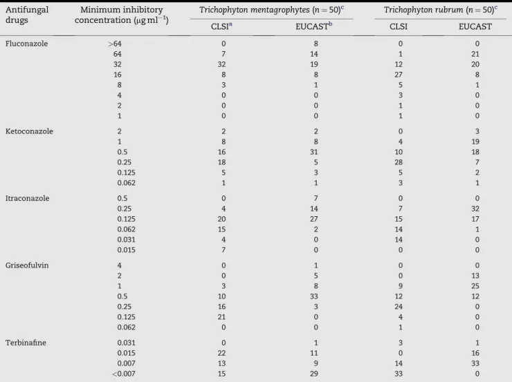
MIC values for 100 isolates ofTrichophytonspp. are summarized inTables 1 and 2. Itraconazole and terbinafine had the highest in-hibitory activities with both methodologies; 90 % of the isolates
had MIC values of 0.25mg ml1and 0.015mg ml1, respectively
(Table 2). When the EUCAST method was used, seven isolates of T. mentagrophytes had MICs of 0.5mg ml1for itraconazole
and one had an MIC of 0.031mg ml1for terbinafine (Table 1).
For the drugs ketoconazole and griseofulvin, the MIC90
values were the same using either methodology (1mg ml1;
Table 2). Using the AFST-EUCAST method, one sample of T. mentagrophyteshad a MIC of 4mg ml1for griseofulvin and
none of theT. rubrumisolates had MICs less than 0.5mg ml1.
The same method detected five samples ofT. mentagrophytes and 13 ofT. rubrumwith MICs of 2mg ml1for the same drug
(Table 2).
Fluconazole was the drug with the lowest activity against the isolates. Using both methods the same MIC50and MIC90
values were observed (Table 2). None of theT. mentagrophytes isolates were susceptible to 4mg ml1, nevertheless five
sam-ples ofT. rubrumwere susceptible to4mg ml1when
proto-cols M38-A of CLSI were used and eight samples of T. mentagrophytes had MICs 64mg ml1 when the
AFST-EUCAST protocol was used (Table 1).
Table 1 –In vitroantifungal activities of five drugs tested using two methods against 100 strains ofTrichophytonspp.
Antifungal drugs
Minimum inhibitory concentration (mg ml1)
Trichophyton mentagrophytes(n¼50)c Trichophyton rubrum(n¼50)c
CLSIa EUCASTb CLSI EUCAST
Fluconazole >64 0 8 0 0
64 7 14 1 21
32 32 19 12 20
16 8 8 27 8
8 3 1 5 1
4 0 0 3 0
2 0 0 1 0
1 0 0 1 0
Ketoconazole 2 2 2 0 3
1 8 8 4 19
0.5 16 31 10 18
0.25 18 5 28 7
0.125 5 3 5 2
0.062 1 1 3 1
Itraconazole 0.5 0 7 0 0
0.25 4 14 7 32
0.125 20 27 15 17
0.062 15 2 14 1
0.031 4 0 14 0
0.015 7 0 0 0
Griseofulvin 4 0 1 0 0
2 0 5 0 13
1 3 8 9 25
0.5 10 33 12 12
0.25 16 3 24 0
0.125 21 0 4 0
0.062 0 0 1 0
Terbinafine 0.031 0 1 3 1
0.015 22 11 0 16
0.007 13 9 14 33
<0.007 15 29 33 0
a Document M38-A of the National Committee for Clinical Laboratory Standards (CLSI 2002b).
b Document of the Antifungal Susceptibility Testing Subcommittee of the European Committee on Antimicrobial Susceptibility Testing
For all drugs tested, both methodologies gave similar MIC50
values (Table 2).Table 3 shows that more than 92 % of the strains varied by less than three dilutions in both methods (CLSI and EUCAST). There was no significant difference (P<0.05) between MIC values of both tested species with the two tested methods, considering no dilution interval.
Discussion
Currently, there are published data comparing thein vitro sus-ceptibility of yeasts, especiallyCandidaspp., using documents CLSI M27-A2, AFST-EUCAST and E-test (Chryssanthou & Cuenca-Estrella 2002; Cuenca-Estrellaet al.2002, 2005; Romero
et al.2004), but there is a scarcity of studies including derma-tophytic fungi. In this study we compared two methods of microdilution in liquid media to determine the susceptibility of dermatophytes to antifungal drugs. Five drugs currently used in the treatment of dermatophytosis were tested (Mukherjeeet al.2003).
Kortinget al.(1995)using the method described byGranade & Artis (1980), reported an MIC90for griseofulvin of 10mg ml1
forT. mentagrophytesisolates and of 3mg ml1forT. rubrum.
Here, we observed using both protocols (M38-A of CLSI and AFST-EUCAST) lower MIC90values of 1mg ml1, for both
spe-cies (Table 2). They also obtained very high MIC values for flu-conazole (1024mg ml1). In this study values obtained for
fluconazole were MIC 64mg ml1. An MIC90 of 2mg ml1
was found for fluconazole against dermatophytes by both
Jessupet al.(2000)andGhannoumet al.(2004), which is very different from the MIC found in the present study (MIC90¼32mg ml1). This is possibly due to differences in the
incubation period. In the case of terbinafine, very low values were observed by Kortinget al.(MIC¼0.05mg ml1) and similar
results were obtained in our study (0.015mg ml1).
Our data, by using both methods, were similar to the ones obtained byFerna´ndez-Torreset al.(2000), Ferna´ndez-Torres
et al.(2001) and Serrano-Martino et al.(2003). MIC50 values
for ketoconazole and itraconazole varied by just for one dilu-tion (plus or minus), while for terbinafine both methods were the same.
Gupta & Kohli (2003), working with 68 samples ofT. rubrum and 14 ofT. mentagrophytesand method M27-A (CLSI), found
the MICs90 for ketoconazole, itraconazole and terbinafine
very similar to the ones reported here. Using a different method, and other species of dermatophytes such as Epider-mophyton floccosum, Microsporum canis and Microsporum gyp-seum,Favreet al.(2003)also found the MIC90for fluconazole
similar to the ones reported here, leading us to conclude that fluconazole should not chosen for the treatment of der-matophyte infections.
Overall, our data demonstrate the reproducibility of the methods employed to perform susceptibility tests in derma-tophytes and confirm thein vitroinefficacy of fluconazole, as well as the excellent antifungal activity of terbinafine, and good activity of itraconazole. The methods suggested by the CLSI (document M38-A) and by the AFST-EUCAST can be considered very good protocols to determine MIC values in dermatophytes. Our modifications to adapt the suscepti-bility tests to this group of fungi did not affect the accuracy of the techniques when applied to this specific group. Moreover, the results we have obtained provide greater reproducibility and reliability of susceptibility/resistance
Table 3 – Agreement of the National Committee for Clinical Laboratory Standards (CLSI) and the European Committee on Antimicrobial Susceptibility Testing (EUCAST) methods at the same dilutions for 100 isolates ofTrichophytonspecies
Drugs Dilutionsa Trichophyton
mentagrophytes
Trichophyton rubrum
n¼50b %c n¼50 %
Fluconazole 0 15 30 16 32
1 22 44 14 28
2 9 18 13 26
3 3 6 4 8
4 1 2 1 2
5 0 0 1 2
6 0 0 1 2
Ketoconazole 0 18 36 7 14
1 24 48 17 34
2 4 8 19 38
3 3 6 5 10
4 1 2 2 4
Itraconazole 0 11 22 12 24
1 20 40 14 28
2 9 18 16 32
3 6 12 8 16
4 2 4 0 0
5 2 4 0 0
Griseofulvin 0 9 18 8 16
1 18 36 14 28
2 16 32 16 32
3 4 8 12 24
4 2 4 0 0
5 1 2 0 0
Terbinafine 0 12 24 5 10
1 21 42 31 32
2 17 34 14 28
a 0 – at same dilution; 1, 2, 3, 4, 5, 6 – number of dilutions for up or down, between CLSI and EUCAST methods.
b Number of isolates. c Percentage in agreement.
Table 2 – MICs of five drugs againstTrichophytonspecies
assessed by National Committee for Clinical Laboratory Standards (CLSI)/European Committee on Antimicrobial Susceptibility Testing (EUCAST) methods
Antifungal drugs MIC50(mg ml1)a MIC90(mg ml1)a
NCCLSb EUCASTc NCCLS EUCAST
Fluconazole 32 64 32 64 Ketoconazole 0.25 0.5 1 1 Itraconazole 0.125 0.25 0.25 0.25 Griseofulvin 0.25 0.5 1 1 Terbinafine 0.007 0.007 0.015 0.015
a - MICs at which 50 % and 90 % of the isolates tested were in-hibited, respectively.
determination than previous methods. In the future, we hope that standardization of these methodologies will allow clinical-laboratory studies to better correlate MIC values with clinical outcomes.
Acknowledgements
We are grateful to Walquı´ria Lopes Borges, Bernardo Drumond Matias, and Taˆnia Mara de Gomes Pinho for excellent techni-cal assistance.
r e f e r e n c e s
AFST-EUCAST, 2002.Reference Method for Determination of Minimal Inhibitory Concentration (MIC) by Broth Dilution of Fermentative Yeasts. Discussion document E.Dis. 7.1 ESCAMID. Subcommittee of Antifungal Susceptibility Testing of the European Commit-tee on Antibiotic Susceptibility Testing of the European Society of Clinical Microbiology and Infectious Diseases. Taufkirchen.
Bradley MC, Leidich S, Isham N, Elewski BE, Ghannoum MA, 1999. Antifungal susceptibilities and genetic relatedness of serial Trichophyton rubrumisolates from patients with onychomyco-sis of the toenail.Mycoses42: 105–110.
Chryssanthou E, Cuenca-Estrella M, 2002. Comparison of the an-tifungal susceptibility testing subcommittee of the European committee on antibiotic susceptibility testing proposed stan-dard and E-test with the CLSI broth microdilution method for voriconazole and caspofungin susceptibility testing of yeast species.Journal of Clinical Microbiology40: 3841–3844.
CLSI (NCCLS), 2002a.Reference Method for Broth Dilution Antifungal Susceptibility Testing of Yeast; Approved Standard document M27-A2, 2nd edn. National Committee for Clinical Laboratory Standards, Wayne, PA.
CLSI (NCCLS), 2002b.Reference Method for Broth Dilution Antifungal Susceptibility Testing of Filamentous Fungi; Approved Standard document M38-A. National Committee for Clinical Laboratory Standards, Wayne, PA.
Cuenca-Estrella M, Gomez-Lopez A, Mellado E, Rodriguez-Tudela JL, 2005. Correlation between the procedure for anti-fungal susceptibility testing forCandidaspp. of the european committee on antifungal susceptibility testing (EUCAST) and four commercial techniques.Clinical Microbiology and Infection
11: 486–492.
Cuenca-Estrella M, Lee-Yang W, Ciblak MA, Arthington-Skaggs BA, Mellado E, Warnak OW, Rodriguez-Tudela JL, 2002. Comparative evaluation of CLSI M27-A and EUCAST broth microdilution procedures for antifungal susceptibility testing ofCandidaspecies.Antimicrobial Agents and
Chemotherapy46: 3644–3647.
Cuenca-Estrella M, Moore CB, Barchiese F, Bille J, Chryssanthou E, Denning DW, Donnelly JP, Dromer F, Dupont B, Rex JH, Richardson MD, Sancak B, Verweij PE, Rodriguez-Tudela JL, AFST Subcommittee of the European Committee on Antimi-crobial Susceptibility Testing, 2003. Multicenter evaluation of the reproducibility of the proposed antifungal susceptibility testing method for fermentative yeasts of the Antifungal Susceptibility Testing Subcommittee of the European Com-mittee on Antimicrobial Susceptibility Testing (AFST-EU-CAST).Clinical Microbiology and Infection9: 467–474.
Favre B, Hofbauer B, Hildering KS, Ryder NS, 2003. Comparisson of in vitroactivities of 17 antifungal drugs against a panel of 20
dermatophytes by using a microdilution.Journal of Clinical Microbiology41: 4817–4819.
Ferna´ndez-Torres B, Va´zquez-Veiga H, Llovo X, Pereiro Jr M, Guarro J, 2000.In vitrosusceptibility to itraconazole, clotri-mazole, ketoconazole and terbinafine of 100 isolates of Trichophyton rubrum.Chemotherapy46: 390–394.
Ferna´ndez-Torres B, Carrillo AJ, Martı´n E, Del Palacio A, Moore MK, Valverde A, Serrano M, Guarro J, 2001.In vitro activities of ten antifungal drugs against 508 dermatophyte strains.Antimicrobial Agents and Chemotherapy45: 2524–2528. Ghannoum MA, Chatuverdi V, Espinel-Ingroff A, Pfaller MA,
Rinaldi MG, Lee-Yang W, Warnock DW, 2004. Intra- and in-terlaboratory study of a method for testing the antifungal susceptibilities of dermatophytes.Journal of
Clinical Microbiology42: 2977–2979.
Granade TC, Artis WM, 1980. Antimycotic susceptibility testing of dermatophytes in microcultures with a standardized frag-mented mycelial inoculum.Antimicrobial Agents and Chemo-therapy17: 725–729.
Gupta AK, Kohli Y, 2003.In vitrosusceptibility testing of ciclo-pirox, terbinafine, ketoconazole and itraconazole against dermatophytes and nondermatophytes, andin vitroevalution of combination antifungal activity.British Journal of Dermatol-ogy149: 296–305.
Gupta AK, Shear NH, 2000. A risk–benefit assessment of the newer oral antifungal agents used to treat onychomycosis. Drugs Safety22: 33–52.
Heinlaid K, Naaber P, Jarv H, 2003. Evaluation of media to enhance conidial production of dermatophytes.Clinical Microbiology and Infection9: 335.
Jessup CJ, Warner J, Isham N, Hasan I, Ghannoum MA, 2000. An-tifungal susceptibility testing of dermatophytes: establishing a medium for inducing conidial growth and evaluation of susceptibility of clinical isolates.Journal of Clinical Microbiology
38: 341–344.
Korting HC, Ollert M, Abeck D, German Collaborative Dermato-phyte Drug Susceptibility Study Group, 1995. Results of ger-man multicenter study of antimicrobial susceptibilities of Trichophyton rubrumandTrichophyton mentagrophytesstrains causingtinea unguium.Antimicrobial Agents and Chemotherapy
39: 1206–1208.
Marty JP, Lambert J, Jackel A, Adjadj L, 2005. Treatment costs of three nail lacquers used in onychomycosis.Journal of Derma-tological Treatment16: 299–307.
Meletiadis J, Mouton JW, Meis JFGM, Bouman BA, Verweij PE, EUROFUNG N, 2002. Comparison of the Etest and Sensititre colorimetric methods with the NCCLS proposed standard for antifungal susceptibility testing ofAspergillusspecies.Journal of Clinical Microbiology40: 2876–2885.
Mock M, Monod M, Baudraz-Rosselet F, Panizzon RG, 1998.Tinea capitisdermatophytes: susceptibility to antifungal drugs tested in vitroandin vivo.Dermatology197: 361–367.
Mukherjee PK, Leidich SD, Isham N, Leitner I, Ryder NS, Ghannoum MA, 2003. ClinicalTrichophyton rubrumstrain ex-hibiting primary resistance to terbinafine.Antimicrobial Agents and Chemotherapy47: 82–86.
Niewerth M, Korting HC, 2000. The use of systemic antimycotics in dermatotherapy.European Journal of Dermatology10: 155–160.
Petrikkou E, Rodrı´guez-Tudela JL, Cuenca-Estrella M, Go´mez A, Molleja A, Mellado E, 2001. Inoculum standardization for an-tifungal susceptibility testing of filamentous fungi pathogenic for humans.Journal of Clinical Microbiology39: 1345–1347. Pujol I, Guarro J, Llop C, Soler L, Ferna´ndez-Ballart J, 1996.
Com-parison study of broth macrodilution and microdilution anti-fungal susceptibility tests for the filamentous fungi.
Antimicrobial Agents and Chemotherapy40: 2106–2110.
Romero M, Ca´nton E, Pe´man J, Gobernado M, 2004. Estudio de la actividadin vitrode caspofungin sobre especies de leva-duras diferentes deCandida albicans, determinada por dos me´todos: M27–A2 y EUCAST.Revista Espan˜ola de Quimiotera-pia17: 257–262.
Santos DA, Hamdan JS, 2005. Evaluation of broth microdilution antifungal susceptibility testing conditions forTrichophyton rubrum.Journal of Clinical Microbiology43: 1917–1920.
Santos DA, Barros MES, Hamdan JS, 2006. Establishing a method of inoculum preparation for susceptibility testing of Tricho-phyton rubrumandTrichophyton mentagrophytes.Journal of Clin-ical Microbiology44: 98–101.