ORIGINAL ARTICLE
Fluoride releasing and enamel demineralization
around orthodontic brackets by fluoride-releasing
composite containing nanoparticles
Mary A. S. Melo&Weslanny A. Morais&Vanara F. Passos& Juliana P. M. Lima&Lidiany K. A. Rodrigues
Received: 18 December 2012 / Accepted: 24 July 2013 / Published online: 22 August 2013
#Springer-Verlag Berlin Heidelberg 2013
Abstract
Introduction Fluoride-containing materials have been suggested to control enamel demineralization around orthodontic brackets during the treatment with fixed appliances. The improvement of their properties has been made through innovations, such as the application of nanotechnology by incorporation of nanofillers. Objective This in vitro study evaluated the capacity of fluo-ride releasing and enamel demineralization inhibition of fluoride-releasing nanofilled cement around orthodontic brackets using an artificial caries biofilm model.
Materials and methodsForty bovine enamel discs were select-ed by evaluating surface microhardness and randomizselect-ed into four groups (n=10): non-fluoride-releasing microfilled compos-ite, fluoride-releasing microfilled composcompos-ite, resin-modified glass ionomer cement (RMGI), and fluoride-releasing nanofilled composite (FN). After brackets bonding in each disc, the spec-imens were subjected to a cariogenic challenge through a Streptococcus mutans biofilm model. After the experimental period, the biofilm formed around the brackets was collected for fluoride analysis and the mineral loss around the brackets was determined by integrated demineralization via cross-sectional microhardness measurement at 20 and 70μm from the bracket margin. Additionally, samples of each group were subjected to energy-dispersive X-ray spectroscopy (EDX) anal-ysis examined under a scanning electron microscopy (SEM). ANOVA followed by Tukey test were applied for fluoride concentration and mineral loss data, respectively.
Results At both distances, only RMGI statistically differed from the other groups presenting the lowest demineralization, although there was a trend to a lower demineralization of enamel around brackets in FN group. Similar condition was found to fluoride concentration and EDX/SEM analysis. Conclusions Under the cariogenic exposure condition of this study, the fluoride-releasing nanofilled material had similar performance to fluoride-releasing microfilled materials. Clinical relevance The presence of nanofillers in the fluoride-releasing materials studied did not promote further benefits against caries lesion development around brackets and presented inferior demineralization inhibition than the resin-modified glass ionomer material.
Keywords Nanotechnology . Biofilm . Fluoride-releasing materials . Orthodontic brackets . EDX element analysis
Introduction
One of the main problems related to orthodontic treatment is the development of demineralized areas around brackets dur-ing fixed orthodontic therapy [1], which is reported to occur in short time and with high rates among patients [2]. The fixed orthodontic appliance components as brackets, arch wires, O-ring, and metal ligature ties do create additional retentive sites, encouraging biofilm accumulation [3]. Microbiological changes after brackets placement towards increased levels of cariogenic species such asStreptococcus mutans[4], which may result clinically in a higher enamel demineralization adjacent to orthodontic appliances. Over time, this condition associated with patient's caries risk factors may result in active white spot lesions, and if untreated, cavitated caries lesions can occur [5].
Adjunctive preventive strategies to avoid the increase of caries risk for patients with fixed orthodontic treatment in-volve the use of fluoride, a well-documented anticaries agent, M. A. S. Melo
:
W. A. Morais:
J. P. M. Lima:
L. K. A. Rodrigues (*)
Post-graduate Program in Dentistry, Faculty of Pharmacy, Dentistry and Nursing, Federal University of Ceará, 944 Cap Francisco Pedro Street—Rodolfo TeófiloFortaleza Ceará 60430-170, Brazil e-mail: [email protected]
V. F. Passos
School of Dentistry, University of Fortaleza, Washington Soares Avenue,1321Fortaleza Ceará 60811-905, Brazil
able to promote remineralization of early caries and to inhibit demineralization [6,7]. One of these preventive methods is focused on development of various fluoride-releasing mate-rials to be used as orthodontic bonding agents [8]. This is particularly interesting since these materials with sustained release of fluoride ions will be applied adjacent to the brackets, an area with great association to enamel demineral-ization. This way, materials for the adhesive fixation of the brackets also were formulated to release fluoride, typically by the inclusion of fluoride-containing compounds such as fluoroaluminosilicate glasses, yttrium fluoride, stannous fluo-ride, or organic amine fluorides [9].
Nowadays, there are numerous available fluoride-containing orthodontic cements in the dental market including glass ionomers, resin-modified glass ionomer cements, polyacid-modified composites (compomer), and composites [10,11]. The glass ionomer materials are highlighted by their anticariogenic properties due to their capacity to release and store fluoride [12]. However, the disadvantage of glass ionomer material for orthodontic use is its low bond strength to dental substrate leading to more bracket debond failure [12]. The inclusion of filler in composite for bonding ortho-dontic brackets has been done in order to increase the shear bond strength to the enamel structure without reducing the low flowability, well-known characteristic of these materials [13].
The application of nanotechnology has changed the size scale in dental research materials. Dental materials ap-proaching nanoscale dimensions exhibit unusual properties with numerous applications [14]. The nano-sized material particles can be dispersed in higher filler concentrations and polymerized into the resin system to increase filler loading of composites. It has led to the optimization of individual mate-rial characteristics and claims impart distinct advantages, such as superior polishability and better mechanical and wear prop-erties [15]. Additionally, the distribution of nanoparticles re-sults in a smoother, satisfactory consistency, and adequate flowability of the material [16]. Due to the benefits from the nanofiller incorporation, the manufacturers of the recently commercially available nanofilled resin-based orthodontic bonding agent claims that it is a material with ideal flowability able to show significantly improved surface smoothness and high immediate shear bond strength values of metal brackets bonded to bovine enamel. Those entire features combined with fluoride release capacity.
The ability to release fluoride is quite a complex process, and different factors such as the fluoridated particle type and size, different matrices, setting mechanisms, as well as silane treatment may influence the process of ion migration [17,18]. It is possible that the adoption of nanofiller loading might have implications on fluoride-releasing mechanism of bonding or-thodontic brackets due to specific features promoted by pres-ence of nanofiller. Numerous studies on the amount of fluoride
provided by fluoride-releasing materials and its effect on enamel demineralization have been published [19,20], but an insight into fluoridated composite resins containing nanofillers is still lacking. The purpose of this study was to investigate the concentrations of fluoride found in a S. mutans cariogenic biofilm grown around orthodontic brackets bonded with fluo-ridated composite containing nanofillers and to compare it to a fluoride-releasing microfilled composite, a non-fluoride-releasing microfilled composite and a resin-modified glass ionomer cement. Another aim was to evaluate the extent of enamel demineralization around the brackets after these mate-rials had been used.
Materials and methods
Four commercially available materials, including a non-fluoride-releasing microfilled composite (MC) that was cho-sen to be the negative control group for fluoride release, a fluoride-releasing microfilled composite (FM), a resin-modified glass ionomer cement (RMGI) as a positive control group, and a fluoride-releasing nanofilled composite (FN) were used in the experiment (Table1).
Specimen preparation
Forty polished enamel discs (diameter 5.0 mm and thick 2.0 mm) were prepared from the labial surface of 90 freshly extracted, sound bovine mandibular incisors (Fig. 1). The bovine teeth were stored in 0.01 % (w/v) thymol solution at 4 °C, and they were used within 1 month after extraction. The discs were prepared using a water-cooled diamond core drill (trephine) and a bench-type drilling machine (Schulz S/A, Joinville, SC, Brazil). The samples were serially flattened with water-cooled abrasive discs (320, 600, and 1,200 grit Al2O3
papers; Buehler, Lake Bluff, IL, USA) and polished with felt paper and diamond spray (1 μm; Buehler) mounted in a polishing machine (Arotec SA, Cotia, SP, Brazil). After sam-ple preparation, the discs were kept in refrigerated and humid-ity environment prior the experiments.
The surface Knoop hardness number (KHN) was deter-mined by performing five indentations (FM 100, Future Tech, Tokyo, Japan) in the center of the enamel surface, with pa-rameters set at 50 g and 5 s for selection and randomization distribution purposes. Enamel discs presenting mean microhardness values (302.4±22.1 KHN) were randomly assigned in each group (n=10) according to a computer gen-erated randomization list.
material (Table1). Except for RMGI group, which was not etched, a conventional etching procedure with 37 % phos-phoric acid was performed and the bonding materials were applied to the bracket base and pushed against the enamel surface. In order to standardize bonding pressure and cement thickness, a 453.6-g Gillmore needle was held vertically on
the bracket while excessive material around the bracket base was removed with a clinical probe and the material was then light-activated for 20 s from the mesial and the distal sides with a LED (Optilight LD Max (Gnatus, Ribeirão Preto, SP, Brazil) with a power density of 600 mW/cm [2] at a fixed distance and angle to the surface.
Table 1 Commercial materials used in this study
Group/material Type Composition Batch no. Manufacturer
MC/natural ortho Non-fluoride-releasing microfilled composite
BisGMA, glass borosilicate, UDMA, silic,
barium glass, PEGMA, DMAEMA, camphorquinone, hidroxitoluene, and pigments
10111434 Novo DFL, Rio de Janeiro, RJ, Brazil
FM/orthodontic fill magic
Fluoride-releasing microfilled composite
BisGMA, methacrylic acid esters, fluorosilicate glass 016/10 Vigodent AS, Rio de Jainero, RJ, Brazil RMGI/vitremer Resin-modified glass
ionomer cement
Polyacrylic-itaconic acid with pendant methacrylate, water, HEMA, fluoroaluminosilicate glass
0707500147 3M ESPE, Denatal products, Saint Paul, MS, USA FN/orthocem Fluoride-releasing
nanofilled composite
Methacrylate monomers such as BisGMA, TEGDMA, phosphate methacrylates, stabilizers, fluorosilicate glass, camphorquinone, co-initiator and silicium dioxide nanoparticles
040410 Dentscare Ltda., Joinville, SC, Brazil
HEMA hydroxyethyl methacrylate, BisGMA bisphenol glycidyl methacrylate, UDMA urethane dimethacrylate, TEGDMA tetraethylene glycol dimethacrylate;PEGMApoly(ethylene glycol) dimethacrylate,DMAEMAdimethylaminoethyl methacrylate
Fig. 1 Schematic diagram illustrating the set up for this study.Aenamel disc samples (5 mm diameter);Bbonding procedures.CBrackets bonded on central area.DFixation of the slabs in the device (5-day microbial
S.mutansmicrobial biofilm model
The enamel bracket set, which provide minimal bonding materials beneath the brackets and similarly mimics the clin-ical situation, was prepared for this study [10]. The sets were fixed in the lids of glass container vessels with plastic rods, immersed in sterile distilled water, and sterilized prior to inoculation for biofilm growth as described by Zanin et al. [21].S.mutansstrain UA159 was first grown in an overnight culture of Brain Heart Infusion medium (BHI, Difco, Sparks, MD, USA) in a 5 % CO2 atmosphere (Thermo Fisher
Scientific Inc., Waltham, MA, USA) to prepare the inoculum to grow mono-specie biofilms. Each enamel disc was placed into an individual container, which was filled with 5 mL of BHI, supplemented with 5 % fresh prepared sucrose (w/v) [22]. Inoculation of each BHI-containing recipient was performed only once on the first day. Every 24 h, the medium was replaced with pre-warmed fresh BHI with 5 % sucrose during 5 days. The inspection for contamination of the cul-tures in the media was verified everyday using Gram staining and plating samples onto a new fresh BHI agar media.
Concentrations of acid-soluble fluoride in the biofilm
After experimental period, the biofilms grown around the brackets were removed for fluoride analysis. The fluoride content in the plaque samples was measured according to the method described by Cury et al. [23]. Briefly, their wet weight was verified after completely removing the biofilms with a sterile scalpel, placing them in pre-weighed microcentrifuge tubes. The plaque samples and microcentrifuge tubes were weighed and final weights registered. The weights were obtained by subtracting the weight of the microcentrifuge tubes from the final value. Following this step, 0.5 M hydrochloric acid solution—HCl (0.50 mL/10 mg of wet biofilm) was appended. After extrac-tion for 3 h at room temperature via agitaextrac-tion, the samples were centrifuged and TISAB II solution (containing 20 g NaOH/L) was added to supernatant in order to maintain pH 5.0 and to eliminate the interference effect of complexion ions [24]. The amount of acid-soluble fluoride (F) was determined electrochemically using a fluoride-sensitive electrode (model 96–09; Orion Research, Cambridge, MA, USA) and an ion analyzer Orion EA-940. The standard solutions were used to plot the calibration graph. All determinations were performed in triplicate and the fluoride concentration expressed as mi-crograms per milligram of biofilm.
Cross-section microhardness analysis
Enamel discs were longitudinally sectioned through the center of the bracket. One of the remaining halves of each slab was randomly selected to be analyzed. It was embedded in acrylic
resin, the cut surface being exposed, for subsequent flattening and polishing, respectively. Microhardness was measured using a Knoop indenter with 25 g load for 5 s. Each three lanes of 15 indentations was made in duplicate (each side of bracket), which the lanes were 20 and 70μm distant from the bracket margin [25]. The 50μm distance between the lanes was established to minimize interactions between neighboring marks. The first ten indentations were spaced 10μm from the previous one and the last ones at 20 μm intervals. Integrated demineralization (ΔS) was calculated according to Sousa et al. [25].
SEM/EDX analysis
In order to investigate alterations in chemical composition (element quantities) contained in the enamel sample, scanning electron microscopy (SEM)/energy-dispersive X-ray spec-trometer (EDX) analysis was performed for all groups. After biofilm removal, the demineralized samples were rinsed and stored in a desiccator with silica gel for at least 2 h to remove remnants of water. After that, samples were mounted on SEM stubs with a conductive double-sided adhesive carbon tape previously on the analysis and inserted into a vacuum cham-ber. Five points in each surface were selected around the bracket. The system used in this study presents one EDX detector used for qualitative and quantitative microanalysis. The EDX detector is the third generation, the X-flash-6® that does not require liquid nitrogen cooling and is about 10 times faster than the conventional Si (Li) detectors. The X-rays have to pass the radiation entrance window of the detector before entering the active volume of detector. Only this detector was used in EDX analysis. Normalized high resolution spectra of the main elements' concentration in weight percent were performed and later calculated by an EDX using the backscattered electron collector attached to a scanning elec-tron microscope (TESCAN Model VEGA II\XMU, Brno, Czech Republic) operating at 30 kV and working distance of 20 mm. Data acquisition and analysis were performed using Quantax 800 software (Bruker AXS, Karlsruhe, Baden-Württemberg, Germany).
Statistical analysis
Results
Concentrations of acid-soluble fluoride in biofilm
Group RMGI exhibited significantly higher fluoride amounts in biofilm compared to the other tested materials (p<0.05), as expected. The FN group released more fluoride compared to the FM, but afterward, both materials exhibited similar fluo-ride concentrations, which were not significantly different (p>0.05) (Table2). Group MC expressed the lowest value.
CSMH
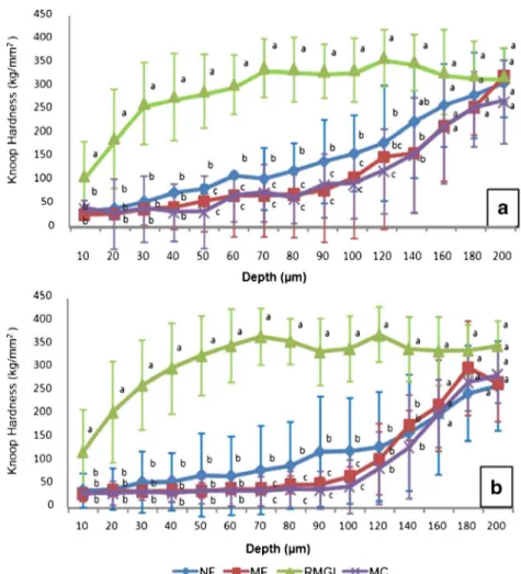
ΔS related to RMGI group was statistically lower (p<0.05) in relation to all groups. The findings show that there was a trend to lower demineralization of enamel around brackets fixed by the FN group comparing to FM and MC groups, but the difference among them was not statistically significant (Table 2). The microhardness data at each depth from the enamel surface are expressed in Fig.2.
SEM/EDX analysis
In the EDX analysis, the main elements observed in the enamel were calcium (Ca), phosphorus (P), oxygen (O), and fluorine (F). Minor quantities of other elements: sodium (Na), magnesium (Mg), chlorate (Cl), silica (Si), and carbon (C) were also observed in the present study. Elemental mappings showed calcium and phosphorus primarily detected, while fluorine and oxygen were identified in reduced amounts. Means and standard deviation of percentage of elemental concentration in weight (weight percent) detected in the sam-ples are summarized in Fig.3. A quantitative element analysis revealed no statistically significant differences in Ca, P, and O
content among the groups (p>0.05); exception to F, which presented a significantly higher amount in RMGI group.
Discussion
The incorporation of nanoparticles in resin material may allow the production of materials for the adhesive fixation of brackets with better flowability and higher mechanical prop-erties than for microfilled materials. Also, this process could alter characteristics of the ion-releasing materials [17,26]. The in vitro study mainly showed that the fluoride-releasing com-posite containing nanoparticles for bonding orthodontics brackets exert similar performance of the microfilled compos-ite in relation to fluoride-release and anticaries effect. This showed that nanofiller incorporation did not improve the fluoride release and consequently did not alter the anticaries potential of this material.
In the present study, the microfilled control material used in this study (natural ortho) was chosen since it presents similar composition to microfilled control material refereed in previ-ous orthodontic studies (Transbond XT). Besides, although microradiography is considered as gold standard method for determining mineral loss, microhardness analysis was used due to its great correlation demonstrated by excellent linear relation between square root of KHN and the mineral volume percent [27]. Additionally, microhardness presents sensitivity to detect the demineralization on enamel subsurface areas as the artificially caries lesions produced in the present study. It is important to remember that even though polishing procedures change natural enamel surface, roughness is important for the exact hardness measurement being a requirement for performing surface microhardness analysis, which was carried out for standardization and selection of the samples. Thus, polishing procedures were submitted to all samples, conse-quently applying this variable to all groups not interfering in the present results.
According to KHN data, enamel demineralization in vitro was inhibited to a certain degree in our study. Among the tested materials, the control group RMGI exhibited signifi-cantly higher performance in all performed analysis compared to the other tested materials. In agreement with other studies [28,29], our results have demonstrated that resin-modified glass ionomer materials showed a fluoride-releasing capacity sufficient to promote lower enamel mineral loss over the tested composite resin materials [30]. This was an expected outcome since the total amount of fluoride released differs significantly between the resin-modified glass ionomer ce-ments and the composite resins. It has been suggested that this superior performance of RMGI is attributed to some factors as the acid–base setting reaction between the fluoride-containing aluminosilicate glass powder base and the polyacid liquid results in the liberation of fluoride ions
Table 2 Mean and standard deviations of fluoride concentration (micro-gram per (micro-gram wet biofilm) and demineralization at studied distances for each tested material
Group F (μg/g wet biofilm)
∆S at distance from bracket base
20μm 70μm
MC (n=10) 0.27(0.08)b 5,320.44(1,241.06)a 4,661.94(1,615.80)a FM (n=9) 0.91(0.56)b 5,204.19(1,761.33)a 4,573.67(122.77)a RMGI (n=8) 10.50(4.20)a 876.55(440.80)b 1,392.37(916.74)b FN (n=10) 1.48(1.24)b 4,017.63(1,872.54)a 4,228.56(1,990.69)a
The same superscript letter indicate no statistically significant difference between the indicated groups (p> 0.05)
[29]. In poly-salt matrix, fluoride ions can be bound in strong complexes with the metal ions, especially aluminum and the pore liquid, in which the fluoride ions are loosely bound and free to move [31].
In our results, KHN for 50–120 μm in depth at both analyzed distances showed significant difference for FN
group expressing a trend to better performance of fluoride-releasing nanofilled composite than microfilled materials. The ΔS and fluoride concentration data from FN group also sup-ported this condition; however, they were not significantly different. Only few previous studies investigated fluoride release from nanomaterials. In a previous study, Kusgöz
Fig. 2 Mean ± standard deviation of the mean values (n=10) of enamel Knoop hardness according to the materials and depth of analysis at 20μm (a) and at 70μm (b) from the edge of the bracket base.Small letters indicate statistically significant difference between groups at each distance (p<0.05)
et al. [32]. compared fluoride release of a nanofilled resin-based fissure sealant and an unfilled resin-resin-based fissure seal-ant. The authors observed that the nanofilled sealant released a bit more fluoride than the unfilled sealant, but these results were not significantly different. These finding are consistent with results of the present study, showing that there is no significant influence on ion fluoride release, but it was able to show higher values than microfilled materials. It can be hypothesized that nanoparticles present in resin matrix of commercial materials can be related to this situation since the studied fluoride-releasing nanofilled composite presents 100 % of its reinforcement particles (silicon dioxide) in nanoscale.
On the other hand, Xu et al. [33]. showed that a nanocom-posite containing calcium fluoride (CaF2) had a higher fluoride
release capacity when compared to the control. This fact may be related to the nano-CaF2surface area, which presented a
surface area nearly 20-fold higher surface area than a tradition-al 1μm CaF2. This clearly suggests that the size of particles
from fluoride source itself presents more relevance for an anticaries effect than the simple presence of nanoparticles in resin matrix.
Fluoride release from composite resin materials is based on the diffusion process where initial leaching of fluoride from glass particles in the surface layer of the material may account for this mechanism. Fluoride ions diffuse through fluid and plaque from the composite to the adjacent enamel surface [34]. The SEM/EDX analysis explored the possibility of different ion diffusion rates from fluoride-containing nanomaterials to the enamel adjacent to bonded bracket and consequently alteration in chemical composition of enamel around the tested materials. The amounts (weight percent) of fluoride found for both fluoride-releasing composite resins, regardless of the presence of nanoparticles, were very low or undetectable. Although other methods might be used to deter-mine firmly bound F formed in enamel such as removing an enamel layer by acid action, the SEM/EDX analysis currently used may be considered reproducible since similar results were found with fluoride-releasing composite resins by Gjorgievska et al. [18] in a study that aimed to determine the extent to which ions released from fluoride-containing dental restorative materials migrated through the enamel. Besides, the use of acid to determine firmly bound F in enamel could introduce bias in microhardness analysis due to demineraliza-tion justifying the use of the nondestructible SEM/EDX anal-ysis. In relation to the calcium and phosphorus content in the surface enamel around the brackets, the values were similar in all groups showing that no material was able to promote a more significant diffusion of ions in the enamel surface.
Overall, the present evidences that RMGI present anticaries effect are based onΔS data obtained by microhardness anal-ysis. This way, our results showed that under the conditions of this investigation, only RMGI was capable of decreasing
demineralization around orthodontic brackets and the inclu-sion of nanofillers fluoridated composite resin did not provide greater inhibition of demineralization.
Future research should focus in applying the nanotechnol-ogy to preventive dentistry in attempt to improve fluoride-releasing capacity of dental materials. Until then, oral hygiene and other regular fluoride sources should be suggested for the orthodontic patient.
Acknowledgments The authors gratefully thank José Júnior da Alves da Silva for his assistance with SEM/EDX analysis and Institute of Research, Development and Innovation (IPDI) for providing the SEM equipment. The first and second authors have received scholarships during this study from CNPq (#141791/2010-1) and CNPq/PIBIC/ UFC, respectively.
Conflict Of Interest Statement None declared.
References
1. Mitchell L (1992) Decalcification during orthodontic treatment with fixed appliances—an overview. Br J Orthod 19:199–205
2. O’Reilly MM, Featherstone JDB (1987) Demineralization and remineralization around orthodontic appliances: an in vivo study. Am J Orthod Dentofacial Orthop 92:33–40
3. Demling A, Elter C, Heidenblut T, Bach FW, Hahn A, Schwestka-Polly R, Stiesch M, Heuer W (2010) Reduction of biofilm on ortho-dontic brackets with the use of a polytetrafluoroethylene coating. Eur J Orthod 32(4):414–418
4. Corbett JA, Brown LR, Keene HJ, Horton IM (1981) Comparison of Streptococcus mutansconcentrations in non-banded and banded orthodontic patients. J Dent Res 60(12):1936–1942
5. Al Mulla AH, Al Kharsa S, Kjellberg H, Birkhed D (2009) Caries risk profiles in orthodontic patients at follow-up using cariogram. Angle Orthod 79(2):323–330
6. Derks A, Kuijpers-Jagtman AM, Frencken JE, Van't Hof MA, Katsaros C (2007) Caries preventive measures used in orthodontic practices: an evidence-based decision. Am J Orthod Dentofacial Orthop 132(2):165–170
7. Benson PE, Parkin N, Millett DT, Dyer FE, Vine S, Shah A (2004) Fluorides for the prevention of white spots on teeth during fixed brace treatment. Cochrane Database Syst Rev. (3):CD003809.
8. Cacciafesta V, Sfondrini MF, Tagliani P, Klersy C (2007) In vitro fluoride release rates from 9 orthodontic bonding adhesives. Am J Orthod Dentofacial Orthop 132(5):656–662
9. Mystkowska J, Marczuk-Kolada G, Leszczyńska K, Ryszard Dąbrowski J, KaraśJ (2009) Fluoride release and antibacterial activ-ity of self-made composite materials for dental fillings. Solid State Phenomena 147–149:801–806
10. Chin MYH, Sandham A, Rumachik EN, Ruben JL, Huysmans M-CDNJM (2009) Fluoride release and cariostatic potential of ortho-dontic adhesives with and without daily fluoride rinsing. Am J Orthod Dentofacial Orthop 136:547–553
11. Rogers S, Chadwick B, Treasure E (2010) Fluoride-containing or-thodontic adhesives and decalcification in patients with fixed appli-ances: a systematic review. Am J Orthod Dentofacial Orthop 4:390.e1–8, discussion 390–1
13. Passalini P et al (2010) Mechanical properties of one and two-step fluoridated orthodontic resins submitted to different pH cycling re-gimes. Braz Oral Res 24(2):197–203
14. Chen MH (2010) Update on dental nanocomposites. J Dent Res 89(6):549–560
15. Sumita BM, Wu D, Brian NH (2003) An application of nanotech-nology in advanced dental material. J Am Dent Assoc 134:1382– 1390
16. Kumar PS, Savadi SKRC (2011) Jins John nanodentistry: a paradigm shift—from fiction to reality. J Indian Prosthodont Soc 11(1):1–6 17. Wiegand A, Buchalla W, Attin T (2007) Review on
fluoride-releasing restorative materials-fluoride release and uptake character-istics, antibacterial activity and influence on caries formation. Dent Mater 23(3):343–362
18. Gjorgievska E, Nicholson JW, Grcev AT (2012) Ion migration from fluoride-releasing dental restorative materials into dental hard tissues. J Mater Sci Mater Med 23(7):1811–1821
19. Xu HHK, Moreau JL, Sun L, Chow LC (2008) Strength and fluoride release characteristics of a calcium fluoride based dental nanocom-posite. Biomaterials 29:4261–4267
20. Asmussen E, Peutzfeldt A (2002) Long-term fluoride release from a glass ionomer cement, a compomer, and from experimental compos-ite resins. Acta Odontol Scand 60:93–97
21. Zanin IC, Lobo MM, Rodrigues LK, Pimenta LA, Höfling JF, Gonçalves RB (2006) Photosensitization of in vitro biofilms by toluidine blue O combined with a light-emitting diode. Eur J Oral Sci 1:64–69
22. Melo MAS, Paula DM, Lima JPM, Borges FMC, Steiner-Oliveira C, Nobre-dos-Santos M, Zanin ICJ, Barros EB, Rodrigues LKA (2010) In vivo photodynamic antimicrobial chemotherapy in dentine con-taminated by cariogenic bacteria. Laser Phys 20(6):1504–1513 23. Cury JA, do Amaral RC, Tenuta LMA, Del Bel Cury AA, Tabchoury
CPM (2010) Low fluoride toothpaste and deciduous enamel demin-eralization under biofilm accumulation and sucrose exposure. Eur J Oral Sci 118:370–375
24. Tenuta LM, Zamataro CB, Del Bel Cury AA, Tabchoury CP, Cury JA (2009) Mechanism of fluoride dentifrice effect on enamel deminer-alization. Caries Res 43(4):278–285
25. Sousa RP, Zanin IC, Lima JP, Vasconcelos SM, Melo MA, Beltrão HC, Rodrigues LK (2009) In situ effects of restorative materials on dental biofilm and enamel demineralisation. J Dent 37(1):44–51 26. Mitra SB, Dong WU, Holmes BN (2003) An application of
nano-technology in advanced dental materials. J Am Dent Assoc 134:1382–1390
27. Kielbassa AM, Wrbas KT, Schulte-Mönting J, Hellwig E (1999) Correlation of transversal microradiography and microhardness on in situ-induced demineralization in irradiated and nonirradiated hu-man dental enamel. Arch Oral Biol 44(3):243–251
28. Ahn SJ, Lee SJ, Lee DY, Lim BS (2011) Effects of different fluoride recharging protocols on fluoride ion release from various orthodontic adhesives. J Dent 39(3):196–201
29. Tay WM, Braden M (1988) Fluoride ion diffusion from polyalkenoate (glass-ionomer) cements. Biomaterials 9:454–456
30. Baliga MS, Bhat SS (2010) Effect of fluorides from various restor-ative materials on remineralization of adjacent tooth: an in vivo study. J Indian Soc Pedod Prev Dent 28(2):84–90
31. Verbeeck RM, De Maeyer EA, Marks LA, De Moor RJ, De Witte AM, Trimpeneers LM (1998) Fluoride release process of (resin-modified) glass-ionomer cements versus (polyacid-(resin-modified) com-posite resins. Biomaterials 19(6):509–519
32. Kuşgöz A, Tüzüner T, Ulker M, Kemer B, Saray O (2010) Conversion degree, microhardness, microleakage and fluoride re-lease of different fissure sealants. J Mech Behav Biomed Mater 3(8):594–599
33. Xu HH, Moreau JL, Sun L, Chow LC (2010) Novel CaF(2) nano-composite with high strength and fluoride ion release. J Dent Res 89(7):739–745