Side-Effects of Irinotecan (CPT-11), the Clinically
Used Drug for Colon Cancer Therapy, Are
Eliminated in Experimental Animals Treated with
Latex Proteins from
Calotropis procera
(Apocynaceae)
‡
Nylane Maria Nunes de Alencar,1* Flávio da Silveira Bitencourt,1
Ingrid Samantha Tavares de Figueiredo,1Patrícia Bastos Luz,1Roberto César P. Lima-Júnior,1 Karoline Sabóia Aragão,1Pedro Jorge Caldas Magalhães,1Gerly Anne de Castro Brito,2 Ronaldo Albuquerque Ribeiro,1†Ana Paula Fragoso de Freitas1and Marcio Viana Ramos3 1Departamento de Fisiologia e Farmacologia/UFC, Coronel Nunes de Melo, 1127 Rodolfo Teófilo, 60430-270, Ceará, Brazil 2Departamento de Morfologia/UFC, Delmiro de Farias S/N, Rodolfo Teófilo, 60416030, Ceará, Brazil
3Departamento de Bioquímica e Biologia Molecular/UFC, Campus do Pici, Caixa Postal 6033, 60451-970, Ceará, Brazil
Intestinal mucositis (IM) is the critical side effect of irinotecan (CPT-11), which is the front-line drug used for the treatment of colorectal cancer. This study aimed to evaluate the effectiveness of latex proteins (LP) from
Calotropis procerato prevent IM and diarrhea in animals. Swiss mice were treated daily with saline or LP (1, 5, or 50 mg/kg, i.v.) 24 h prior to CTP-11 (75 mg/kg/4 days, i.p) and for additional 6 days. Animal survival, body weight variation, and diarrhea were registered. After animal sacrifice (day 7 post first injection of CPT-11), intestinal samples were collected to study morphology and inflammatory parameters. Animals given LP exhibited improved parameters (survival, body weight, and absence of diarrhea) as compared with the CPT-11 control. The severity of IM observed in animals given CPT-11 was reduced in animals treated with LP. Treatment with LP also prevented the reduction in the villus/crypt ratio promoted by CPT-11. The rise in MPO activity and pro-inflammatory cytokines, over-contractility of the smooth muscle, and diarrhea were all abrogated in LP-treated mice. Markedly reduced immunostaining intensity for COX-2, TNF-α, IL-1β, iNOS, and NF-κB was observed in the intestinal tissue of animals treated with LP. The side-effects of CPT-11 were eliminated by LP treatment in experimental animals and improved clinical parameters characteristic of IM All known biochemical pathogenesis. Copyright © 2016 John Wiley & Sons, Ltd.
Keywords: Calotropis procera; cytokines; irinotecan; latex proteins; mucositis.
INTRODUCTION
Drug resistance and the harmful side-effects observed with therapy are generally the main concerns associated with medicines. Both of these issues prevent adequate clinical progress in patients and thus are the main motivating causes of treatment cessation. In an effort to mitigate these side-effects, the use of natural medicines as alternatives to the medicine-based clinical protocols has been encouraged. This study addresses the management of intestinal mucositis (IM) and diarrhea associated with colorectal therapy using CPT-11. A spectrum of signs and symptoms compromises the life quality of patients undergoing cancer therapy. Among them, mucositis is recognized worldwide as a challenge to be handled. Different types of human malignancies, such as advanced metastatic colorectal
cancer, are clinically managed with a potent DNA
topoisomerase I inhibitor known as irinotecan
(CPT-11: 7-ethyl-10[4-[1-piperidino-1-piperidino]
carbonyl-oxycamptothecin). CPT-11 is metabolized vis
carboxylesterase-mediated hydrolysis in the liver and converted to SN-38, the active metabolite (Hebbar
et al., 2009). Treatment based on CPT-11 chemotherapy results in harmful side-effects in patients. CPT-11 induces IM, which is characterized by an inflammatory and ulcerative process of the mucous membranes lining the digestive tract (Sonis, 2004). This status is clinically characterized by abdominal pain, vomiting, and persistent diarrhea, with secondary anti-nutritional consequences and weight loss. These effects are associated with intense IM resulting from SN-38 activity
(Petersonet al., 2011).
The pharmacological potential of latex compounds
has been progressively recognized and globally
validated (Samy et al., 2012). Particularly, the
latex-producing plant Calotropis procera (Apocynaceae),
which is part of the Ayurveda, the Indian natural medi-cal compendium, has had some of its claimed activities
confirmed scientifically (Chaudhary et al., 2015).
Further isolation of latex fractions and discarding the
* Correspondence to: Nylane Maria Nunes de Alencar, Departamento de Fisiologia e Farmacologia/UFC, Coronel Nunes de Melo, 1127 Rodolfo Teófilo, 60430-270 Ceará, Brazil.
E-mail: [email protected]
†
in memoriam
‡Correction added on 21 December 2016, after first online publication on
02 December 2016. Figure 5 has been corrected.
Published online 2 December 2016 in Wiley Online Library (wileyonlinelibrary.com)DOI: 10.1002/ptr.5752
insoluble material (rubber) provides organic and protein fractions, which are both sources of pharmaco-logically active molecules. For instance, secondary metabolites and some of their chemical derivatives have been highlighted as promising anticancer drugs (Van
Quaquebeke et al., 2005). Complementarily, the
involvement of LP in the modulation of some inflamma-tory disorders is now well-established and partially
characterized (Chaudhary et al., 2015). Thus, we
examined the ability of the LP ofC. procerato prevent
oral mucositis induced in 5-fluorouracil treated animals, because LP have been proposed to down-regulate inflammatory mediators and to reestablish homeostasis
in inflamed tissues (Freitaset al., 2012). In the present
study, LP was assessed for its ability to eliminate the adverse clinical signs commonly observed in patients undergoing CPT-11 treatment. For this, experimental animals were used, and a panel of parameters were recorded and evaluated. The results reported in this study show that LP is a strong candidate for phytotherapy.
MATERIAL AND METHODS
Plant material and latex processing.The fresh latex was collected in tubes (1:1, v/v in distilled water) from the
aerial parts of C. procera (Ait.) R.Br. growing in the
vicinity of Fortaleza, Brazil in July 2012. The plant voucher (sample specimen no. 32663) was deposited at Prisco Bezerra Herbarium of Federal University of Ceará, Brazil, where the botanical material was identi-fied by a local taxonomist. Latex/water mixtures were adjusted to a dilution ratio of 1:2 (v/v) and further
proc-essed (Freitaset al., 2012). Water soluble LP used in this
study was lyophilized and stored at 25 °C until use. All known biochemical and pharmacological characteriza-tions of this sample have been extensively reported
(Freitaset al., 2012; Oliveiraet al., 2010). Briefly,
cyste-ine proteases, chitinases, peroxidases, and osmotin have been identified as the major proteins present in LP.
Induction of intestinal mucositis.Adult male Swiss mice (Mus musculus), weighing 233.0 g, were obtained from the Central Animal House of the Federal Univer-sity of Ceará, Brazil. The animals were housed in plastic cages under controlled laboratory conditions
(tempera-ture of 25 °C, humidity 5510% and 12/12 h light/dark
cycles) and provided water and food ad libitum
(commercial sterile diet; Purina, Paulínia, SP, Brazil). All animal experiments were conducted in accordance with the Guide for the Care and Use of Laboratory Animals of the National Institute of Health (NIH 1985), and were approved by the ethical committee of the Federal University of Ceará (protocol n°24/2009). The experimental protocol was based on a previously
described model (Ikuno et al., 1995) and adapted for
the experimental conditions. Irinotecan hydrochloride (CPT-11) (irinotecan, Camptosar®, Pharmacia and Upjohn Co, Kalamazoo, MI, USA, 100-mg ampoule) was administered intraperitonally (i.p.) to healthy animals at a dose of 75 mg/kg, once a day for 4 days.
Experimental design. The animals were randomly
ar-ranged into five experimental groups (n= 8 animals/per
group), as follows: (i) normal group: animals treated
only with sterile saline (5 mL/kg, i.p.); (ii) CPT-11 group: animals treated with saline (5 mL/kg, i.p.) and 24 h later given CTP-11, as described above; and (iii) LP groups: animals treated (i.v.) with the laticifer proteins of
C. procera (LP), dissolved in saline (vehicle), at doses of 1, 5, or 50 mg/kg 24 h prior to initiating CTP-11 treatment and continuing for six consecutive days (single daily dose). The schedule of administration of irinotecan or LP and the evaluated parameters are summarized in Fig. 1. For all experiments, data were recorded from eight animals per group. Reproducibility was confirmed by three independent experiments.
All efforts were made in order to minimize animal suffering. In the survival study, the animals were moni-tored twice daily for 10 days following the first injection of irinotecan. During the experiments, the animals succumb because of the treatment and its consequences, including diarrhea. Those animals that showed signs of
imminent death, including piloerection, reduced
locomotion, inability to maintain upright position, ataxia, tremor, and altered breath frequency were sacrificed by
ketamine/xylazine overdose (>100/10 mg/kg, s.c., União
Química, São Paulo, Brazil) followed by cervical dislocation. Pain relievers or anesthesia were not used in our experiments because those agents directly interfere with the production of inflammatory mediators and/or alter the gastrointestinal transit and mask the diarrheic events in this animal model. At the end of the survival experiment, live animals were sacrificed by
ketamine/xylazine overdose (>100/10 mg/kg, s.c., União
Química, São Paulo, Brazil) followed by cervical dislocation. The experimental protocol, including the mortality aspects of the protocol, was reviewed and approved by the Committee on the Ethics of Animal Experiments of the Federal University of Ceará.
Animals from all experimental groups were sacrificed by cervical dislocation on experimental day 7 after CPT-11 exposure. The proximal intestine (duodenum) of each was harvested, and tissue samples were immediately subjected to morphometric,
immuno-histochemistry analysis, or functional studies (in vitro
contractility of the isolated duodenum). In other
experiments, the proximal intestines were harvested,
weighed, and stored at 70 °C until required for the
myeloperoxidase assay (MPO), measurement of
cytokines (TNF-α and IL-1β), or assayed by Western
blot for iNOS.
Clinical parameters. Animals from all experimental groups were observed daily for mortality, from the beginning of CPT-11 treatment and continuing for 12 days. The results are expressed as the percentage of survival. Throughout the experimental period, the animals were monitored for body weight loss and the severity of diarrhea. Each mouse was weighed at the beginning of the experiment, before treatment and daily throughout the experiment. The values are expressed as the percentage of weight loss and compared to the initial weight. On experimental day 7 after CPT-11 exposure, prior to animal euthanization, the severity of the
diarrhea was scored (Kurita et al., 2000). All
comparative scoring measures were done using a blinded method to prevent observer bias.
Histopathological analysis.The specimens were fixed in 10% (v/v) neutral-buffered formalin, dehydrated, and embedded in paraffin. The samples were cut into 5-μm
sections, stained with hematoxylin–eosin (H&E) and
examined by light microscopy in a blinded manner. All histopathological analyses were performed using random fields per slice. The severity of mucositis was graded using previously described criteria (Woo
et al., 2000). The morphometric study was performed using ImageJ®1.36b software (National Institutes of Health, USA). The height of the villus was deter-mined from the top to the bottom, which corresponds to the crypt/villus junction. The depth of the crypts was defined as the invagination between adjacent villi. An average of 5 to 10 different intestinal crypt/villus measurements per experimental group was taken in the histological section, and the villus-to-crypt length ratios were calculated.
In vitro contractility of duodenum. Duodenum segments (0.6 cm long) were dissected and processed
to investigate in vitro contractility according to the
method of Araújoet al. (2005). A dose–response curve
to the cholinergic agonist acetylcholine was constructed, using increasing and cumulative concentrations ranging
from 10 10 to 10 4M. The data obtained from the
cholinergic dose–response curve were analyzed as
the percentage contractile response in comparison with the mean of two contractions initially observed using the 60 mM KCl standard.
Myeloperoxidase assay (MPO).The intensity of neutro-phil accumulation in intestinal samples was indirectly estimated by the measurement of MPO activity through
colorimetric analysis (Bradleyet al., 1982). The changes
in absorbance were recorded and plotted on a standard curve of neutrophil density. Data were expressed as myeloperoxidase activity (neutrophils/mg of tissue).
Detection of cytokines (TNF-α, IL-1β) in the
duode-num.The tissue was homogenized and processed as
pre-viously described (Safieh-Garabedianet al., 1995). The
measurement of TNF-α and IL-1β concentrations was
performed using murine TNF-α and IL-1β DuoSet®
ELISA Kits (R&D Systems, Inc., Minneapolis, USA). The steps were carried out according to the
manufac-turer’s instructions.
Immunohistochemistry. Detection of TNF-α, IL-1β, iNOS, COX-2, and NF-κB was developed using the
streptavidin–biotin–peroxidase method in
formalin-fixed, paraffin-embedded tissue sections (4μm thick),
mounted on poly-L-lysine-coated microscope slides (Hsu et al., 1981). Primary antibodies against TNF-α (Sigma), IL-1β, iNOS, COX-2, or nuclear localization sequence (NLS) of the NF-kB p50 subunit (Santa Cruz)
were used at a 1:200 dilution in PBS–BSA. The avidin–
biotin–horseradish peroxidase conjugate (Strep ABC
complex; Santa Cruz Biotechnology) was used
accord-ing to the manufacturer’s protocol. The reaction was
visualized with the chromogen 3,3 diaminobenzidine (DAB; liquid DAB + substrate chromogen system;
Dako). Slides were counterstained with Mayer’s
hematoxylin (Invision Flex-Dako) or methyl green (for NF-κB). A qualitative evaluation using light microscopy
was performed as previously described (Yeohet al., 2005).
Data analyses. Data are expressed as meanstandard error of the mean (SEM) or median (non-parametric data) of at least three independent observations. Analysis
of variance (ANOVA), followed by Bonferroni’s test, was
used to compare the means, and the Kruskal–Wallis test,
followed by Dunn’s test, was used to compare medians.
Statistical significance was accepted when P<0.05. All
data were analyzed using GraphPad Prism software version 5.0 (Graph-Pad Software, Inc., La Jolla, CA, USA).
RESULTS
LP improves the clinical parameters of intestinal mucositis in mice
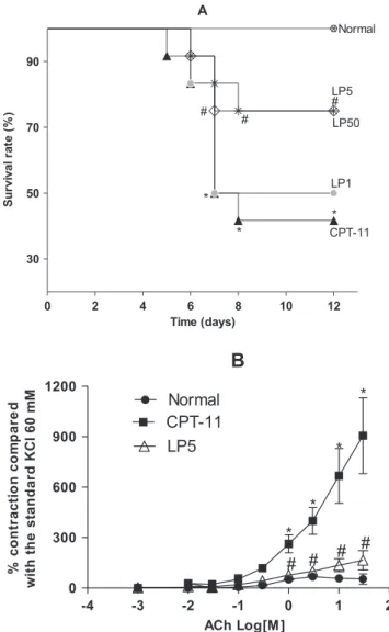
Fig. 2A shows increasing mortality over time in animals given CPT-11. The survival of animals in this group reached a minimum (41.67%) at day 12. As expected, the survival rate in untreated animals (normal control) was 100%. Therefore, the vehicle used to dissolve the latex did not affect the clinical parameters. The perfor-mance of animals given CPT-11 with LP at 1 mg/kg was quite similar to that documented in the CPT-11 group. However, the groups given CPT-11 as a preventative treatment with LP at 5 or 50 mg/kg statistically differed from the CPT-11 group; the survival rate in these groups was above 70%. Animals in the normal group progres-sively gained body weight until day 12. No significant differences were observed between the groups given CPT-11 and CPT-11 followed by LP treatment in terms of body weight (data not shown). Table 1 presents the scores according to the occurrence and intensity of diarrhea in animals. As observed, the data are coherent with those shown in Fig. 2A. While animals receiving LP at the lower concentration almost replicated the
performance of animals given CPT-11 (p<0.05),
exhibiting strong and persistent diarrhea, this condition was mitigated in animals given CPT-11 but treated with LP at the higher concentrations, differing from CTP-11
group (p<0.05) and being similar to the normal
Damage to the intestinal mucosa induced by CPT-11 is prevented by LP treatment
As shown in Table 2 and Fig. 3, the use of CPT-11 induced severe injury to the intestinal mucosa of animals. The intense diarrhea observed in the animals
of this group is thought to be, at least in part, a conse-quence of this histological damage. The full integrity of the intestinal mucosa observed in animals in the control group was confirmed. The parameters used to assess the intestinal mucosa of animals treated with LP at 1 mg/kg group were statistically similar to those in the group given CPT-11 alone. Of note, the histological integrity of the intestinal mucosa of animals given CPT-11 and treated with LP at 5 or 50 mg/kg was preserved and comparable to that of animals given only sterile saline. Further, the histological integrity of the mucosa in animals given LP was probably the main factor driving
the observed homeostasis in the in vitro contractility
assays. The data in Fig. 2B show that the responsiveness of the intestinal mucosa of animals from the normal control and the group given CPT-11 followed by LP treatment were both statistically different to that of animals receiving only CPT-11. These findings also corroborate the important reduction in diarrhea in these groups.
LP modulates pro-inflammatory mediators in CTP-11-induced mucositis
Fig. 4A shows the differences in MPO activity observed in the duodenal tissue of animals. The inflammatory process resulting from irinotecan delivery was indicated by increasing levels of myeloperoxidase measured in the duodenum (Fig. 4A). MPO activity indicates the presence of activated neutrophils, found only in inflamed tissues. The inflammatory response stimulated by CPT-11 was reduced in animals given CPT-11 and treated with LP at 5 or 50 mg/kg, that is, by 83% and 77.21%, respectively. This is the first experimental evidence that the beneficial effects of LP treatment on animals given CPT-11 may be because of its well-documented ability to modulate inflammatory pro-cesses. This observation was supported by reductions in the levels of pro-inflammatory mediators, that is,
TNF-α (Fig. 4B) and IL-1β (Fig. 4C), as well as the
decrease in immunostaining for both markers (Fig. 4D) in intestinal tissues of animals given CPT-11 and treated with LP.
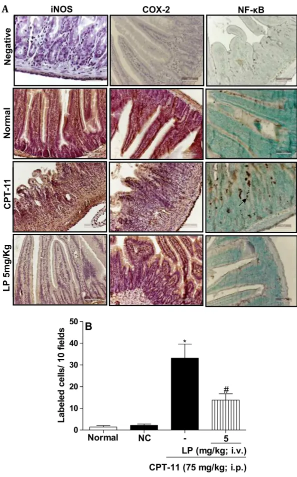
Reduced immunohistochemical detection of iNOS and COX-2 in LP-treated animals
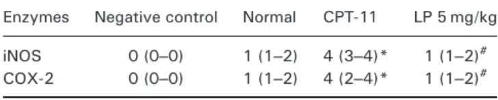
Fig. 5A provides images of the intestinal tissues of ani-mals given CPT-11 and aniani-mals given CPT-11 followed by treatment with LP. Table 3 presents the histological scores in each group. Immunohistochemical detection of both iNOS and COX-2 were statistically comparable to the control and different to that in the group given CPT-11 alone. The increase in iNOS and COX-2 detected in the duodenal tissues of animals given only CPT-11 indicates an inflammatory process in progress. The reduced detection of iNOS accompanied by previ-ous observation that fewer activated neutrophils were found in animals given CPT-11 followed LP treatment
(western blot analysis also suggested this profile—data
not shown). However, the reduced detection of COX-2 in animals given CPT-11 followed by LP treatment as compared to the level of COX-2 in samples from
Figure 2. Treatment with LP improved the survival rate of mice with intestinal mucositis (A). Values are expressed as percentage of the median from three independent observations. *p<0.05 versus normal group and#p<0.05 versus CPT-11 group; (n= 8 animals per group; Mantel–Cox log rank test). Inhibitory effect of LP on mice duodenal contractilityin vitro(B). Values are expressed as meanstandard error mean (S.E.M.) from three independent determinations.*p<0.05 versus normal group and#p<0.05 versus CPT-11 group (n= 8 animals per group; ANOVA—Bonferroni test).
Table 1. The severity of irinotecan-induced diarrhea is alleviated in LP-treated mice. Values are expressed as medians
Group Diarrhea assessment scores
Normal 0 (0–0)
CPT-11 3 (2–3)*
LP—1 mg/kg 2.5 (1–3)*
LP—5 mg/kg 1 (0–2)#
LP—50 mg/kg 1 (0–2)#
*p<0.05versus the normal group.
#
p<0.05versus the CPT-11 group.
animals given CPT-11 only provides evidence of the ability of LP to robustly modulate CPT-11-induced IM.
NF-κB expression is stimulated in animals given CPT-11 but down-regulated by LP treatment
Immunostaining for NF-κB in tissues collected from an-imals given only CPT-11 was increased by almost 10-fold. The NLS of the NF-kB p50 subunit is markedly in-creased in CPT-11-injected mice and attenuated in LP5-treated animals. This increase was statistically reduced by half in animals given CPT-11 and treated with LP (Fig. 5A, B). The variation in the immunoexpression for each inflammatory marker is to be mainly observed
in the submucosal layer. In the study of Collet et al.
(2008) early epithelial changes associated with altered responsiveness to bacteria precede increased permeabil-ity and mucosal inflammation in a model of colitis. Normal and damaged epithelial cells in the intestine express the studied markers, because the epithelial surface is continuously exposed to antigens from the
intestinal lumen (Collett et al., 2008). However, during
mucositis the submucosa is exposed to translocating bacteria and resident cells become activated by pathogen-associated molecular patterns and damage associated molecular patterns, which signal to induce
pro-inflammatory cytokine expression (Wong et al.,
2015; Lima-Júnioret al., 2014).
DISCUSSION
Persistent episodes of diarrhea constitute a central ad-verse concern of almost all patients undergoing chemo-therapy for malignancy. It must be kept in mind that the diarrhea resulting from chemotherapy protocols is clinically different of other etiological causes, such as infections (Gibson and Stringer, 2009). Therefore, the rationality of its clinical management is also different.
Late diarrhea provoked by use of CPT-11 is secretory, because of the reduced intestinal absorption of fluids, the presence of exudate components, and also because of the lack of specificity of the drug and its metabolically
active derivative, SN-38 (Ikuno et al., 1995). The
Table 2. Histopathological damage to the intestinal mucosa in mice is prevented by LP treatment in irinotecan-induced mucositis
Groups Histological grading Villus/crypt ratio Histological description
Normal 0 (0–0) 2.70.15# Healthy intestinal mucosa
CPT-11 4 (1–4)* 1.60.17* CPT-11 promoted intestinal crypt destruction, shortened villi (vertical arrows), and led to intense infiltration of inflammatory cells in the lamina propria (slanted arrows) and vacuolization of the epithelial cell lining (horizontal arrow).
LP 1 mg/kg 3.5 (1–4)* 1.80.21*
LP 5 mg/kg 1 (0–2)# 2.80.17# LP (5 and 50 mg/kg) prevented the histopathological damage induced by irinotecan (CPT-11) on the intestinal mucosa.
LP 50 mg/kg 1 (0–3)# 2.7
0.09#
*p<0.05 versus the normal group.
#
p<0.05 versus the CPT-11 group.
n= 8 animals per group; Kruskal–Wallis test followed by Dunn’s test or ANOVA followed by the Bonferroni test.
The values of histological grading are expressed as medians. The values of the villi/crypt ratio are expressed as meanstandard error of the mean (SEM). Magnification (× 100).
unspecific activity of currently used drugs, such as CPT-11, not only severely impairs the natural rate of cellular replacement, but also disrupts any regenerative process. This explains mucositis and anemia as the first impair-ments faced by patients treated with anticancer drugs. Bearing in mind this current scenario, even aggravated by the poor perspective regarding the immediate avail-ability of new and more specific medicinal drugs for the management of neoplasia and other malignancies,
the search for complementary and alternative
supporting protocols to current therapies is appreciated. For more than a decade, we have dedicated efforts to scientifically validate the folk use of the latex of
C. procera. In the beginning, we decided to clean the latex of organic soluble compounds in order to study the soluble protein fraction, because all available litera-ture at that time reported data on the whole dried latex or organic extracts obtained after extraction of the whole dried latex, including toxicology (Kumar and Shivkar, 2004).
In our initial studies, LP, the laticifer proteins, were shown to inhibit inflammation artificially induced by phlogistic agents such as carrageenan and dextran,
reducing peritonitis and paw edema (Alencar et al.,
2004). Later, we showed that LP could suppress tumor
growth (in vivo), prevent septic shock in lethally
infected animals (Oliveira et al., 2010), and treat
arthritic animals (Kumar et al., 2011). These activities
were accompanied by down-regulation of
pro-inflammatory cytokines and other inflammatory
mediators, the preservation of tissue architecture, and the maintenance of oxidative homeostasis (Chaudhary
et al., 2015). Even though LP has demonstrated important antiinflammatory features, it was still surprising to document the ability of LP to abolish oral
mucositis in animals subjected to 5-fluorouracil
treatment (Freitas et al., 2012). It was found that LP
inhibited the expression of iNOS, COX-2, and the
pro-inflammatory mediators IL-1β and TNF-α. These
pharmacological characteristics of LP and the later find-ings on oral mucositis were therefore the justification to investigate the presumed protective effect of LP on IM. In previous studies, Indian researchers evaluated the
effect of the dried latex (500 mg/kg) ofC.procerain rats
orally treated with castor oil (Kumar et al., 2001). The
main findings included a decrease in the frequency of
defecation and the prevention of castor oil-induced enteropooling. Thus, the present data, showing a reduction in diarrhea and the reduced responsiveness of the duodenum to stimulation with acetylcholine,
corroborate the study reported by our Indian
colleagues. It is also indicative that LP may have multiple independent activities, because it was able to retard defecation independently of IM, because castor oil does not induce inflammation. In the clinical setting, physicians use diarrhea as a clinical sign of IM. However, we have some evidence that the modulation of diarrhea with loperamide does not interfere with underlying inflammatory response associated with IM
(Lima-Júnioret al., 2012). In addition, the modulation
of inflammatory parameters not always prevents
diarrheic events (Wong et al., 2015). Those findings
suggest that the underlying mechanisms of diarrhea and IM might be partially different, although diarrhea is sometimes consequent to intestinal injury (Murray and Rubio-Tapia, 2012). Interestingly, LP prevented both diarrhea and intestinal injury. Thus, in the present study, LP may have played a dual role by
down-regulating key pro-inflammatory mediators and
inhibiting overexcited duodenal contractility driven by the effects of CPT-11.
The failure of LP to prevent body weight loss, a characteristic observed in animals given CPT-11, needs to be approached in a different way. In the present study, the duodenum was chosen for analyses because of the length of villus, which would allow us to quantify the extension of injury more easily, as also reported in
previous studies (Lima-Júnioret al., 2012). Even though
LP contributed to the preservation of intestinal tissue structure and down-regulated pro-inflammatory media-tors, the body weight progress in the LP groups was as bad as that in animals give. Even though LP contributed to the preservation of intestinal tissue structure and down-regulated pro-inflammatory mediators, the body weight progress in the LP groups was as bad as that in animals given CPT-11 alone. In other studies, the treat-ment of animals with glutamine and alanyl glutamine, aimed at protecting animals against IM induced by 5-fluorouracil, preserved tissue architecture but did not
prevent body weight loss (Carneiro-Filho et al., 2004).
These findings indicate the multifactorial negative effects associated with CPT-11 administration and are in line with the lack of specificity in the mechanism of action of this drug. Even if LP or other compounds can overcome IM, the broad effects of CPT-11 on general metabolism probably play a decisive role on the negative performance of animals in terms of weight gain. As a whole, IM and diarrhea seem to be only part of the harmful effects observed in animals given CPT-11,
and thus the loss of body weight observed in animals given CPT-11 even when treated with IM regenerative drugs suggests the high degree of metabolic catabolism promoted by CPT-11 and its toxic metabolites. For instance, the treatment of animals with thalidomide or pentoxifylline reduces the histopathological damage induced by CPT-11 in the intestinal mucosa. However,
only pentoxifylline reduces diarrhea (Meloet al., 2008).
In the present study, we used the expression of pro-inflammatory cytokines and enzymes as putative markers of intestinal inflammation associated with
irinotecan injection. It is expected that an
antiinflammatory drug is capable of attenuating the expression of most of these inflammatory markers, because the inflammatory reaction is orchestrated in a
sequence of events as a cascade (Lima-Júnior et al.,
2012). As described here, LP significantly attenuated the immunoexpression of these markers. The precise mechanism through which LP controls the inflammatory reaction is still a matter of debate. However, one possible explanation might involve the direct reduction of neutrophil influx to the inflammatory foci (Alencar
et al., 2004).
LP has been characterized in terms of its protein content and related activities. Although it is currently impossible to determine the role played by any latex protein (LP) on inflammation, potential proteins involved in the modulation of inflammatory processes have emerged. Chitinases, proteases, and osmotins are now recognized as the major proteins found in LP
(Ramos et al., 2010). To a lesser extent, anti-oxidant
enzymes are also found (Freitas et al., 2007). The
fractionation of chitinases and proteases plus osmotins
suggests that the antiinflammatory activity persists in
both fractions (Ramos et al., 2009). As far as we are
concerned, there are no data on chitinases having antiinflammatory effects. Also, the data associating pro-teases and the modulation of inflammatory processes are sparse. Recent literature, however, proposes an unex-pected effect of osmotins on inflammatory events (Freitas
et al., 2011). We have recently purified the latex osmotin of C. procera and characterized the protein in some
detail (Ramoset al., 2015). It is now our primary goal to
determine whether this protein plays any role on the mit-igation of the side-effects induced by CPT-11 treatment.
During IM, inflammatory cytokines induce tissue injury and mediate complications, such as diarrhea. The severity and maintenance of injuries are associated with high levels of cytokines such as IL-1β, IL-6, TNF-α, and IL-2 (Williams, 2001). Overall, the mechanistic events supporting the beneficial effects of LP observed in animals exposed to IM are indicated in the present
study. Accordingly, the inhibition of TNF-α and IL-1β
release was characteristic of LP treated animals.
According to Melo et al., 2008 on day 7 after
irinotecan-induced mucositis, high levels of TNF-α, IL-1β, and KC are involved with the pathogenesis and
the inflammatory response. A study by Freitas et al.
(2012) also demonstrated in 5-fluorouracil-induced oral mucositis that LP (5 mg/kg) was able to reduce the immunoreactivity for TNF-α, IL-1β, iNOS, and COX-2 in the oral mucosa of hamsters. In addition to the immu-nohistochemistry results, a suggestive reduction in iNOS expression by Western blot confirmed the involvement of nitric oxide in the antiinflammatory effects of LP in irinotecan-induced mucositis (data not shown).
Table 3. The immunoexpression of iNOS and COX-2 is attenuated in LP-treated mice subjected to irinotecan-induced intestinal mucositis
Enzymes Negative control Normal CPT-11 LP 5 mg/kg
iNOS 0 (0–0) 1 (1–2) 4 (3–4)* 1 (1–2)# COX-2 0 (0–0) 1 (1–2) 4 (2–4)* 1 (1–2)#
*p<0.05 versus the normal group.
#
In conclusion, our results demonstrate that the inflammatory response in irinotecan-induced mucositis
was modulated by the laticifer proteins of C. procera
(LP) in mice. The morphological alterations as well the functional and inflammatory disorders associated with mucositis were suppressed by treatment with LP. This study shows that proteins endogenously expressed in
the latex of the medicinal plant C.procera are able to
impair the establishment of IM in animals subjected to irinotecan treatment. The results of this study and the literature reporting the pharmacological properties of LP strongly support it as a reliable and promising material for the development of new phytotherapeutics.
Acknowledgements
The biochemical, functional, and applied studies of the latex of
C. procera are financed by grants from the following Brazilian agencies: Conselho Nacional de Desenvolvimento Científico e Tecnológico (CNPq: Universal/RENORBIO); Coordenação de Aperfeiçoamento de Pessoal de Nível Superior (CAPES), and Fundação Cearense de Apoio ao Desenvolvimento Científico e Tecnológico (FUNCAP). This study is part of the consortium ‘Molecular Biotechnology of Plant Latex’.
Conflict of Interest
The authors declare that there are no conflicts of interest.
REFERENCES
Alencar NMN, Figueredo IST, Vale MR,et al.2004. Anti-inflamma-tory effect of the latex from Calotropis procera in: three different experimental models: Peritonitis, Paw edema and Hemorrhagic cystitis.Planta Med7: 1144–1149.
Araújo PV, Clemente CM, da Graça JRV,et al.2005. Inhibitory effect of sildenafil on rat duodenal contractilityin vitro: putative cGMP involvement.Clin Exp Pharmacol Physiol32: 191–195. Bradley PP, Christensen RD, Rothstein G. 1982. Cellular and
extra-cellular myeloperoxidase in pyogenic inflammation.Blood60: 618–622.
Carneiro-filho BA, Oria RB, Wood RK,et al.2004. Alanyl-glutamine hastens morphologic recovery from 5-fluorouracil-induced mucositis in mice.Nutrition20: 934–941.
Chaudhary P, Viana CA, Ramos MV, Kumar VL. 2015. Antiedematogenic and antioxidant properties of high molecu-lar weight protein sub-fraction ofCalotropis proceralatex in rat.J Basic Clin Pharm6: 69–73.
Collett A, Higgs NB, Gironella M,et al.2008. Early molecular and functional changes in colonic epithelium that precede increased gut permeability during colitis development in mdr1a( / ) mice.Inflamm Bowel Dis14(5): 620–31. Freitas APF, Bitencourt FS, Brito GAC,et al.2012. Protein fraction
of Calotropis procera latex protects against 5-fluorouracil-induced oral mucositis associated with downregulation of pivotal pro-inflammatory mediators. Naunyn Schmiedebergs Arch Pharmacol10: 981–990.
Freitas CDT, Oliveira JS, Miranda MR, et al. 2007. Enzymatic activity and protein profile of latex ofCalotropis procera.Plant Physiol Biochem45: 781–789.
Freitas CDT, Nogueira FCS, Vasconcelos IM, Oliveira JTA, Domont GB, Ramos MV. 2011. Osmotin purified from the latex ofCalotropis procera: biochemical characterization, biological activity and role in plant defense.Plant Physiol Biochem49: 738–743. Gibson RJ, Stringer AM. 2009. Chemotherapy-induced diarrhoea.
Curr Opin Support Palliat Care3: 31–35.
Hebbar M, Ychou M, Ducreux M. 2009. Current place of high-dose irinotecan chemotherapy in patients with metastatic colorectal cancer.J Cancer Res Clin Oncol135: 749–752.
Hsu SM, Raine L, Protein A. 1981. Avidin, and biotin in immuno-histochemistry.J Histochem Cytochem29: 1349–1353. Ikuno N, Soda H, Watanabe M, Oka M. 1995. Irinotecan (CPT-11)
and characteristic mucosal changes in the mouse ileum and caecum.J Natl Cancer Inst87: 1876–1888.
Kumar S, Dewan S, Sangraula H, Kumar VL. 2001. Anti-diarrhoeal activity of the latex ofCalotropis procera.J Ethnopharmacol
76: 115–118.
Kumar VL, Shivkar YM. 2004.In vivoandin vitroeffect of latex of Calotropis procera on gastrointestinal smooth muscles. J Ethnopharmacol93: 377–379.
Kumar VL, Chaudhary P, Ramos MV,et al.2011. Protective effect of proteins derived from the latex ofCalotropis proceraagainst inflammatory hyperalgesia in monoarthritic rats. Phytother Res25: 1336–1341.
Kurita A, Kado S, Kaneda N, Onoue M, Hashimoto S, Yokokura T. 2000. Modified irinotecan hydrochloride (CPT-11) administra-tion schedule improves inducadministra-tion of delayed-onset diarrhea in rats.Cancer Chemother Pharmacol46: 211–220.
Lima-Júnior RCP, Figueiredo AA, Freitas HC,et al.2012. Involve-ment of nitric oxide on the pathogenesis of irinotecan-induced intestinal mucositis: role of cytokines on inducible
nitric oxide synthase activation.Cancer Chemother Pharmacol
69: 931–942.
Lima-Júnior RCP, Freitas HC, Wong DVT, et al. 2014. Targeted inhibition of IL-18 attenuates irinotecan-induced intestinal mucositis in mice.Br J Pharmacol171: 2335–2350. Melo MLP, Brito GAC, Soares RC,et al.2008. Role of cytokines
(TNF-α, IL-1βand KC) in the pathogenesis of CPT-11-induced intestinal mucositis in mice: effect of pentoxifylline and thalidomide.Cancer Chemother Pharmacol61: 775–784. Murray JA, Rubio-Tapia A. 2012. Diarrhoea due to small bowel
diseases.Best Pract Res Clin Gastroenterol26: 581–600. Oliveira JS, Costa-Lotufo LV, Bezerra DP, et al. 2010. In vivo
growth inhibition of sarcoma 180 by latex proteins from Calotropis procera. Naunyn Schmiedebergs Arch Pharmacol
382: 139–149.
Peterson DE, Bensadoun RJ, Roila F. 2011. Management of oral and gastrointestinal mucositis: ESMO clinical practice guidelines.Ann Oncol22: vi78–vi84.
Ramos MV, Grangeiro TB, Freire EA,et al.2010. The defensive role of latex in plants: detrimental effects on insects. Arthropod-Plant Interact4: 57–67.
Ramos MV, Oliveira JS, Figueiredo JG,et al.2009. Involvement of NO in the inhibitory effect ofCalotropis proceralatex protein fractions on leukocyte rolling, adhesion and infiltration in rat peritonitis model.J Ehtnopharmacol125: 387–392.
Ramos MV, Oliveira RSB, Pereira HM,et al.2015. Crystal structure of an antifungal osmotin-like protein fromCalotropis procera and its effects on Fusarium solani spores, as revealed by atomic force microscopy: Insights into the mechanism of action.Phytochemistry119: 5–18.
Safieh-Garabedian B, Poole S, Allchorne A, Winter J, Woolf CJ. 1995. Contribution of interleukin-1 beta to the inflammation-induced increase in nerve growth factor levels and inflamma-tory hyperalgesia.Br J Pharmacol115: 1265–1275. Samy RP, Rajendran P, Li F,et al.2012. Identification of a novel
Calotropis proceraprotein that can suppress tumor growth in breast cancer through the suppression of NF-κB pathway. PLoS One7: e48514.
Sonis ST. 2004. The pathobiology of mucositis.Nat Rev Cancer4: 277–284.
Van Quaquebeke E, Simon G, Dewelle AJ,et al.2005. Identifica-tion of a novel cardenolide (2″-Oxovoruscharin) fromCalotropis proceraand the hemisynthesis of novel derivatives displaying potentin vitroantitumor activities and highin vivotolerance: structure–activity relationship analyses. J Med Chem 48: 849–856.
Williams DA. 2001. Inflammatory cytokines and mucosal injury. J Natl Cancer Inst Monographs29: 26–30.
Woo PCY, Ng WF, Leung HCH, Tsoi HW, Yuen KY. 2000. Clarithromycin attenuates cyclophosphamide-induced muco-sitis in mice.Pharmacol Res41: 526–532.
Wong DVT, Lima-Júnior RCP, Carvalho CBM, et al. 2015. The adaptor protein Myd88 is a key signaling molecule in the pathogenesis of irinotecan-induced intestinal mucositis.PLoS One10(10): e0139985.