OBSERVATIONS
ON THE EPIZOOTIOLOGY
OF VAMPIRE BAT RABIES’
Rexford
D. Lord, B.A., MS., Sc.D.? Eduardo Fuenzalida, D.V.M.;’
Horatio
Delpietro, D.V.M.;3 Oscar P. Larghl, .-* Ana M. 0. de D&z;* and Luis L6zaro2
Field studies indicate that rabies virus behaves similarly in vampire bat populations to other infectious agents in other hosts. In the work reported here, virus was isolated from vampires only during and shortly before outbreaks in cattle. Rabies antibody appeared infrequently in vampire sera taken before bovine outbreaks; during outbreaks low antibody rates were detected, and after outbreaks higher antibody rates were found.
Introduction
After the dog, one of the more important hosts and vectors of rabies virus in the Americas is Desmodus rotundus, the common vampire bat. This vampire species is found from Mexico to northern Argentina; it is regularly abundant wherever domestic stock are available and where there is an adequate distribution of suitable roosting sites such as caves, mines, hollow trees, large open wells, etc. Bovine losses from vampire-transmitted rabies have been esti- mated at between 500,000 and 1 ,OOO,OOO head per year (1,2). As more land is opened up for cattle raising, future increases in cattle popula- tions may result in even greater losses. Human
mortality from vampire-transmitted rabies
remains low because the bats do not often prey on man; nevertheless, as of a few years ago a total of about 150 human deaths had been reported (3).
Rabies virus is probably transmitted between vampire bats in their roosts, by either bite or
‘Also appearing in Spanish in the Boletin de la Oficina Sanitaria Panamericana, 1976.
*Pan American Health Organization, Pan American Zoonoses Center, Casilla 23, Ramos Mejia, Province of Buenos Aires, Argentina.
3Health Campaigns Service (SELSA), Ministry of Agriculture and Livestock, Buenos Aires, Argentina.
aerosol (4,5). Neutralizing antibody found in the sera of vampire bats from which virus could not be isolated (7) indicates that some bats apparently survive exposure to rabies virus (6).
What normally call attention to the presence of a rabies epizootic among vampire bats are paralytic rabies cases in cattle. Though other domestic animals, wild animals, and man are also attacked, cattle are the vampires’ preferred source of blood (8). For this reason the course of a rabies epizootic in vampire bats is usually followed through observations of bovine rabies (9). Such a procedure runs the risk of confusing vampire-transmitted rabies with rabies transmit- ted by other animals such as dogs, foxes, or skunks (10). Usually, however, inquiry about rabies in other local animals and a sampling of the local vampire bat population is sufficient to establish the source of virus in a bovine rabies epizootic.
No meaningful pattern in the epizootiology of vampire bat rabies emerged until the descrip- tion published in 1969 by Lopez, et al. (9).
Through detailed examination of voluminous data obtained in northern Argentina, these
authors showed how vampire-transmitted
bovine rabies entered the country from the north on several occasions at different points,
FIGURE 1-A map of northern Argentina showing regions where two widely separated epizootics of vampire-transmitted bovine rabies occurred (shaded sectors). Arrows show where vampire bats were captured in the course of the work reported here.
and afterwards spread slowly southward. Using an outbreak originating in the Province of Salta on the Bolivian border in 1959, they traced this
southward progress. The estimated average
distance travelled annually was about 40 kilo- meters. This pattern of rabies virus movement through vampire bat populations has been called a migrating epizootic (10).
In 1972, Delpietro, et al. (7) described different patterns of rabies neutralizing anti- body and rabies virus in vampire bat sera from areas which they classified as “epizootic,” “recession,” and “rabies-free.” They obtained rabies virus from eight of 33 bats examined in the “epizootic” sector but found rabies neutral- izing antibodies in only one. On the other hand, they discovered antibody in 24 per cent of the sera from the “recession” sector, but found no virus. In the “rabies free” sector neither anti- body nor virus was found.
During our own study of various aspects of vampire bat ecology in Argentina, populations were sampled for rabies virus and rabies neutral-
izing antibody. By chance, different popula- tions were sampled before, during, and at varying intervals after outbreaks of bovine rabies. These opportunistic observations, while not perfectly satisfactory, have provided the
useful information presented here on the
epizootiology of vampire bat rabies.
Materials and Methods
Lord, et al.
.
VAMPIRE BATRABIES191
PHOTO 1
-Setting a Japanese nylon mist net to capture bats.
I:5 or greater were considered positive. The bat carcasses were thawed; brain, salivary gland, and brown fat tissues were removed, ground with standard diluent (7), and inoculated into the brains of newborn mice for virus isolation. The brains of mice dying after the fourth day
of observation were tested for rabies by im- munofluorescence, using equipment, reagents, and methods described elsewhere (12).
Results
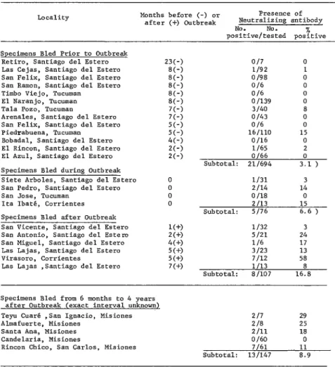
A total of 1,024 vampire bat sera were tested for antibody; of these, 57 yielded titers of 1:s or more and were therefore consi’dered positive. Table 1 classifies 877 of these sera according to the location of captured bats, the time between capture and the occurrence of a rabies outbreak in local cattle, and the presence or absence of neutralizing antibody.
The other 147 vampire sera tested do not appear at the top of the table because the time of local bovine rabies outbreaks could not be determined with sufficient accuracy. It is known, however, that in every case the most recent outbreak had occurred from 6 months to 4 years before the bats were bled. The results obtained with these 147 sera, shown in the lower part of Table 1, indicate that 13 sera (9 per cent) were positive for rabies neutralizing antibody.
All of the 1,024 bats tested for antibody were also tested for rabies virus, but no virus was detected.
Besides these bats captured with mist nets, 83 vampire bats were captured in the Province of Santiago de1 Ester0 during fumigation of their roosts. When tested for rabies virus, 11 were found to have the virus in one or more
bovine rabies was reported in the area. The other nine positive bats were part of a sample of 50 specimens exterminated in roosts located in an area which was experiencing an epizootic of bovine rabies (13).
Discussion
Examination of the results in Table 1 and other data (7) show that, with one exception, neutralizing antibody has not appeared fre- quently in vampire serum samples obtained before the occurrence of rabies outbreaks in local cattle. The one exception (the Piedra- buena samples listed in Table 1) may illustrate problems inherent in depending on cattle rabies as an indicator of vampire rabies. A low but slightly higher overall percentage of samples containing rabies neutralizing antibody were obtained from bats captured during bovine rabies outbreaks, and varying but generally higher percentages of samples with antibody were obtained from bats captured one to seven months after bovine outbreaks.
The only virus isolated in the course of this study was obtained from bats captured just before or at the same time as bovine rabies cases occurred.
These results agree with and supplement those of Delpietro, et al. (7). Together with the epidemiologic information published by Lopez, et al. (19), they point to a rabies virus epizootiology in vampire bats similar to that of many infectious agents in other hosts. It appears that on entering a vampire bat popula- tion, rabies virus successfully infects much of it, possibly killing some individuals (3) and causing
Lord, et al. . VAMPIREBATRABIES 193
TABLE l-Rabies neutralizing antibody in vampire sera, grouped according to the capture site and the time between capture and occurrence of a local bovine rabies outbreak.
Locality Months before (-) or Presence of after (+) Outbreak Neutralizing antibody
NO. NO. %
positive/tested positive
Specimens Bled Prior to Outbreak Retiro, Santiago de1 Ester0 Las Cejas, Santiago de1 Estero San Felix, Santiago de1 Estero San Ramon, Santiago de1 Estero Timbo Viejo, Tucuman
El Naranjo, Tucuman Tala Pozo, Tucumao
Arenales, Santiago de1 Estero San Felix, Santiago de1 Estero Piedrabuena, Tucuman
Bobadal, Santiago de1 Estero El Rincon, Santiago de1 Estero El Azul, Santiago de1 Estero Specimens Bled during Outbreak Siete Arboles, Santiago de1 Estero San Pedro, Santiago de1 Ester0 San Jose, Tucuman
Ita Ibatb, Corrientes
Specimens Bled after Outbreak San Vicente, Santiago de1 Estero San Antonio, Santiago de1 Esters San Miguel, Santiago de1 Estero Las Lajas, Santiago de1 Estero Virasoro, Corrientes
Las La~as ,Santiago de1 Ester0
23(-) SC-) EC-) 8C-j SC-) 8(-j 7(-) 7(-) 5(-) 5(-) 4(-) 2(-j 2(-) I(+) 2(+) 4(+) 5(+) 5(+) 7(+)
O/7 0
l/92 1 O/98 0
O/b 0
O/b 0
0 I139 0 3140 8 o/43 0
O/b 0
lb/110 15 O/lb 0 l/b5 2 O/b6 0 Subtotal: 211694 3.1 )
l/31 3 Z/14 14 0 I18 0 2113 15 Subtotal: S/76 6.6 )
l/32 3 5121 24 l/b 17 3/23 13 7/12 58 l/13 8 Subtotal: a/107 16.8
Specimens Bled from 6 months to 4 years after Outbreak (exact interval unknown) Teyu CuarO ,San Igoacio, Misiones Almafuerte, Misiones
Santa Ana, Misiones Candelaria, Misiones
Rincon Chico, San Carlos, Misiones
217 29
Z/8 25
2111 18
0 lb0 0
7/61 11
Subtotal: 13/147 8.9
sufficient susceptibles in these ways the epi- zootic presumably subsides or moves on to neighboring populations.
Such findings are not surprising and in fact serve to explain bovine rabies phenomena seen throughout the range of the vampire bat. Periodic outbreaks occur because time is needed to build up a sufficient density of susceptibles in the bat population to reach or exceed the required threshold of contagion.
Most vampire bat populations are in contact,
tissues from these bats plus 83 others were tested for virus.
Neutralizing antibody only rarely appeared in vampire serum samples taken before bovine rabies outbreaks, and only low percentages of samples positive for rabies antibody were obtained from bats captured during bovine outbreaks. In contrast, varying percentages of positive samples (including some high percent- ages) were taken from bats captured at various
in vampire populations the way diverse infec- tious agents typically behave in other hosts. That is, the virus infects many individuals; some die and other s survive to demonstrate their exposure through the appearance of antibody. The disease disappears from the bat population in time and does not return until a sufficient number of susceptible bats have re-entered the population.
ACKNOWLEDGMENTS
The authors are grateful to Drs. Ramon for their support during the course of this Rodriguez T., Boris Szyfres, and Ruben A. study. Mr. Abel Fornes (SELSA), who lost his Lombard0 of the Pan American Zoonoses life while working on other aspects of vampire Center, and to Dr. Jorge R. Valotta (SELSA)4 bat ecology, assisted frequently during the
‘Health Campaigns Service, Ministry of Agriculture
capture and processing of bats. Mrs. N. Per- domo and Mr. J.C.F. Areitio assisted in labora-
and Livestock, Buenos Aires, Argentina. tory studies.
REFERENCES
(I) Acha, P. N. Epidemiology of paralytic bovine rabies and bat rabies. Bull Of Int Epizoot 67: 343-382, 1967.
(2) Steele, J. H. International aspects of veterinary medicine and its relation to health, nutrition and human welfare. Milit Med 131: 165-778, 1966.
(3) Constantine, D. G. Bat rabies: Current know- ledge and future research. In: Rubies. Edited by Y. Nagano and F. M. Davenport. Univer- sity Park Press, Baltimore, Md., 1971, p. 253. (4) Constantine, D. G. Bats in relation to the health, welfare, and economy of man. In: Biology of Buts, Volume 2. Edited by W. A. Wimsatt.
Academic Press, New York, 1970, p. 319.
(5) Villa, R. B. “The ecology and biology of vampire
bats and their relationship to paralytic rabies: Report to the Government of Brazil.” United Nations Development Program/Food and Agriculture Organization, Rome, 1969. (Report TA 2656.)
(6) Pawan, J. L. Rabies in the vampire bat of Trinidad, with special reference to the clinical course and the latency of infection. Ann Trop Med Parasitoi 30: 401422, 1936. (7) Delpietro, H., A.M.C. de Diaz, E. Fuenzalida,
and J. F. Bell. Determination de la tasa de ataque de rabia en murcielagos. Bol Of Sani? Panam 73: 222-228,1972.
Lord, et al. . VAMPIRE BAT RABIES 195
(9) L6pez Adaros, H., M. Silva, and M. La Mata. Rabia paralitica en el Norte Argentino. In: Seminario sobre Rabin para 10s Pa&es de In Zona IV-Bolivia, Colombia, Ecuador, Perti- Lima, 6-II de octubre, 1969, pp. 161-203. Pan American Health Organization, Washing- ton, D.C., 1969.
(IO) Johnson, H. N. General epizootiology of rabies. In: Rabies. Edited by Y. Nagano and F. M. Davenport. University Park Press, Baltimore, Md., 1971, p. 237.
(II) Atanasiu, P. Quantitative assay and potency test of antirabies serum. In: Laboratory Tech- niques in Rabies, Second Edition. World Health Organization, Geneva, 1966, p. 167. (WHO Monograph Series, No. 23.)
(12) Larghi, 0. P., and E. Jimenez Ch. Methods for accelerating the fluorescent antibody test for rabies diagnosis. ApplMicrobiol21: 611-613, 1971.
(13) Fornes, A., R. D. Lord, M. L. Kuns, et al. Control of bovine rabies through vampire bat control. J WiidlDis 310-316,1974.
(14) Mitchell, G. C., R. Flores Crespo, R. J. Burns, et al. Vampire bats: Rabies transmission and livestock production in Latin America. 1971 Annual Report, Palo Alto, Mexico, Field Station, Denver Wildlife Research Center, U.S. Bureau of Sports Fisheries and Wildlife, in cooperation with USAID and the Institute National de Investigaciones Pecua- rias, Secretaria de Agricultura y Ganaderia de Mixico.
(15) Young, A. M. Foraging of vampire bats @es- modus rotundus) in Atlantic wet lowland Costa Rica. Rev Biol Trap 18: 73-88, 1971. (16) Lazaro, L., R. D. Lord, M. L. Kuns, H. Delpietro,
et al. Recaptures of banded vampire bats. (Manuscript in preparation.)
WHO RECEIVES MILLION-DOLLAR VOLUNTARY CONTRIBUTION
On 15 August the Japan Shipbuilding Industry Foundation made a
contribution to the World Health Organization of 300 million yen (US$I ,016,949). This sum, the largest voluntary contribution from a
nob-governmental source ever received by WHO, was donated in
support of WHO’s programs for control of leprosy and global eradication of smallpox.
Mr. Ryoichi Sasakawa, President of the Japan Shipbuilding Indus- try Foundation and the Sasakawa Memorial Health Foundation, presented the check to Dr. Francisco J. Dy, WHO Regional Director for the Western Pacific. The ceremony took place at the Japanese Ministry for Health and Public Welfare in the presence of the Minister, Mr. Masami Tanaka.
Mr. Sasakawa said: “Because of my belief in the brotherhood and sisterhood of mankind, I and my Foundation wish to do everything possible to combat two of the most dreaded diseases, smallpox and
leprosy.” (Source: World Health Organization Press Release WH0/28,