Progressive massive fibrosis in silica-exposed workers.
High-resolution computed tomography findings*
ÂNGELA SANTOS FERREIRA1, VALÉRIA BARBOSA MOREIRA2, HEVÂNIA MARA VAZ RICARDO3, RENATA COUTINHO3, JOSÉ MANOEL GABETTO4, EDSON MARCHIORI4
* Study conducted at the Pulmonary Outpatient Clinic and at the Radiology Service of the Antônio Pedro University Hospital (APUH) of the Universidade Federal Fluminense (UFF, Fluminense Federal University) - Niterói, Brazil.
1. Adjunct Professor of Pulmonary Medicine at the Universidade Federal Fluminense (UFF, Fluminense Federal University) -Niterói, Brazil
2. Masters degree in Pulmonary Medicine from the Universidade Federal Fluminense (UFF, Fluminense Federal University) -Niterói, Brazil - Physician at the Hospital Universitário Antônio Pedro (HUAP, Antônio Pedro University Hospital) - --Niterói, Brazil.
3. Undergraduate student at the Universidade Federal Fluminense (UFF, Fluminense Federal University) School of Medicine - Niterói, Brazil
4. Full Professor of Radiology at the Universidade Federal Fluminense (UFF, Fluminense Federal University) - Niterói, Brazil Correspondence to: Ângela Santos Ferreira. Rua Mario Alves, 78/1.902 BII, Icaraí - CEP: 24220-270 Niterói, RJ, Brasil. Tel: 55 21 2610-2692. Email: [email protected]
Submitted: 28 November 2005. Accepted, after review: 7 March 2006.
ABSTRACT
Objective: To evaluate the radiological characteristics of conglomerate masses using high-resolution computed tomography
of the chest. Methods: From among the patients treated between 1986 and 2004 at the Antonio Pedro University Hospital, 75 patients with silicosis and massive fibrosis, most working in the field of sandblasting, were selected for study. These patients were submitted to a clinical evaluation, chest X-ray and high-resolution computed tomography of the chest. Results: In more than half of the patients with accelerated silicosis, the chest X-ray revealed large type B and C opacities, denoting the severity of the disease in those patients. In 1 case, a unilateral mass simulating lung cancer was observed. High-resolution computed tomography scans of the chest were acquired for 44 patients. In most cases (88.6%), the masses were located in the superior and posterior thirds of the lung. Common findings within the masses included air bronchograms (in 70.4%) and calcifications (in 63.6%). A history of tuberculosis was reported by 52% of the patients.
Conclusion: In the vast majority of cases, the masses were bilateral, predominantly located in the superior and posterior regions of the lung, featuring air bronchograms and interposed calcifications. Concomitant calcification of the mediastinal and hilar lymph nodes was another common finding. Exposure to high concentrations of dust and having a history of tuberculosis were considered significant risk factors for the development of progressive massive fibrosis.
Keywords: Pulmonary fibrosis; Silicosis; Silicon dioxide; Occupational diseases; Environmental exposure;
INTRODUCTION
Silicosis is a pulmonary disease contracted through the inhalation of crystalline silica, which induces a fibrogenic tissue response.(1) Silicosis can
be clinically categorized into three forms, based on the course of the disease: chronic, accelerated and acute.(2) The chronic form is the most common,
occurring after years of exposure to relatively low levels of dust. Accelerated silicosis is a clinical term applied to define a condition in which the of progression from acute silicosis to the classic chronic nodular disease occurs at an intermediate rate of speed, requiring, on average, five to ten years of exposure to dust for radiological alterations to surface.(3) In its acute form, the disease
is quite rare, striking workers exposed to exceptionally high concentrations of fine particles of recently crushed crystalline silica as occurs in sandblasting and stone grinding.(4)
Radiological manifestations of silicosis vary among the different clinical forms. Simple silicosis is characterized by the presence of multiple nodules ranging from 1 to 10 mm in diameter and distributed predominantly in the superior and posterior regions of the upper lobes. Nodules tend to be delineated, of uniform density and generally symmetrical. These nodules occasionally calcify and tend to be found in the subpleural region. Lymph node enlargement is common and can precede the appearance of diffusely distributed nodules. Peripheral hilar and mediastinal lymph nodes can become calcified, a condition known as eggshell calcification.(5)
Accelerated silicosis is characterized by the presence of large opacities with homogenous consolidation areas of nonsegmental distribution, principally affecting the upper and middle zones of the lung. This results from the confluence of small fibrotic nodules, which grow to over 10 mm in diameter. Accelerated silicosis is also known as progressive massive fibrosis (PMF), a term initially used by Fletcher in 1948 to refer to pneumoconiosis that evolved from the simple to the complex form.(6)
Typically occurring in black lung disease and silicosis, PMF is rarely seen in other pneumoconiosis. From a pathological point of view, by definition, the lesion exhibits a diameter of at least 2 cm in histological tissue sections. However, the International Classification of Radiographs of
Pneumoconiosis devised by the International Labour Office considers PMF any pulmonary opacity that exceeds a diameter of 1 cm on a chest X-ray.(7)
The main symptom of accelerated silicosis is progressive, disabling dyspnea. During the advanced phases of the disease, a scenario of evident respiratory insufficiency, weight loss and feebleness can arise, therewith rendering the patient susceptible to infections of the lower respiratory tract. More frequently, accelerated silicosis patients evolve to present a more complex form of the disease, in which large fibrous masses form.(8)
Tuberculosis is the principal complication of silicosis. The susceptibility to tuberculosis seems to result from a combination of factors, chief among which is a possible chemical effect that silica has on the multiplication of bacilli, together with macrophage toxicity and a greater permanence of bacilli in lung tissue due to inefficient lymphatic drainage.(9)
The incidence of tuberculosis is higher among silicosis patients presenting PMF than among those presenting the simple form of the disease. The combination of silicosis and tuberculosis further accelerates the evolution of pneumoconiosis.(10)
The objective of this paper was to evaluate the radiological characteristics of conglomerated masses seen on high-resolution computed tomography (HRCT) scans of the chest.
METHODS
A review was made of the 312 records on file at the Occupational Pulmonary Diseases Outpatient Clinic of the Antônio Pedro University Hospital for patients with a history of exposure to silica and treated during the period from January of 1986 to July of 2004.
Of the 189 patients in whom silicosis was confirmed based on their occupational history and radiological alterations compatible with the disease, 75 cases were selected. These cases displayed the accelerated form of the disease, which is characterized by a high number of opacities, typically larger than 1 cm in diameter, in the lung parenchyma.
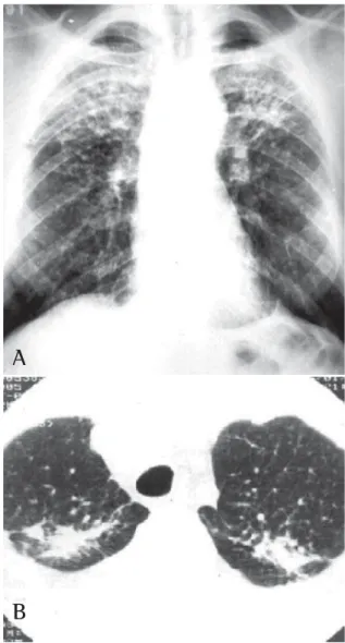
Figure 1 - A) Typical case of accelerated silicosis in a 53-year old patient, a sandblaster at a dockyard, with an 18-year exposure to silica. Chest X-ray: small nodules and large opacities in the upper lobes; B) High-resolution computed tomography scan of the chest: large opacities in the posterior segments of the upper lobes
A
B
between the focal plane and the film was 180 cm (International Labour Office, 1980). The HRCT scans were acquired using GE CETEMAX 640 or Toshiba Asteion tomographs.
The 1980 International Labour Office International Classification of Radiographs of Pneumoconiosis,(11) has been used for the
classification and comparative interpretation of the X-rays against the standards provided. Large opacities have been classified into the following groups: type A, in which opacity diameters are from 1 to 5 cm or there is more than one opacity with a diameter equal to or greater than 1 cm, but the sum of the diameters is less than 5 cm; type B, in which the combined diameter of one or more opacities is greater than 5 cm, but the total area is smaller than the upper third of the right lung; and type C, in which the area presenting the large opacities is greater than the upper third of the right lung.
For patients who underwent HRCT, we analyzed the following characteristics of the mass: location, cavitations, air bronchograms and internal calcification. Emphysematous lesions adjacent to the masses, as well as lymph node calcification, were also assessed.
Consideration was also given to other variables such as age, profession, time of exposure to silica dust and concomitance with tuberculosis.
RESULTS
All 75 patients with accelerated silicosis were male. The mean age was 43 years (range, 28-76 years). The mean duration of exposure was 14.2 years (range, 7 months-38 years). Of the 75 patients, 62 (82.6%) were sandblasters, 11 (14.6%) were blacksmiths, 1 (1.4%) was a welder, and 1 (1.4%) was a metal polisher. In reference to concomitant tuberculosis, 39 patients reported a history of pulmonary tuberculosis (52%). Of those, 37 developed the disease during their exposure to silica.The diagnosis was confirmed in 31 cases: 24 through direct sputum smear microscopy; 3 through direct mycobacteria culture of the sputum; 1 through mycobacteria culture of the bronchoalveolar lavage fluid; and 3 through histopathology. In 8 patients, there was clinical and radiological suspicion of tuberculosis based on a response to specific treatment. The radiological classification of the large opacities seen on the chest X-rays was as follows: Type A in 23
patients (30.7%); Type B in 25 (33.3%); and Type C in 27 (36%) (Figures 1A and 1B). Of the cases analyzed, 74 presented bilateral conglomerate masses, whereas only 1 presented unilateral lesion (Figures 2A and 2B).
A
B
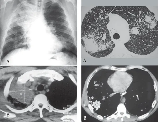
Figure 2 - A) Atypical case of accelerated silicosis case in a 45-year old patient, a blacksmith with a 10-year exposure to silica, complaining of chest pain in the right hemithorax, coughing with hemoptysis and weight loss. Chest X-ray: opacity with volumetric reduction in the upper right lobe. Open lung biopsy: silicosis; B) High-resolution computed tomography scan of the chest: voluminous opacity with ill-defined borders in the upper right lobe
A
B
Figure 3 - A) Accelerated silicosis in a 43-year old patient, a sandblaster at a glassware factory, with a 6-year exposure to silica. High-resolution computed tomography scan of the chest: (window for the parenchyma): presence of small nodules and large opacities in the upper lobes; B) High-resolution computed tomography scan of the chest (mediastinal window): calcifications within the mass and in the mediastinal lymph nodes
in 70.4% and internal calcification in 63.6%. Emphysematous lesions surrounding the masses were observed in 72.7%, and lymph node calcification was seen in 81.8% (Figures 3A and 3B).
DISCUSSION
Silicosis is the most prevalent pneumoconiosis in Brazil and in the world at large. Interest in this disease was sparked when a high incidence was reported among longshoremen working on the docks at the port of Guanabara Bay, in the city of Niterói, who had sought medical attention in
regional hospitals. These patients, typically exposed to exceptionally high concentrations of silica particles, had developed a form of the disease that was accelerated and more frequently evolved to the complex form, accompanied by the formation of large fibrous masses that distort and destroy to the lung architecture.(12)
Certain authors(10) evaluated 92 cases of PMF
and also reported the importance of the quantity of dust in the development of this severe form of pneumoconiosis. Other authors,(13) upon having
analyzed 733 silicosis PMF patients, determined that the most significant risk factors associated with this form of disease were high levels of exposure to dust, a history of tuberculosis, and a high profusion of small opacities on the chest X-ray. In the present study, the combination of tuberculosis and PMF was found in 52% of the patients. This confirms the importance of tuberculosis infection as a risk factor for the development of PMF. It is known that silicosis is a risk factor for tuberculosis.(14) Some authors(15) have
reported a high (34%) prevalence of tuberculosis among sandblasters with silicosis.
It can be extremely difficult to establish a diagnosis of tuberculosis in patients with silicosis, especially when the chest X-ray reveals the complex form of the disease. The presence of systemic symptoms should raise the suspicion of tuberculosis (despite the fact that accelerated silicosis can also provoke such symptoms). Tuberculosis should also be suspected when chest X-rays reveal the rapid emergence of new opacities, together with pleural effusion and cavitation, all of which are suggestive of a diagnosis of tuberculosis.(16)
On a simple chest X-ray, accelerated silicosis is characterized by large opacities (over 1 cm in diameter), with nonsegmentally distributed, homogenous areas of consolidation, principally affecting the upper fields. The lesions tend to form on the periphery, migrating toward the hilum, creating a zone of emphysematous lung tissue between the consolidation and the pleural surface. The borders of the large opacities are irregular and poorly defined. As the disease progresses, the masses tend to shrink. Emphysematous lesions and bullae then appear surrounding the masses and at the lung bases. The lungs gradually lose volume. The large masses seen in accelerated silicosis are capable of causing cavitation due to the resultant ischemia and necrosis. Nevertheless, cavitation is a relatively uncommon finding in silicosis. When it does occur, one should first consider tuberculosis. The pleural thickening frequently observed in tuberculosis is also commonplace in PMF, although pleural effusion is extremely rare.(17)
The greater the profusion of nodules on the
chest X-ray (categories 2 and 3), the greater are the odds of evolving to PMFThe chest X-ray generally shows large lung opacities against a background of diffuse pulmonary nodule formation. However, PMF has been reported in the absence of pulmonary nodule formation, since the silicosis nodules present reduced visibility due to their incorporation within the fibrous mass.(18)
The chest X-rays of more than half of the accelerated silicosis patients showed large type B or C opacities, denoting the seriousness of the disease in these patients.
Of the 75 cases analyzed, only 1 presented a large unilateral opacity mimicking a tumor. The diagnosis of silicosis was made via thoracotomy with open lung biopsy. Isolated PMF lesions are frequently mistaken for lung cancer. Given the higher incidence of lung cancer in these patients, it is important to take a diagnostic approach in order to effectively differentiate malignant pneumoconiosis lesions from benign ones.(19)
In the present study, chest HRCTs revealed that the large opacities were predominantly (in 88.6% of the cases) located in the upper and posterior thirds of the lung.Air bronchograms and calcifications within the masses were found in over half of the patients. Calcified hilar and mediastinal lymph nodes were observed in 81% of the cases, 25% being of the eggshell type. Only 8 cases displayed cavitation within the masses, 6 presenting concomitant tuberculosis.
Some authors(20) have described sparse
calcifications within silicosis-related PMF masses, whereas others(21) have reported lymph node
calcification in 26 of 49 silicosis cases studied, 12.2% being of the eggshell type, which was found to be much more frequent in accelerated silicosis than in the simple nodular form.
Other authors(22) used serial X-rays to study the
radiological evolution of 141 silicosis patients and found that radiological progression occurred more often in those presenting large opacities on the initial X-rays than in those who had had tuberculosis.
Silicosis is a progressive fibrosing occupational disease, and patients with accelerated silicosis are more likely to present radiological progression, altered respiratory function and premature death. Therefore, it is essential to avoid risk factors for the development of this form of the disease.
REFERENCES
1. Wagner GR. Asbestosis and silicosis. Lancet. 1997;349(9061): 1311-5.
2. Brasil. Ministério da Saúde. Fundação Nacional de Saúde. Manual de normas para o controle das pneumoconioses: silicose, pneumoconiose dos trabalhadores de carvão e pneumoconiose por poeiras mistas. Brasília, Fundação Nacional de Saúde, 1997.
3. Ziskind M, Jones RN, WeilL H. Silicosis. Am Rev Respir Dis. 1976;113(5):643-65.
4. Ferreira AS. Silicose aguda. Pulmão RJ. 1999;8(4):349-58. 5. Mcloud TC. Occupational lung disease. Radiol Clin North
Am. 1991;29(5):931-41.
6. Fletcher CM. Pneumoconiosis of coal-miners. Br Med J. 1948;1:1065-74.
7. Diseases associated with exposure to silica and nonfibrous silicate minerals. Silicosis and Silicate Disease Committee. Arch Pathol Lab Med. 1988;112(7):673-720.
8. Ferreira AS. Silicose. In: Aidé MA, Cardoso AP, Rufino R, David F, Carvalho SR, Lucas VS, Zamboni MM, editores. Pneumologia: aspectos práticos e atuais. Rio de Janeiro: Revinter; 2001. p. 383-90.
9. Algranti E. Slateworkers, pneumoconiosis. Cardiff: University of Wales; 1982.
10. Souza Filho AJ, Alice SH. Fibrose maciça pulmonar progressiva. J Pneumol. 1991;17(4):147-53.
11. International Labour Office. Guidelines for the use of ILO International classification of radiographs of pneumoconiosis. ed.rev. Geneva: ILO; 1980. (Occupational Safety and Health Series, 22).
12. Ferreira AS, Moreira JS, Caetano R, Gabetto JM, Santos TQ. Caracterização imunofenotípica das subpopulações de linfócitos do lavado broncoalveolar de pacientes com silicose. J Pneumol. 2000;26(3):107-12. 13. Ng TP, Chan SL. Factors associated with massive fibrosis
in silicosis. Thorax. 1991;46(4):229-32.
14. Ding M, Chen F, Shi X, Yucesoy B, Mossman B, Vallyathan V. Diseases caused by silica: mechanisms of injury and disease development. Int Immunopharmacol. 2002;2(2-3):173-82.
15. Ferreira AS, Moreira VB, Souza AM, Gabetto JM, Clemente CC, Aidé MA. Silicotuberculose: análise de 82 casos. J Pneumol. 2000;26(S3):43-4.
16. Bailey WC, Brown M, Buechner HA, Weill H, Ichinose H, Ziskind M. Silico-mycobaterial disease in sandblasters. Am Rev Resp Dis. 1974; 110: 115-125.
17. Al-Kassimi FA. Pleural efusion in silicosis of the lung. Br J Indian Med. 1992;49(6):448-50.
18. McCloskey M, Cook N, Cameron D, Summers Q. Progressive massive fibrosis in the absence of lung nodulation. Austral Radiol. 1997;41(1):63-4. 19. Carneiro APS, Santos MAM, Maia PV, Barreto SM. Câncer
de pulmão em trabalhadores expostos à sílica. J Bras Pneumol. 2002;28(4):233-6.
20. Marchiori E, Dantas MCH, Nobre LF. Silicose: correlação da tomografia computadorizada de alta resolução com a anatomopatologia. Radiol Bras. 2001;34(1):1-6. 21. Moreira VB, Ferreira AS, Gabetto JM, Marchiori E, Lourenço
PMC. Estudo comparativo da tomografia computadorizada de alta resolução com a radiografia de tórax na silicose. Rev Port Pneumol. 2003;9(1):33-40.