Carcinoma basocelular
Lectinas
Análise de imagem
Neoplasia cutânea y
Digital image analysis of skin neoplasms evaluated by
lectin histochemistry: potential marker to biochemical
alterations and tumour differential diagnosis
Análise digital de imagens de neoplasias da pele avaliadas pela histoquímica com lectinas: marcador potencial
para alterações bioquímicas e diagnóstico diferencial de tumores
Mario R. Melo-Júnior1; Jorge Luiz S. Araújo-Filho2; Vasco José Ramos M. Patu3; Marcos Cezar F. de Paula Machado2; Eduardo I.C. Beltrão4; Luiz B. Carvalho Jr.5
key words
unitermos
abstract
The present study aims to evaluate, through lectin histochemistry, the alterations in the expression of cell surface carbohydrate between benign and malignant lesions of skin using computer image analysis. Skin fragments were obtained through biopsies and diagnosed as basal cell carcinoma (BCC), epidermoid carcinoma (EpC), trichoepithelioma (TE), keratoacanthoma (KA), seborrheic keratosis (SK) and actinic keratosis (AK). Lectins Con A, WGA, PNA, UEA-I and LTA were used in histochemistry study.
Image analysis was carried out in a workstation using OPTIMASTM software system. PNA strong binding
pattern to studied tumours evidenced the high expression of D-galactose residues in the epidermal neoplasms when compared to other sugars recognized by the other lectins. Among benign neoplasms,
KA presented a high expression of glucose/mannose, α-fucose and D-galactose residues evidenced by
the intense staining of Con A (94.7%), LTA (84.2%) and PNA (89.4%), respectively. Malignant tumours showed distinct binding patterns. EpC presented signiicant binding only by PNA lectin. BCC was diffe-rentially stained in comparison to the staining pattern observed in benign lesions such as TE. Qualitative (lectin histochemistry) and quantitative (digital image analysis) data obtained in this study evidenced those lectins are potential markers to biochemical alterations in skin neoplasms.
Basal cell carcinoma
Lectins
Image analysis
Skin neoplasms
resumo
O presente estudo objetivou avaliar, através da histoquímica com lectinas, as alterações na expressão dos carboi-dratos da superfície celular entre lesões benignas e malignas da pele usando análise de imagens computadorizadas. Fragmentos de pele foram obtidos através de biópsias e diagnosticados como carcinoma basocelular (CBC), carci-noma epidermóide (EpC), tricoepitelioma (TE), cerato acantoma (KA), ceratose seborreica (CS) e ceratose actínica (CA). As lectinas Con A, WGA, PNA, UEA-I e LTA foram usadas no estudo histoquímico. A análise de imagens foi realizada numa estação de análise usando o sistema OPTIMASTM de análises. A PNA tem sido largamente utilizada
no estudo de tumores, evidenciando a expressão de D-galactose nas neoplasias epidérmicas; esse açúcar apresenta alta expressão quando comparado com os outros reconhecidos pelas demais lectinas. Entre as neoplasias benignas, KA apresentou alta expressão glucose/manose; resíduos de α-fucose e D-galactose apresentaram intensa marcação com ConA (94,7%) LTA (84,2%) e PNA (89,4%), respectivamente. Os tumores malignos mostraram marcações distintas: EpC apresentou marcação significativa somente com a lectina PNA; CBC apresentou diferente padrão de marcação quando comparado ao observado nas lesões benignas assim como no TE. Os resultados qualitativos (análise de imagens) e quantitativos (histoquímica com lectinas) desse estudo evidenciaram que as lectinas têm grande potencial como marcadores de alterações bioquímicas nas neoplasias da pele.
Primeira submissão em 26/01/06
Última submissão em 14/09/06
Aceito para publicação em 18/09/06
Publicado em 20/12/06
1. Professor-titular de Patologia da Associação Caruaruense de Ensino Superior (ASCES). 2. Mestrando em Patologia pelo Centro de Ciências da Saúde (CCS) da UFPE. 3. Mestrando em Ciências Biológicas pelo Centro de Ciências Biológicas (CCB) da UFPE. 4. Professor-adjunto do Departamento de Bioquímica do CCB/UFPE.
5. Professor-titular do Departamento de Bioquímica do CCB/UFPE.
Introduction
The dificulty in diagnosing many skin neoplasias is related to a variety of factors such as a huge spectrum of tumours and their variants and the lower differentiation and complicated nomenclature(43). Besides that, many
studies are inconclusive to afirm whether the cellular origin of some malignant lesions is derived from benign tumour differentiation and pre-neoplasic tumours or from normal cell(42).
In routine histology, there is not a precise staining pattern to differentiate all skin tumours. Epidermis neoplasic lesions such as squamous cell carcinoma and keratoacanthoma are many times dificult to be differentiated from each other using routine histology analysis(7). Trichoepithelioma and
basocellular carcinoma; or Bowen disease, actinic keratosis and seborrheic keratosis histology differentiation can be also dificult in small tissue samples(16, 32).
Immunohistochemistry techniques(33) and lectin
staining(3, 27) have been tested as supporting tools for
differential diagnosis for skin lesions.
Investigation of lectin/carbohydrate interactions helps in the understanding of histogenetic origin and tumour behaviour during its differentiation(14) and conirms the
hypothesis that biochemistry changes in the cell are events that can signalize cell morphodifferentiations(36).
Glycosylation and glycoconjugate expression are usually altered in tumour cells in comparison to their normal counterpart(4, 24).
The results of the very few histochemical studies that have been performed so far on the lectin-binding proile of normal human epidermis are mostly controversial; thus, the carbohydrate residue composition of the cell surface in the latter still remains in dispute, and the possible alterations in the epidermal lectin-binding proile are unknown(11).
In comparison with normal human skin, epidermal lectin-binding pattern in the tumour cells showed considerable qualitative and quantitative variations(13).
Many methods of analysis have been used to translate in objective and numerically representative standards the transformations occurred in tumour cells(10). Such methods
include colorimetric analysis of neoplasic cell culture(1),
automatic cytometry(28), nuclear morphometric variation
analysis(35) and image analysis of tumour lectin staining(22, 25).
Digital image analysis has given more accurate quantitative and qualitative data of histomorphologic aspects of neoplasms(8, 23). From this perspective the present study
aims to evaluate the differences of cell surface carbohydrate expression between benign and malignant lesions of the skin using lectin staining and digital image analysis.
Material and methods
Case selection
Skin fragments were obtained through biopsies and diagnosed as basal cell carcinoma (BCC, n = 35), epidermoid carcinoma (EpC, n = 18), trichoepithelioma (TE, n = 12), keratoacanthoma (KA, n = 19), seborrheic keratosis (SK, n
= 10) and actinic keratosis (AK, n = 18). Normal human epidermis (NE, n = 25) samples were obtained from the same anatomical site on the opposite side of the tumours. Patients (male and female), whose ages varied from 45 to 80 years old, agreed to participate in this study and signed a consent declaration. The delineation of the study was a retrospective analysis based on parafin blocks from archived cases (Laboratório de Imunopatologia Keizo Asami [LIKA] of Universidade Federal de Pernambuco [UFPE]).
Tissue preparation
Biopsies were ixed in 10% buffered formalin, submitted to histological routine, and parafin embedded. Slices (4µm) were adhered in slides treated with 3-amine-propyltriethoxy-silane (APES [Sigma, USA]).
Lectin histochemistry
performed using the corresponding lectin speciic sugar: methyl-α-D-mannoside for Con A; N-acetyl-glucosamine for WGA; α-L-fucose for UEA-I and LTA; and D-galactose for PNA, at inal concentration of 300mM.
Digital image analysis
Images of slices of the stained cells (magniication 100x) were obtained using a digital video camera (Sony, Japan) connected to a microscope and processed using OPTIMASTM, version 6.1 (Optimas Corporation, USA).
Fields were chosen randomly from various section levels to ensure objectivity of sampling. The iles were then opened in Adobe Photoshop (Adobe Inc., Mountain View, CA) and further processed to assess stained cells. To accomplish this, acquired color images were irst split into red, green, and blue channels (Optimas Commander: Split channels).
The images were binarized to black and white and a common threshold level was chosen for representing correct stained cells and minimizing background noise levels (Optimas Commander: Adjust Threshold). To investigate changes in lectin binding patterns revealed by the DAB-peroxidase reaction we employed the following method: red channel images were initially blurred with a Gaussian ilter (one pixel for all sample images) to reduce electronic and background noise (Optimas Commander: Filter: Blur: Gaussian blur); eight-bit grayscale levels were expanded linearly so that the whitest point fell at a grayscale value of 255 and the blackest point at 0; the images were then so that all points above the chosen grayscale value were included for measurement and those below were excluded.
However, a common threshold level was chosen for all analyzed images to prevent intra-assay variations. We then inverted the images, which changed all white pixels to black ones and vice versa (Optimas Commander: Adjust: Invert),
and employed the image processing for further analysis. Staining control (tissues treated with sugar-inhibited lectins and normal skin samples) was obtained to minimize distortions in values due to the presence of non-marked cells, thus a correction factor (CF) was applied according to the equation CF = s/S, in which s means relative area of the surface and S, the total measured area(24).
Analyses were developed in three areas, randomly chosen, of the stained tumour tissue where the mean area (µm2) and the number of cells per area (total area: 12234
µm2) were calculated. Dermal and hypodermal vessel areas
were omitted. Obtained data were compared using Mann-Whitney U test and Student’s t test (p < 0.05) through PRISMA 3.0 software (Texas, USA).
Results
Tissue samples presented a homogeneous characterization related to morphologic features related to sex and age (Table 1). Among benign neoplasias, KA presented a high expression of glucose/mannose, α-fucose and D-galactose residues evidenced by the intense staining of Con A (94.7%), LTA (84.2%) and PNA (89.4%), respectively. AK exhibited positive staining to PNA and WGA, being negative or irregular (< 5%) for the other lectins staining. The lowest staining was observed for trichoepithelioma.
Lectins presented a varied staining pattern. Speciic staining was observed to Con A, WGA and PNA while LTA and UEA-I presented non-speciic staining. Epidermal cornea layer was stained by all used lectins. The strong binding pattern of studied tumours by PNA evidenced the high expression of D-galactose residues in the epidermal neoplasms when compared to other sugars recognized by the other lectins (Figure 1).
General aspects of the lesions from patients with skin tumours
Tumor*
Age (years)**
Tumor size (cm
2)**
Total biopsies
Male
Female
Male
Female
Male
Female
BCC 55.2 ± 4.8 58.1 ± 3.7 1.06 ± 0.21 1.37 ± 0.86 12 10
TE 61.3 ± 2.3 47.5 ± 2.1 0.64 ± 0.21 1.83 ± 1.13 11 10
SK 57.1 ± 7.8 44.8 ± 4.5 0.86 ± 0.21 1.24 ± 0.15 10 10
KA 53.2 ± 5.6 52.7 ± 2.2 2.05 ± 1.21 1.63 ± 0.96 10 9
AK 54.8 ± 3.5 53.9 ± 3.4 2.31 ± 2.01 2.4 ± 0.52 12 10
EpC 55.6 ± 5.8 59 ± 5.1 0.97 ± 0.33 0.88 ± 0.16 12 10
*According to Elder et al. 1997(9). The main anatomical sites were neck, arm and head; **statistic analysis (Tukey test; p = 0.73; r =1.25).
Malignant tumours showed distinct binding patterns. EpC presented signiicant binding only to PNA lectin. Basocellular carcinoma was differentially stained in comparison to the staining pattern observed in benign lesions such as TE.
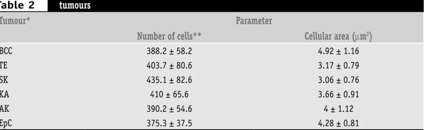
Statistically signiicant variations were not observed in the image analysis data of area and number of cells in the evaluated skin tumours (Table 2).
Discussion
Qualitative and quantitative changes in the glycoconjugates of cell membranes are normal during development and progression of many neoplasic processes(6, 39, 44). In neoplasic
cells the increase on the content and/or expression of surface carbohydrates have been documented using lectin histochemistry(14, 40). Lectins are sensible, stable and
easy-to-use tools to differentiate transformed and non-transformed cells(4, 9, 24, 37).
Image analysis data of lectin histochemistry in the area and number of cells stained on skin
tumours
Tumour*
Parameter
Number of cells**
Cellular area (
µ
m
2)
BCC 388.2 ± 58.2 4.92 ± 1.16
TE 403.7 ± 80.6 3.17 ± 0.79
SK 435.1 ± 82.6 3.06 ± 0.76
KA 410 ± 65.6 3.66 ± 0.91
AK 390.2 ± 54.6 4 ± 1.12
EpC 375.3 ± 37.5 4.28 ± 0.81
*Basal cell carcinoma (BCC, n = 35), epidermoid carcinoma (EpC, n = 18), trichoepithelioma (TE, n = 12), keratoacanthoma (KA, n = 19), seborrheic keratosis (SK, n = 10) and actinic keratosis (AK, n = 18); **average of cells that stained positively ± standard deviation.
Table 2
Figure 1 – Results of image analysis of stained cells area obtained to lectin histochemistry of skin’s neoplasms. *Basal cell carcinoma (BCC, n = 35), epidermoid carcinoma (EpC, n = 18), trichoepithelioma (TE, n = 12), keratoacanthoma (KA, n=19), seborrheic keratosis (SK, n = 10) and actinic keratosis (AK, n = 18). Lectins: Con A (concanavalin A), WGA (wheat germ agglutinin), PNA (peanut agglutinin), UEA-I (Ulex europaeus agglutinin) and LTA (Tetragonolobus purpurea agglutinin). Total area analyzed per slice: 12,234µm2.
µ
0 20 40 60 80 100
BCC TE SK KA AK EpC
Tumours*
P
o
s
it
iv
e
c
a
s
e
s
(%
)
ConA LTA UEA-I PNA WGA
WGA and PNA stained similarly the cornea layer and collagen tissue of both benign and malignant tumours. Previous studies demonstrated the presence of glycoproteins in these tissues(14, 15).
Many lectins, such as Helix pomatia agglutinin (HPA) and PNA, recognize tumour cells of malignant melanomas(42),
indicating the association of surface carbohydrates with the metastatic potential of skin cancers(45). The differentiated
expression of sugars during transformation of melanocytes into keratocytes can be used as a distinct marker between benign and malignant skin lesions(27, 29).
In our study PNA presented a high binding proile to BCC, EpC, SK and KA. Other studies concluded that PNA presented intense binding pattern to KA and squamous cell carcinoma(17, 18). In both cases, aberrant glycosylation may
be associated to a differential cell surface expression of extra cellular matrix receptors in the transformed cell(5).
The differentiation between BCC and TE is sometimes difficult due to the histology of these lesions(26). This
distinction is clinically very important since BCC must be completely excised because of its aggressive behaviour to local recurrence while TE is a benign tumour, which can be treated with a supericial excision, not requiring another treatment or intervention(38). Our results demonstrated
that specially Con A and PNA can be used as markers for distinction between BCC and TE indicating pathobiochemical speciic alterations originated from these neoplasms.
Similar studies to evaluate the cell surface glycoconjugates using PNA, WGA, Con A, LTA and UEA lectins did not observe total accordance concerning lectin-binding(12, 13). On the
Quantitative image analysis of the number and area of cells confirmed that the histological differentiation in tumours seem to be also related to changes in the expression of surface carbohydrates in cells(21, 25, 34). Aberrant
glycosylation of tumour cells, deined by the expression of tumour associated carbohydrate antigens (TACAs), is even more frequently found in tumours than activation of certain oncogenes, such as ras. Thus, the TACAs have been used as markers for prognosis of human tumours(5, 30).
Cell surface glycoconjugates have an important hole in cell/cell interaction, and changes in these glycoconjugates in cancer cells are apparently associated to the altered cell adhesion and development of tumour invasive features(31).
The inal composition of cell glycoconjugates is determined by the combined activities of cell glycosyltransferase and glycosidases. The genetic regulation of these groups of enzymes is still poorly understood, but quantitative and qualitative differences in the glycosylation have been considered to relect differences in gene expression(19).
Our results get together to the indings of other authors who investigated similar and different diseases, which
used Con A, PNA, WGA and UEA-I to characterize tumour parenchyma in BCC(20), Paget disease by WGA and PNA(41)
and epithelioid hemangioendothelioma by UEA-I(2).
Despite the results, it can be concluded that, in general, the more anaplasic (undifferentiated) a cell becomes the more intense the staining of some lectins can be. This observation indicates that the distribution and expression of glyconjugates in the surface were altered(40). The Con A,
LTA and PNA lectins were useful and distinguished among benign and malignant epidermal neoplasms.
Qualitative (lectin histochemistry) and quantitative (image analysis) data obtained in this study evidence that lectins are potential markers and tools to biochemical alterations in skin neoplasms.
Acknowledgments
We thank Carmelita de Lima Bezerra Cavalcanti and Valdir Bandeira Silva, M.D. (Universidade Federal de Pernambuco, Brazil) for technical and scientiic assistance, respectively. This work was supported by CNPq (CTPETRO nº 463655/001).
1. ALLEY, M. C. et al. Morphometric and colorimetric analysis of human tumor-cell line growth and drug sensitivity in soft agar culture. Cancer Res, v. 51, n. 4, p. 1247-56, 1991.
2 . A R N O L D, G . ; K L E I N , P. J . ; F I S H E R , R . E p i t h e l i o i d hemangioendothelioma. Report of a case with immuno-lectinhistochemical and ultrastructural demonstration of its vascular mature. Virchows Arch (Pathol Anat), v. 408, p. 435-43,1986.
3. BASARAB, T.; ORCHARD, G.; RUSSELL-JONES, R The use of immunostaining for bcl-2 and CD 34 and the lectin peanut agglutinin in differentiating between basal cell carcinomas and trichoepitheliomas. Am J Dermatopathol, v. 20, n. 5, p. 448-52, 1998.
4. BELTRÃO, E. I. C.; CARVALHO Jr., L. B. Parkia pendula lectin as histochemistry marker for meningothelial tumour. Eur J Histochem, v. 47, n. 2, p. 139-42, 2003.
5. CHAMMAS, R. et al. Carbohydrate-binding proteins in cell-matrix interactions. Braz J Med Biol Res, v. 27, p. 2169-79, 1994. 6. CHAN, F. L.; CHOI, H. L.; HO, S. M. Analysis of glycoconjugate
patterns of normal and hormone-induced dysplastic noble rat prostates and an androgen-independent noble rat prostate tumor by lectin and protein blotting. Prostate, v. 46, n. 1, p. 21-32, 2001.
7. CRIBIER, B.; ASCH P. H.; GROSSHANS, E. Differentiating squamous cell carcinoma from keratoacanthoma using
References
histopathological criteria. Is it possible ? A study of 296 cases. Dermatol, v. 199, n. 3, p. 208-12, 1999.
8. DEMIRKAYA, O. et al. Automated identification of stained cells in tissue sections using digital image analysis. Anal Quant Cytol Histol, v. 21, n. 2, p. 93-102, 1999.
9. ELDER, D. et al. Laboratory methods. In: ELDER, D. et al. Lever’s Histopathology of the skin. 8. ed. New York: Lippincort-Raven,
1997. Cap. 4, p. 1073.
10. FRANCIS, I. M. et al. Manual versus image analysis estimation of PCNA in breast carcinoma. Anal Quant Cytol Histol, v. 22, n. 1, p. 11-6, 2000.
11. GEORGIOW, S. et al. Age-related alterations in the carbohydrate residue composition of the cell surface in the unexposed normal human epidermis. Gerontol, v. 51, p. 155-60, 2005. 12. GHERI, G. et al. Changes in expression of the oligosaccharides in
the human fetal skin. Ann Anat, v. 179, n. 1, p. 49-56, 1997. 13. GHERI, G. et al. The oligosaccharidic component of the
glycoconjugates in lichen planus, granuloma annulare, seborrheic keratosis and plamoplantar keratoderma: lectin histochemical study. Histol Histopathol, v. 14, n. 3, p.
697-704, 1999.
14. HERLING, M. et al. Glycohistochemical monitoring of chemically induced sarcomas at different stages of tumorigenesis. In Vivo, v. 14, n. 4, p. 499-506, 2000.
Mailing address
Mario Ribeiro de Melo Júnior
Laboratório de Imunopatologia Keizo Asami (LIKA), Setor de Patologia, Universidade Federal de Pernambuco (UFPE)
Av. Prof. Morais Rêgo s/n, Campus Universitário CEP 50670-910 – Recife-PE
e-mail: [email protected] specific carbohydrate configurations in human skin using
fluorescein-labelled lectins. Brit J Dermatol, v. 100, p. 237-45, 1979.
16. ISHIDA, H. et al. Comparative histochemical study of Bowen’s disease and actinic keratosis: perserved normal basal cells in Bowen’s disease. Eur J Histochem, v. 45, n. 2, p. 177-90, 2001.
17. KANITAKIS, J. et al. Nucleolar organizer region enumeration in keratoacanthomas and squamous cell carcinomas of the skin. Cancer, v. 69, n. 12, p. 2937-41, 1992.
18. KANNON, G.; PARK, K. Utility of peanut agglutinin (PNA) in the diagnosis of squamous cell carcinoma and keratoacanthoma. Am J Dermatopathol, v. 12, p. 31-6, 1990.
19. KROGERUS, L.; ANDERSON, L. C. Different lectin binding patterns in primary breast cancers and their metastases. Cancer, v.66, p. 1802-9, 1990.
20. KRÜGER, K.; BLUME-PEYTAVI, U.; ORFANOS, C. E. Basal cell carcinoma possibly originates from the outer root sheat and/or the bulge region of the vellus hair follicle. Archiv Dermatol Res, v. 291, n. 5, p. 253-9, 1999.
21. LALWANI, A. K. et al. Lectin binding characteristics of squamous cell carcinomas of the head and neck. Acta Otolaryngol, v. 116, p. 125-31, 1996.
22. LATHAM, V. H.; OPPENNHEIMER, S. B. A simple image analysis method for evaluating cell binding to derivatized beads. Acta Histochem, v. 101, n. 3, p. 263-70, 1999.
23. LEE, J. S.; JUNG, J. J.; KIM, J. Quantification of angiogenesis by a computerized image analysis system in renal cell carcinoma. Anal Quant Cytol Histol, v. 22, n. 6, p. 469-74, 2000. 24. LITYNSKA, A. et al. Comparison of the lectin-binding pattern
in different human melanoma cell lines. Melan Res, v. 11, n.
3, p. 205-12, 2001.
25. MELO-JÚNIOR, M. R. et al. Altered lectin-binding sites in normal colon and ulcerative colitis. J Bras Patol Med Lab, v. 40, n. 2, p. 123-5, 2004.
26. MILLER, S. J. Biology of basal cell carcinoma (Part 1). J Am Acad Dermatol, v. 24, p. 1-13, 1991.
27. MINWALLA, L. et al. Inhibition of melanosome transfer from melanocytes to keratinocytes by lectins and neoglycoproteins in vivo and in vitro model system. Pig Cell Res, v. 14, n. 3, p. 185-94, 2001.
28. MOMMERS, E. C. M. et al. Malignancy-associated changes in breast tissue detected by image cytometry. Anal Cell Pathol, v. 20, n. 4, p. 187-95, 2000.
29. MONASTIRLI, A. et al. Lectin-binding pattern of primary malignant melanomas and melanocytic nevi. J Cut Pathol, v. 27, n. 3, p. 103-7, 2000.
30. MURAMATSU, T. Carbohydrate signals in metastasis and prognosis of human carcinomas. Glycobiol, v. 3, p. 291-6, 1993.
31. OKUYAMA, T. et al. Interrelation between tumor-associated cell surface glycoprotein and host immune response in gastric carcinoma patients. Cancer, v. 82, n. 8, p. 1468-75, 1998. 32. PETTER, G.; HAUSTEIN, U. F. Rarely occurring and newly
described histological variants of cutaneous squamous cell carcinoma. Classification based on histopathology, cytomorphology and biologic behavior. Hautarzt, v. 52, n. 4, p. 288, 2001.
33. PONIECKA, A. W.; ALEXIS, J. B. An immunohistochemical study of basal cell carcinoma and trichoepithelioma. Am J Dermatopathol, v. 21, n. 4, p. 332-6, 1999.
34. REANO, A. et al. Lectins as markers of human epidermal cell differentiation. Differentiation, v. 22, p. 205-10, 1985. 35. ROELS, S. L. M. F. et al. DNA ploidy and nuclear morphometric
variables for the evaluation of melanocytic tumors in dogs and cats. Am J Vet Res, v. 61, n. 9, p. 1074-9, 2000.
36. SALZMAN, N. H. et al. Enteric defensin expression in necrotizing enterocolitis. Pedriatr Res, v. 44, p. 20-6, 1998.
37. SCHUMACHER, D. V. et al. Is the binding of the lectin Helix pomatia agglutinin (HPA) of prognostic revelance in tumours of the upper aerodigestive tract ? Eur J Surg Oncol, v. 22, n. 6, p. 618-20, 1996.
38. SMOLLER, B. R. et al. bcl-2 expression reliably distinguishes trichoepitheliomas from basal cell carcinomas. Brit J Dermatol, v. 131, p. 28-31, 1994.
39. TAKAHASHI, T. et al. Alpha 1,6 fucosyltransferase is highly and specifically expressed in human ovarian serous adenocarcinomas. Int J Cancer, v. 88, n. 6, p. 914-9, 2000. 40. TAKANO, Y. et al. Lymph node metastasis-related carbohydrate
epitopes of gastric cancer with submucosal invasion. Surgery Today, v. 30, n. 12, p. 1073-82, 2000.
41. TAMAKI, K. et al. Lectin-binding sites in Paget’s disease. Brit J Dermatol, v. 113, p. 17-24, 1985.
42. THIES, A. et al. Lectin binding to cutaneous malignant melanoma: HPA is associated with metastasis formation. Brit J Cancer, v. 84, n. 6, p. 819-23, 2001.
43. WEYERS, W. et al. Spiradenomas in Brooke-Speigler syndrome. Am J Dermatopathol, v. 15, p. 156-60, 1993.
44. YU, L. G. et al. Opposite effects on human colon cancer cell
proliferation of two dietary Thomsen-Friedereich antigen-binding lectins. J Cell Physiol, v. 186, n. 2, p. 282-7, 2001. 45. ZEBDA, N. et al. Expression of PNA-binding sites on specific