Accuracy of mammographic findings in breast
cancer: correlation between BI-RADS classification
and histological findings*
Acurácia dos achados mamográficos do câncer de mama: correlação da classificação BI-RADS e achados histológicos
José Hermes Ribas do Nascimento1, Vinícius Duval da Silva2, Antônio Carlos Maciel3
OBJECTIVE: The present study was aimed at evaluating the BI-RADS® classification accuracy in mammography. Additionally, the frequency of different findings was described and the interobserver agreement was evaluated. MATERIALS AND METHODS: Mammographic images of 115 patients were independently and blindly reviewed by two specialists in compliance with BI-RADS recommendations, and later compared with histological data. The BI-RADS accuracy in mammography was evaluated. The interobserver agreement was analyzed with the Cohen’s kappa (κκκκκ) test, and the differences between groups were evaluated with the chi-squared test. RESULTS: The present study demonstrated that the mammographic accuracy ranged from 75% to 62% in the differentiation between benign and malignant lesions with the utilization of the BI-RADS classification. Statistically significant interobserver agreement was observed in the description of masses margins (κκκκκ = 0.66). A low agreement rate was identified in the description of masses borders (shape) (κκκκκ = 0.40) and calcifications, both in relation to their distribution (κκκκκ = 0.24) and morphology (κκκκκ = 0.36). CONCLUSION: The present study demonstrated the BI-RADS accuracy in the differentiation between benign and malignant lesions. The interobserver agreement was poor in the analysis of calcifications morphology and distribution, but a progressive increase in the positive predictive values was observed in the subcategory 4.
Keywords: Breast cancer; Mammography; Histopathology; Accuracy; BI-RADS; Ultrasonography.
OBJETIVO: A proposta deste estudo foi avaliar a acurácia da classificação BI-RADS® na mamografia. Os pontos secundários foram descrever a frequência de apresentação dos diferentes achados mamográficos e avaliar a concordância entre observadores. MATERIAIS E MÉTODOS: Os exames de 115 pacientes, encami-nhados para core biopsy, foram reavaliados independentemente por dois médicos especialistas, cegados, utilizando a recomendação do BI-RADS. Posteriormente, os exames foram comparados com a histologia. A acurácia da classificação BI-RADS na mamografia foi avaliada. A concordância entre os médicos foi calcu-lada pela estatística kappa (κκκκκ) de Cohen e as diferenças nos grupos de comparação foram analisadas com teste qui-quadrado. RESULTADOS: Esta pesquisa demonstrou que a acurácia mamográfica oscilou de 75% a 62% na diferenciação entre lesões benignas de malignas com o uso do BI-RADS. Houve importante con-cordância na descrição das margens dos nódulos (κκκκκ = 0,66). Baixa concordância foi identificada na descri-ção dos contornos (formas) dos nódulos (κκκκκ = 0,40) e na descrição das calcificações, tanto em relação à sua distribuição (κκκκκ = 0,24) como também em relação à morfologia (κκκκκ = 0,36). CONCLUSÃO: O presente estudo demonstrou que o método é acurado na diferenciação de lesões benignas de malignas. A concordância foi fraca na análise das calcificações quanto a morfologia e distribuição, no entanto, identificou-se elevação progressiva dos valores preditivos positivos nas subcategorias 4.
Unitermos: Neoplasia da mama; Mamografia; Histologia; Acurácia; BI-RADS; Ultrassonografia.
Abstract
Resumo
* Study developed at Clínica de Radiodiagnóstico Imagem Ltda., Santo Ângelo, RS, Brazil.
1. Master, MD, Radiologist, Director of the Clínica de Radio-diagnóstico Imagem Ltda., Professor at Division of Imaging Diag-nosis, Instituto Cenecista de Ensino Superior Santo Ângelo, Santo Ângelo, RS, Brazil.
2. PhD, Associate Professor, Pontifícia Universidade Católica do Rio Grande do Sul (PUCRS), Porto Alegre, RS, Brazil.
3. PhD, MD, Radiologist at Hospital de Clínicas e Irmandade da Santa Casa de Misericórdia, Porto Alegre, RS, Brazil.
Mailing address: Dr. José Hermes Ribas do Nascimento. Rua Marechal Floriano, 774, Meller Sul. Santo Ângelo, RS, Brazil, 98801-650. E-mail: [email protected]
duction of BI-RADS for ultrasonography and magnetic resonance imaging, with the objective of standardizing the assessment and reporting of breast lesions, and provid-ing mastologists with guidance on the probability of malignancy of a given lesion by helping to conduct the investigation(1,2),
thus minimizing confusion in the images description and interpretation, and facilitat-ing the elaboration of the final reports of
Nascimento JHR, Silva VD, Maciel AC. Accuracy of mammographic findings in breast cancer: correlation between BI-RADS classification and histological findings. Radiol Bras. 2010;43(2):91–96.
INTRODUCTION
The Breast Imaging Reporting and Data System (BI-RADS®) developed by the
American College of Radiology (ACR) was published in 1993 for mammography, and it was updated in 2003, with the
breast studies. This system comprises a specific vocabulary for describing each lesion and, as a report conclusion, the study result is classified into categories ranging from 0 to 6 according to the degree of sus-picion of the findings, based on the posi-tive predicposi-tive value (PPV) of the imaging study for breast cancer.
The BI-RADS is structured in four sec-tions: section I – breast imaging lexicon; section II – reporting systematization; sec-tion III – follow-up and outcome monitor-ing; section IV – creation of a nationwide database (1).
A mammography is considered as nega-tive for breast cancer when classified into BI-RADS categories 1, 2 and 3, and posi-tive in the remaining categories. At cat-egory 1, there is no significant finding, the breasts are symmetrical, with no calcifica-tions, masses, asymmetries, focal distor-tions or other alteradistor-tions. At category 2, definitely benign findings are described, and at category 3 findings with < 2% chance of malignancy are described, with recommendation of a six-month follow-up evaluation. Category 0 corresponds to an incomplete study, requiring a complemen-tary imaging study or even comparison with previous images. This is almost al-ways recommended in a screening situa-tion(2).
Category 4 is reserved for those findings that do not present the classical malignancy appearance, but do present a wide spectrum of malignancy probability that is greater than that of category 3 lesions. According to BI-RADS, category 4 comprises lesions with a malignancy probability ranging from 3% to 94%, while those with a probability of 95% or more are classified as category 5. The approach recommended in category 4 cases is the request for cytological or his-tological investigation, while for cases in category 5, surgery is mandatory(1,2).
The fourth BI-RADS edition was issued in 2003, and brought an update on the le-sions descriptors (lexicon). The morpho-logical description of microcalcifications was broken into the following categories that predict malignancy or benignity: a) typically benign; b) intermediate; c) high probability of malignancy(3). Pleomorphic
microcalcifications were subdivided into coarse heterogeneous (with intermediate
degree of concern) and fine pleomorphic linear (category with high malignancy probability)(2,3). Heterogeneous
microcal-cifications are irregular, generally larger than 0.5 mm, and are considered as inter-mediate degree of concern, as well as amor-phous or indistinct microcalcifications(3).
Fine pleomorphic microcalcifications vary in size and shape, are usually smaller than 0.5 mm in diameter, and are considered as high malignancy probability, as well as the fine linear branching microcalcifications(3,4).
Punctate calcifications (smaller than 0.5 mm) have been associated to less than 2% malignancy, and can be classified as prob-ably benign, depending on their distribu-tion. Fine linear or fine linear branching calcifications are considered as highly sus-picious, particularly when in segmental or linear distribution(3), being associated with
malignant lesions in 81% to 92% of cases. According to Liberman et al.(4),
approxi-mately 41% of fine pleomorphic calcifica-tions are associated to malignancy. Amor-phous microcalcifications, in this BI-RADS edition indicated as a morphology of intermediate suspicion degree, presented a malignancy rate between 20% and 26%, specially associated with the segmental and linear distribution(5,6).
It was therefore necessary to character-ize microcalcifications according to their morphology, taking into account their dis-tribution, and then classify them into BI-RADS categories. It is possible to observe that three subdivisions were suggested for category 4, with likely subjectivity in the choice between categories 4A, 4B and 4C, as there are two microcalcification groups with suspicious morphologies: those with intermediate suspicion (amorphous or in-distinct and coarse heterogeneous) and those with a high probability of malignancy (fine pleomorphic and fine linear or fine linear branching).
Based on the knowledge of the predic-tive values of the different categories, the BI-RADS system determines that manage-ment recommendations should be sug-gested(1,7).
The current recommendations advocate A PPV between 25% and 40% for breast cancer considering the lesions that are re-ferred for biopsy(8). The results of
mam-mography sensitivity measurements range
from 68% to 88%(9,10). According to
Kerli-kowske et al., sensitivity achieved 98% in fat containing breasts, decreasing to 63% in extremely dense breasts(10). In the study
developed by Kolb et al., mammography accuracy was 98.6%(8).
It is a known fact that the accuracy of breast imaging studies may be affected by a number of factors, such as technical as-pects, differences related to the character-istics of the population under study, patient’s age, radiologist experience, use of double-reading technique or computer-aided detection systems– CADS), as well as the variability in the interpretation by the radiologist utilizing the BI-RADS(11–13).
Objective
The objective of the present study is to evaluate the accuracy of the BI-RADS clas-sification in mammography, more specifi-cally in what concerns the differentiation of benign lesions from malignant masses, description of frequency of the different mammographic findings, and evaluation of interobserver agreement.
MATERIALS AND METHODS
Interobserver agreement for the final categories as a whole, and separately for each category, was calculated by means of the Cohen’s kappa (κ) test, and the differ-ences in the comparison groups were ana-lyzed by means of the chi-squared test for category variables.
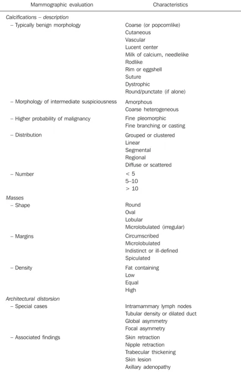
The observers described each lesion using the terminology from the fourth BI-RADS edition (Table 1) and the final mam-mography categorization included in the new BI-RADS subcategory 4 (Table 2). The radiologists did not receive any spe-cific training on the use of BI-RADS, thus the criteria adopted by each of the radiolo-gists were subjective, based on their previ-ous knowledge of BI-RADS guidelines as well as on their individual experience. Once the lesions were duly described, they were classified as shown on Table 2.
Category 3 was included in the group of benign lesions and classes 4 (probably be-nign) and 5 were brought together as ma-lignant. PPV and NPV were calculated for each class and description.
RESULTS
The present study population included 113 women and 2 men. The patients’ ages ranged from 37 to 61 years with a mean age of 49 years (± 12 years).
Biopsies of 115 breast masses detected at mammography were performed. Sixty-seven of these lesions (58.3%) were benign and 48 (41.7%) were malignant.
Based on the BI-RADS for mammog-raphy, the cases were thus classified by the observer A as follows: 66 (57.4%) category 3, 30 (26.1%) category 4, and 19 (16.5%) category 5. The observer B classified the cases as follows: 36 (31.3%) category 3, 54 (47.0%) category 4 and 25 (21.7) category 5. None of the cases were classified as cat-egories 0, 1, 2 and 6.
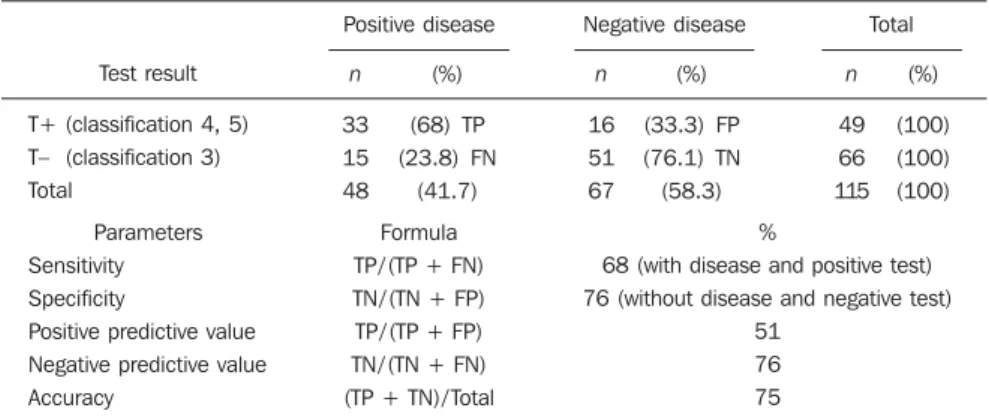
For the observer A, the NPV was 76% and PPV was 51%. The sensitivity was 68%, specificity 76% and accuracy 75% (Table 3). For observer B, NPV was 83% and PPV 53%. Sensitivity was 87%, speci-ficity 44% and accuracy 62% (Table 4).
Mammographic characteristics
The criteria described in the fourth BI-RADS edition were considered in the
Table 2 BI-RADS fourth edition – final categories(2,4).
Category
1 0 2 3 4A 4B 4C 5
Definitions
Negative
Additional evaluation required Benign findings
Probably benign findings Low malignancy suspicion Intermediate malignancy suspicion Moderate malignancy suspicion Highly suggestive of malignancy
Table 1 BI-RADS fourth edition terminology(2).
Mammographic evaluation
Calcifications – description
– Typically benign morphology
– Morphology of intermediate suspiciousness
– Higher probability of malignancy
– Distribution
– Number
Masses
– Shape
– Margins
– Density
Architectural distorsion
– Special cases
– Associated findings
Characteristics
Coarse (or popcornlike) Cutaneous
Vascular Lucent center
Milk of calcium, needlelike Rodlike
Rim or eggshell Suture Dystrophic
Round/punctate (if alone)
Amorphous
Coarse heterogeneous
Fine pleomorphic Fine branching or casting
Grouped or clustered Linear
Segmental Regional
Diffuse or scattered
< 5 5–10 > 10
Round Oval Lobular
Microlobulated (irregular)
Circumscribed Microlobulated Indistinct or ill-defined Spiculated
Fat containing Low Equal High
Intramammary lymph nodes Tubular density or dilated duct Global asymmetry
Focal asymmetry
described as round, vascular or punctate, 2 (2.9%) as amorphous, 5 (7%) as coarse heterogeneous, 4 (5.8%) as fine branching, and 17 (25%) as fine linear pleomorphic. The NPV for round calcifications was 65%. Of the two amorphous calcifications, one was benign and the other, malignant. Among the five calcifications described as coarse heterogeneous, two were malignant and three were benign, for a PPV of 40%. Those described as fine branching pre-sented a PPV of 75% and for the fine lin-ear pleomorphic ones the PPV was 94.7%. Evaluation of calcifications distribution – The calcifications were described as be-ing grouped by observer A in 13 cases, with 8 cases classified as BI-RADS 4 and 5, with a PPV of 45%. Regional calcifications were identified in 12 cases, with 7 malig-nant and 5 benign, with a PPV of 58%. Scattered or diffuse calcifications pre-sented a NPV of 42.8%. Segmental distri-bution was described in six cases, four of them malignant, with PPV of 66.6%. No case was described as linear ductal. Re-gional calcifications were described by observer B in 16 cases, with 9 being benign and 7 malignant, with a PPV of 45.7%. Calcifications were described as clustered in 20 cases, 14 of them included in BI-RADS categories 4 and 5, and 10 being malignant, with a PPV of 40%. Diffuse or scattered calcifications presented a NPV of 53.8%. Segmental distribution was de-scribed in nine cases, with five being ma-lignant and four benign, with a PPV of 55%. Architectural distortion – In the present study, the evaluation of architectural distor-tion (special cases and associated findings) could not be secondarily evaluated as the authors considered the number of presented cases as being insufficient.
Interobserver variability in mammography
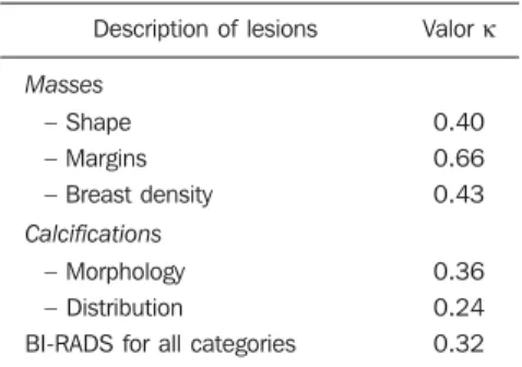
The interobserver variability analysis for the description of mammographic le-sions, using the Cohen’s κ test, is shown on Table 5.
Evaluation of masses on mammography
There was a low global agreement (κ = 0.40) in the description of mass margins. Similarly, a low agreement rate was ob-served in the description of microlobulated Table 3 Distribution of false-positive, true-positive, false-negative and true-negative results based on
the pathological diagnosis and indicated diagnosis (observer A).
Test result
T+ (classification 4, 5)
T– (classification 3) Total
Parameters
Sensitivity Specificity
Positive predictive value Negative predictive value
Accuracy
Positive disease Negative disease Total
n
33 15
48
n
16 51
67
n
49 66
115 (%)
(68) TP (23.8) FN
(41.7)
(%)
(33.3) FP (76.1) TN
(58.3)
(%)
(100) (100)
(100)
Formula
TP/ (TP + FN) TN/ (TN + FP)
TP/ (TP + FP) TN/ (TN + FN)
(TP + TN)/ Total
%
68 (with disease and positive test) 76 (without disease and negative test)
51 76
75
T+, positive test (lesion classified as BI-RADS 4 or 5); T–, negative test (lesion classified as BI-RADS 3). TP, true-positive; FP, false-true-positive; FN, false-negative; TN, true-negative.
Table 3 Distribution of false-positive, true-positive, false-negative and true-negative results based on the pathological diagnosis and indicated diagnosis (observer B).
Test result
T+ (classification 4, 5)
T– (classification 3) Total
Parameters
Sensitivity Specificity
Positive predictive value Negative predictive value
Accuracy
Positive disease Negative disease Total
n
42 6
48
n
37 30
67
n
79 36
115 (%)
(53.2) TP (16.7) FN
(41.7)
(%)
(46.8) FP (83.3) TN
(58.3)
(%)
(100) (100)
(100)
Formula
TP/ (TP + FN) TN/ (TN + FP)
TP/ (TP + FP) TN/ (TN + FN)
(TP + TN)/ Total
%
87 (with disease and positive test) 44 (without disease and negative test)
53 83
62
T+, positive test (lesion classified as BI-RADS 4 or 5); T–, negative test (lesion classified as BI-RADS 3). TP, true-positive; FP, false-true-positive; FN, false-negative; TN, true-negative.
evaluation of masses and calcification demonstrated by mammographic images.
Evaluation of breasts density – The glo-bal agreement for the evaluation of breast density was moderate (κ = 0.43). The PPV for heterogeneously dense breasts was 43.8% for observer A and 39.6% for ob-server B.
Evaluation of lesions margins and shape – According to observer A, the round shape presented NPV of 75% and for the oval lesions, 71%. Lobular lesions pre-sented a PPV of 70% and the microlobu-lated lesions presented a PPV of 90%. For observer B, the round shape presented a NPV of 70% and oval lesions, 66.7%. Lobular lesions presented a PPV of 75% and microlobulated lesions, 80%.
Evaluation of lesion margins – Accord-ing to observer A, the NPV for circum-scribed margins was 84.2%, while the PPVs for indistinct and spiculated margins
were 24.5% and 90%, respectively. Ac-cording to observer B, the NPV for circum-scribed margins was 80.2% and the PPVs for indistinct and spiculated margins were 25.4% and 83.3% respectively.
Evaluation of calcifications morphol-ogy – According to observer A, of the 76 reported calcifications, 23 (30%) were de-scribed as round, vascular or punctate, 6 (8%) as amorphous, 2 (2.6%) as coarse heterogeneous, 32 (42%) as fine branching and 12 (18%) as fine linear pleomorphic. The NPV for round calcifications was 56.5%. Amorphous calcifications pre-sented a NPV of 66.6%. Calcifications described as fine branching presented a PPV of 72.7% and those described as fine linear pleomorphic presented a PPV of 91.6%. The two calcifications described as coarse heterogeneous were benign.
margins (κ = 0.38) and oval shaped masses (κ = 0.32).
A significant global agreement (κ = 0.66) was observed in the evaluation of mass margins, particularly for spiculated margins (κ = 0.70). Global agreement for masses density was moderate (κ = 0.43).
Mammographic evaluation of calcifications
Agreement was almost almost perfect as the presence of calcifications was evalu-ated (κ = 0.88). The observers demon-strated low global agreement in the descrip-tion of calcificadescrip-tions morphology (κ = 0.36). The use of the terms “amorphous” and “fine branching” resulted in a moder-ate agreement (κ =0.41 and κ = 0.43, re-spectively). Agreement was poor for the use of the terms “coarse heterogeneous” (κ = 0.23) and fine pleomorphic (κ = 0.25). Poor agreement was also observed in cal-cifications distribution evaluation (κ = 0.24) (Table 5). Agreement was also poor in the evaluation of presence of architec-tural distortion (κ = 0.23).
Interobserver agreement in relation to the presence of associated findings and special cases could not be secondarily evaluated, considering the low number of cases with architectural distortion.
Final categories evaluation
Poor agreement was observed in the evaluation of final categories.
The highest agreement rate was ob-served for lesions classified with high like-lihood of malignancy, or category 5 (κ = 0.42). Poor agreement was observed for categories 3 (κ = 0.30), 4A (κ = 0.15), 4B (κ = 0.13) and 4C (κ = 0.16). For catego-ries 4, even when grouped (κ = 0.27), poor agreement was observed.
For the final BI-RADS categories, poor interobserver agreement was observed (κ = 0.32) (Table 5).
DISCUSSION
In the present study, the utilization of criteria for breasts density, masses margins, shape, calcifications morphology and dis-tribution was evaluated.
Mammography sensitivity ranged from 68% to 87% between the observers
(iden-tification of malignant lesions in patients with breast cancer), and the NPV was high, ranging between 76% and 83% (identifica-tion of negative findings in cancer-regard-ing characteristics described by the BI-RADS. BI-RADS presented specificity between 76% and 44% (patients without the disease, with negative tests). The PPV (number of cancer cases for mammo-graphic characteristics) ranged between 51% and 53% between the two observers, a rate not distant from results in the stud-ies developed by Burnside et al.(3,6) and
Kerlikowske et al.(10).
The mammographic accuracy ranged between 75% and 62% in the differentia-tion between benign and malignant lesions with the use of BI-RADS. The NPV for category 3 ranged between 76.1% and 83% between both observers, close to the values described by Roveda Junior et al.(14)
It is a known fact that there is a direct association between the increased mammo-graphic density and an increase in the risk for development of breast cancer(15,16). In
the present study, the PPV for heteroge-neously dense breasts were 43.8% for ob-server A and 39.6% for obob-server B. Mod-erate interobserver agreement (κ = 0.43) was observed in the evaluation of breasts density, differently from the findings de-scribed in the study developed by Nichol-son et al.(13), in which the interobserver
agreement in the evaluation of breasts den-sity was 78.4% for extremely dense breasts, and 51.2% for heterogeneously dense breasts, probably because of the different apparatuses utilized for images processing. The present study suggests that the mass margins are useful in the prediction of malignancy, with a lower probability for carcinomas in lesions with well-defined
margins, and high probability in lesions with spiculated margins (non circum-scribed), with NPV between 80% and 84% and PPV between 90% and 93%, respec-tively, for observers A and B, as described by Kestelman et al.(17). It is known that,
according to Nascimento et al.(18), the
sonographic method has also presented high PPV, at 82.4%, in the description of mass margins(19).
As regards to round and oval shapes, these were associated to a high NPV, be-tween 75% and 71% for observer A and between 70% and 66.7% for observer B. Microlobulated and lobular shapes pre-sented a high PPV, between 90% and 70% for observer A, and 80% and 75% for ob-server B. In the present study a moderate interobserver agreement was observed in the global description of mass margins (κ =0.66), in agreement with the findings re-ported by Kerlikowske et al.(10).
In the present study, the observer A iden-tified a high PPV in the description of fine branching microcalcifications and in fine linear pleomorphic microcalcifications (91,6%), a NPV of 56.5% for calcifications described as round, vascular or punctate and 66.6% for the amorphous ones. Ob-server B identified a PPV of 75% for fine branching calcifications, and 94.7% for fine linear pleomorphic calcifications, NPV of 65% for calcifications described as round, vascular or punctate, and 50% for the amorphous ones. For observer B, coarse heterogeneous microcalcifications pre-sented a PPV of 40%. The present study is in agreement with the study developed by Melhado et al.(20), which demonstrated a
progressive increase of PPV in BI-RADS categories 4A, 4B and 4C, suggesting that this subdivision contributes in a more pre-cise manner in the indication of suspicious lesions.
However, poor interobserver agreement was observed for the description of calci-fications morphology (κ = 0.36) and distri-bution at mammography (κ =0.24) were low, as were those described in the litera-ture according to Berg et al.(11) and Lazarus
et al.(12).
In the present study, the poor interob-server agreement in the evaluation of cat-egories 4A (κ = 0.15), 4B (κ = 0.13), 4C (κ = 0.16) and combined categories 4 (κ = Table 5 Interobserver variability in the
descrip-tion of mammographic lesions.
Description of lesions
Masses
– Shape
– Margins – Breast density
Calcifications
– Morphology – Distribution
BI-RADS for all categories
Valor κ
0.40
0.66 0.43
0.36 0.24
0.27) were possibly associated to the high number of offered categories. A higher interobserver agreement was observed in category 5 (κ = 0.42).
CONCLUSION
The present study demonstrated that breast evaluation by mammography, utiliz-ing the BI-RADS classification, is an ac-curate method in the differentiation be-tween benign and malignant lesions. The most frequent findings related to neoplasias were masses with spiculated margins, microlobulated (irregular) shape, lobular mass, fine branching microcalcifications and linear fine pleomorphic calcifications. A high interobserver agreement was not achieved in the analysis of calcifications morphology and distribution, possibly be-cause of the high number of offered catego-ries. However, a progressive increase was observed in the PPVs in subcategories 4A, 4B and 4C, suggesting that such break-down contributes in a more detailed man-ner for the identification of suspiciously malignant lesions. Such stratification may be useful for the communication of suspi-ciousness levels to physicians and patients, who may benefit from this information in their decision making processes.
It must also be highlighted that breast lesions related to BI-RADS category 3 pre-sented a high NPV which should be con-sidered as an relevant factor in the conser-vative management of such lesions with the purpose of avoiding unnecessary biopsies.
REFERENCES
1. American College of Radiology. Mammography. Illustrated Breast Imaging Reporting and Data System (BI-RADS). 4th ed. Reston: American College of Radiology; 2003.
2. Vizcaíno I, Gadea L, Andreo L. Short-term follow-up results in 795 nonpalpable probably benign lesions detected at screening mammography. Radiology. 2001;219:475–83.
3. Burnside ES, Ochsner JE, Fowler KJ, et al. Use of microcalcification descriptors in BI-RADS 4th edition to stratify risk of malignancy. Radiology. 2007;242:388–95.
4. Liberman L, Abramson AF, Squires CB, et al. The Breast Imaging Reporting and Data System: posi-tive predicposi-tive value of mammographic features and final assessment categories. AJR Am J Roent-genol. 1998;171:35–40.
5. Berg WA, Arnoldus CL, Teferra E, et al. Biopsy of amorphous breast calcifications: pathologic outcome and yield at stereotactic biopsy. Radiol-ogy. 2001;221:495–503.
6. Burnside ES, Rubin DL, Fine JP, et al. Bayesian network to predict breast cancer risk of mammo-graphic microcalcifications and reduce number of benign biopsy results: initial experience. Radiol-ogy. 2006;240:666–73.
7. Godinho ER, Koch HA. Submissão às recomen-dações do BI-RADS® por médicos e pacientes: análise preliminar de 3.000 exames realizados em uma clínica particular. Radiol Bras. 2004;37:21–3. 8. Kolb TM, Lichy J, Newhouse JH. Comparison of the performance of screening mammography, physical examination, and breast US and evalu-ation of factors that influence them: an analysis of 27,825 patient evaluations. Radiology. 2002; 225:165–75.
9. Kerlikowske K, Smith-Bindman R, Ljung BM, et al. Evaluation of abnormal mammography results and palpable breast abnormalities. Ann Intern Med. 2003;139:274–84.
10. Kerlikowske K, Grady D, Barclay J, et al. Vari-ability and accuracy in mammographic interpre-tation using the American College of Radiology Breast Imaging Reporting and Data System. Natl Cancer Inst. 1998;90:1801–9.
11. Berg WA, Campassi C, Langenberg P, et al. Breast Imaging Reporting and Data System: inter- and intraobserver variability in feature analysis and final assessment. AJR Am J Roentgenol. 2000; 174:1769–77.
12. Lazarus E, Mainiero MB, Schepps B, et al. BI-RADS lexicon for US and mammography: interobserver variability and positive predictive value. Radiology. 2006;239:385–91.
13. Nicholson BT, LoRusso AP, Smolkin M, et al. Accuracy of assigned BI-RADS breast density category definitions. Acad Radiol. 2006;13: 1143–9.
14. Roveda Junior D, Piato S, Oliveira VM, et al. Valores preditivos das categorias 3, 4 e 5 do sis-tema BI-RADS em lesões mamárias nodulares não-palpáveis avaliadas por mamografia, ultra-sonografia e ressonância magnética. Radiol Bras. 2007;40:93–8.
15. Boyd NF, Dite GS, Stone J. Heritability of mam-mographic density, a risk factor for breast cancer. N Engl J Med. 2002;347:886–94.
16. Warner E, Lockwood G, Tritchler D, et al. The risk of breast cancer associated with mammographic parenchymal patterns: a meta-analysis of the pub-lished literature to examine the effect of method of classification. Cancer Detect Prev. 1992;16: 67–72.
17. Kestelman FP, Souza GA, Thuler LC, et al. Breast Imaging Reporting and Data System – BI-RADS®: valor preditivo positivo das categorias 3, 4 e 5. Revisão sistemática de literatura. Radiol Bras. 2007;40:173–7.
18. Nascimento JHR, Silva VD, Maciel AC. Acurá-cia dos achados ultrassonográficos do câncer de mama: correlação da classificação BI-RADS® e achados histológicos. Radiol Bras. 2009;42:235– 40.
19. Arantes Pereira FP. BI-RADS® ultrassonográfico: análise de resultados iniciais [editorial]. Radiol Bras. 2009;42(4):vii–viii.