J Bras Pneumol. 2008;34(1):55-58
55
Case Reports
* Study carried out at the Faculdade de Ciências Médicas da Santa Casa de São Paulo – FCMSCSP, Santa Casa School of Medical Sciences in São Paulo – São Paulo, Brazil.
1. Tenured Professor in the Surgery Department. Faculdade de Ciências Médicas da Santa Casa de São Paulo – FCMSCSP, Santa Casa School of Medical Sciences in São Paulo – São Paulo, Brazil.
2. PhD Professor in the Surgery Department. Faculdade de Ciências Médicas da Santa Casa de São Paulo – FCMSCSP, Santa Casa School of Medical Sciences in São Paulo – São Paulo, Brazil.
3. Coordinator of the Academic Group for the Study of Thoracic Surgery. Faculdade de Ciências Médicas da Santa Casa de São Paulo – FCMSCSP, Santa Casa School of Medical Sciences in São Paulo – São Paulo, Brazil.
4. PhD Professor in the Thoracic Surgery Department. Faculdade de Ciências Médicas da Santa Casa de São Paulo – FCMSCSP, Santa Casa School of Medical Sciences in São Paulo – São Paulo, Brazil.
5. Intern in the Thoracic Surgery Department. Faculdade de Ciências Médicas da Santa Casa de São Paulo – FCMSCSP, Santa Casa School of Medical Sciences in São Paulo – São Paulo, Brazil.
Correspondence to: Roberto Saad Junior. Faculdade de Ciências Médicas da Santa Casa de São Paulo, Rua Dr. Cesário Motta Jr., 112, Santa Cecília, CEP 01221-020, São Paulo, SP, Brasil.
Tel 55 11 2176-7000. E-mail: [email protected] Submitted: 9 August 2006. Accepted, after review: 5 March 2007.
Mediastinal liposarcoma: a case report*
Roberto Saad Junior1, Vicente Dorgan Neto2, Roberto Gonçalves3,
Márcio Botter4, Leticia Cristina Dalledone Siqueira5
Abstract
Here, we describe the case of a 51-year-old female with mediastinal liposarcoma. Liposarcoma is the most common malignant mesenchymal neoplasm in adults, although a mediastinal location is extremely rare. It has a large volume and varied histologic subtypes. It is characterized by the compression of neighboring structures. Computed tomography and magnetic resonance imaging provide useful data for diagnosis. Tissue biopsy and histological typing are very important in determining the treatment and are needed for the final diagnosis. Radiotherapy and chemotherapy are ineffective treatment modalities. According to the literature, surgical resection is the treatment of choice. Long-term follow-up evaluation is indicated since there is a high rate of recurrence.
Keywords: Liposarcoma; Mediastinal neoplasms; Case reports [publication type].
Introduction
Liposarcoma is one of the most common malignant soft tissue tumors seen in adults.(1,2) It is of mesenchymal origin
and is derived from adipocytes. It affects both genders equally and typically appears in the fourth decade of life. It presents slow growth and becomes symptomatic when it assumes large dimensions.(2) Retroperitoneal liposarcoma has
been well documented. However, it appears in other loca-tions in 20% of cases.(3) However, it is uncommon to find
it in the mediastinum, mediastinal liposarcoma accounting for only 0.2% of all cases. According to the World Health Organization, liposarcomas are divided into 5 subtypes: well-differentiated, myxoid, round cell, dedifferentiated, and pleomorphic.(4) In the present study, we present two variants
within the same tumor: well-differentiated ( lipoma-like) liposarcoma and dedifferentiated liposarcoma.
Case report
56 Saad Junior R, Dorgan Neto V, Gonçalves R, Botter M, Siqueira LCD
J Bras Pneumol. 2008;34(1):55-58
located posteriorly to the heart, anteriorly to the aorta, and to the right of the esophagus, with loose adhesions to the structures mentioned, and it was possible to remove the entire tumor through a left thoracotomy and through the opening in the medi-astinal pleurae. Smaller tumors were dissected from the left paravertebral sulcus. Subsequently, chest tubes were inserted, and the incision was closed in layers. The drains were removed on postop-erative day 3, and the patient was discharged on postoperative day 5. In the macroscopic and anato-mopathological examination, the largest sample was seen to have an hourglass shape and found to weigh 994 g, with two lobes: one globular, 10 cm in diameter, firm, whitish, and fasciculated with gelatinous areas; and the other irregular in shape, measuring 14 × 9 × 8 cm, and yellowish upon dissection. An additional eight samples, irregular in shape and weighing 234 g altogether, were exam-ined. Each was partially covered by a smooth and glossy membrane, with foci of hemorrhage, and, upon dissection, was yellow with whitish streaks and firm. In some cases, there was a bony consist-ency. Microscopy (Figure 3) revealed immature mesenchymal neoplasia characterized by the prolif-eration of anaplastic spindle cells with numerous atypical mitoses and bizarre multinucleated giant and normal hemodynamics. Pulmonary
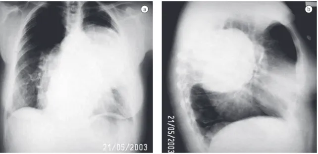
ausculta-tion revealed bilateral breath sounds, which were reduced in the upper third of the left hemithorax, reduced vocal fremitus, underlying fluid density upon percussion, and reduced bronchophony, also in the upper third of the left hemithorax, without adventitious sounds. An anteroposterior X-ray (Figure 1a) revealed that, in two-thirds of the left hemithorax, there was opacity, which extended to the cardiac silhouette, suggesting that the tumor was located anterior to the ascending aorta. In a lateral X-ray (Figure 1b), the image was sugges-tive of a tumor in the paravertebral sulci, extending posteriorly to the heart. A tomography scan of the chest (Figure 2a) revealed a mass in the left paraver-tebral sulcus, extending to the right paraverparaver-tebral sulcus, with central and peripheral calcifications. In order to plan the surgical procedure, we continued the investigation using magnetic resonance imaging (Figure 2b), which revealed a mass in the left para-vertebral sulcus, extending to the right parapara-vertebral sulcus, and maintaining cleavage planes with the adjacent structures. A transparietal puncture biopsy was carried out, and a diagnosis of liposarcoma was made. Resective surgery was then indicated. A left posterior lateral thoracotomy was performed in the fourth intercostal space. We dissected a tumor
a b
Mediastinal liposarcoma: a case report
J Bras Pneumol. 2008;34(1):55-58
57
accompanied by cytogenetic abnormalities and many cellular atypias, including with other mesen-chymal components, such as the cartilaginous and the muscular one.(3,4,10)
Mediastinal liposarcoma presents as a large tumor that exhibits a variety of symptoms, depending on its location and on the compression of neighboring structures.(11) It remains asymptomatic until reaching
a considerable volume, although the symptoms remain nonspecific.(2,8,12) Most patients complain of
dyspnea or chest pain, and some also report cough or weight loss.(12,13)
Although the radiological findings are nonspe-cific, computed tomography and magnetic resonance imaging provide useful data.(7) The tumor usually
presents with invasive characteristics. Biopsy is necessary for the diagnosis. The histological classi-fication, together with the correlation between that classification and the clinical course, is important to determining the therapeutic approach.(11) The
differential diagnosis should include lipoma, angi-olipoma, fibromyxangi-olipoma, and myolipoma.(3,4,14)
Radiotherapy and chemotherapy have limited value and are ineffective treatment modalities.(8,11)
Radiotherapy can be useful in the palliation of unresectable tumors or in the adjuvant treatment of those incompletely resected.(11,15) The role of
adju-vant chemotherapy remains controversial.(12)
The treatment of choice is surgery, and complete resection of the tumor is recommended whenever possible.(1,11,15) According to the literature, aggressive
surgical intervention seems to favor the quality of life of patients and prolong survival, being consid-cells. The remaining samples consisted of
prolifera-tion of adipocytes, forming lobes amidst streaks of fibrosis and containing anaplastic cells with dense, strong chromatin staining. We concluded that it was a case of well-differentiated liposarcoma ( lipoma-like) and dedifferentiated liposarcoma. The patient received adjuvant chemotherapy and under-went outpatient follow-up treatment for 3 years without recurrence.
Discussion
Although liposarcoma is the most common tumor of mesenchymal origin in adults, a medi-astinal location is extremely rare.(1-8) According to
the experience of the Armed Forces Institute of Pathology, in collaboration with the Mayo Clinic, 75% of the cases of liposarcoma develop in deep muscle tissues of the extremities, 20% develop in the retroperitoneum, and the remainder develop at other sites.(3) Primary mediastinal liposarcoma
accounts for only 0.13-0.75% of all mediastinal tumors.(9) In 2003, there were fewer than 100 cases
reported in the international literature, 36 of which occurred in Japan.(8)
These tumors can present as well-differentiated neoplasms with a pseudocapsule, which makes it difficult to characterize malignancy, and it is neces-sary to analyze all of the surgical samples in search of cellular atypias. However, they can manifest as high-grade tumors, which makes prognosis poor. They are divided into histopathological subtypes that behave differently, and the dedifferentiated liposarcoma is the most aggressive variant, being
Figure 2 - Images of the chest: a) High tomographic slices in which the tumor only appears on the left side, adjacent to the paravertebral sulcus (note the various calcifications); and b) Nuclear magnetic resonance imaging revealing tumor in the two paravertebral sulci, giving the impression of a single hourglass-shaped tumor.
58 Saad Junior R, Dorgan Neto V, Gonçalves R, Botter M, Siqueira LCD
J Bras Pneumol. 2008;34(1):55-58
ered the most effective treatment.(8) Surgery can be
curative in most cases.(7,13)
Prognosis depends on the histological subtype and on surgical resection with wide margins of safety.(13,15)
Recurrence is seen in approximately 40% of the cases and has been reported at up to 14 years after the initial surgical procedure.(13) Long-term
post-operative follow-up evaluation is necessary, since there can be an interval between resection and recurrence.(5,16)
We conclude that mediastinal liposarcoma is a rare tumor characterized by its considerable volume. It requires a comprehensive surgical approach, on which the prognosis of patients will depend. The need for effective postoperative follow-up evalua-tion should also be emphasized since there is a high rate of recurrence.
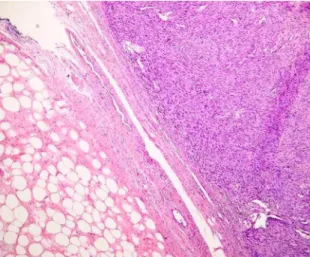
Figure 3 - Photomicrograph showing the border between the two histopathological variants: to the left, the well-differentiated liposarcoma; and, to the right, the dedifferentiated liposarcoma (hematoxylin and eosin staining, magnification of ×100).
References
1. Kiyama H, Tanabe S, Nagasawa S, Irie Y, Ohshima N, Yamada T. [A case of primary anterior mediastinal liposarcoma with a heterotopic mass in the pericardium of the same histology] [Article in Japanese]. Nippon Kyobu Geka Gakkai Zasshi. 1996;44(12):2191-5.
2. Mase T, Kawawaki N, Narumiya C, Aoyama T, Kato S, Nagata Y. Primary liposarcoma of the mediastinum. Jpn J Thorac Cardiovasc Surg. 2002;50(6):252-5.
3. Weiss SW. Liposarcoma. In: Weiss SW, Goldblum JR, Enzinger FM, editors. Enzinger and Weiss’s soft tissue tumors. 4th ed. St Louis: Mosby; 2001. p. 641-94.
4. Wick MR. The mediastinum. In: Sternberg SS, Antonioli DA, editors. Diagnostic Surgical Pathology. Philadelphia: Lippincott Williams and Wilkins; 1999. p. 1147-1208. 5. Okumori M, Mabuchi M, Nakagawa M. Malignant thymoma
associated with liposarcoma of the mediastinum - a case report. Jpn J Surg. 1983;13(6):512-8.
6. Yonehara S, Kou E, Hasegawa K, Kamitsuna A, Katsuta S, Okumichi T, et al. [Liposarcoma of the mediastinum--a case report] [Article in Japanese]. Gan No Rinsho. 1986;32(7):810-4.
7. Rivo JE, Cañizares MA, García-Fontán E, Albort J, González-Piñeiro A, Peñalver R. [Liposarcomas of the mediastinum in atypical locations: report of two cases] [Article in Spanish]. Cir Esp. 2005;77(2):99-101.
8. Teschner M, Lüllig H. [Diagnosis and treatment of primary mediastinal liposarcoma] [Article in German]. Pneumologie. 2003;57(1):22-6.
9. Tanaka F, Kitano M, Tatsumi A, Huang C, Nagasawa M. [A case of mediastinal liposarcoma] [Article in Japanese]. Nippon Kyobu Geka Gakkai Zasshi. 1992;40(7):1125-30. 10. Taira N, Kinoshita S, Miyake T, Hara F, Nakajima T. [Primary
liposarcoma of the anterior mediastinum--case report and review of literature] [Article in Japanese]. Jpn J Thorac Cardiovasc Surg. 1998;46(5):450-4.
11. Doğan R, Ayrancioğlu K, Aksu O. Primary mediastinal liposarcoma. A report of a case and review of the literature. Eur J Cardiothorac Surg. 1989;3(4):367-70.
12. Meyer M, Holzhausen HJ, Neef H, Zerkowski HR. [Primary liposarcomas of the mediastinum] [Article in German]. Langenbecks Arch Chir Suppl Kongressbd. 1998;115:369-73.
13. Bonnette P, Jouan J, Colchen A, Epardeau B, Grapin D, Grapin JP. [Myxoid liposarcoma of the mediastinum] [Article in French]. Rev Mal Respir. 2000;17(1):109-11.
14. Ximenes M, Barbosa JR.Tumores do mediastino. In: Saad Jr R, Ximenes Netto M, editors. Cirurgia torácica. São Paulo: Editora Atheneu; 1997. p. 155-72.
15. Farah M, Abou-Sleiman P, Bahous J. Primary mediastinal liposarcoma: a case report and review of the literature. J Med Liban. 2001;49(3):165-9.