DOI: 10.5935/2359-4802.20170021
177 International Journal of Cardiovascular Sciences. 2017;30(2):177-180
CASE REPORT
Mailing Address: Rodrigo de Moura Joaquim
Rua São Francisco, 217, apt. 701, Centro. Postal Code: 88015-140, Florianópolis, SC – Brazil E-mail rodrigojoaquim@gmail.com
Caseous Necrosis of the Mitral Valve: Imaging Methods Allow the Diagnosis and
Prevent Surgery
Rodrigo de Moura Joaquim, Edileide de Barros Correia, Suéllen Lacerda Bezerra, Ibraim Masciarelli Francisco Pinto, Tiago Senra Garcia dos Santos, Fabiano de Castro Albrecht
Instituto Dante Pazzanese de Cardiologia, São Paulo, SP – Brazil
Manuscript received July 01, 2016; revised manuscript January 17, 2017; accepted January 23, 2017.
Calcinosis; Mitral Valve; Heart Neoplasms; Diagnostic Imaging; Magnetic Resonance Imaging; Multidetector Computed Tomography.
Keywords
Introduction
Calcification of the mitral annulus (CMA) is defined as a chronic degeneration of the mitral annulus fibrous involving mainly the posterior annulus.1 It is a common
condition in the elderly, especially in women, and is associated with hypertension.2 Caseous necrosis of the
mitral valve (CNMV) is a rare and less known CMA variant with a prevalence of 0.64%, which can reach up to 2.7% in autopsy studies of patients with CMA.1-3
On echocardiography, CNMV appears as a large rounded or semilunar echodense mass, with a liquid filling and central echolucency, similar to a periannular tumor.4,5 The mechanism of liquefaction and caseification
are still not well understood, but it is believed to occur due to changes in calcium metabolism.1
Case Report
A 66-year-old female patient with a history of hypertension and chronic renal disease with an atrophic right kidney was referred to the emergency department of our institution after presenting a left atrial tumor mass with atypical characteristics, evidenced on echocardiography at another institution. She reported on the occasion a retrosternal and epigastric pressure-type pain associated with gastric fullness, without relation to physical effort, variable duration from minutes to hours, and without other associated symptoms. She also presented dyspnea on exertion.
T h e p a t i e n t w a s h o s p i t a l i z e d t o u n d e r g o complementary tests and for a possible surgical intervention. The electrocardiogram showed a sinus rhythm and presented criteria for left ventricular overload. Transthoracic echocardiography showed an enlarged left atrium (46 mm) with an indexed volume of 63 mL/m2, moderate concentric left ventricular
hypertrophy, a 76% left ventricular ejection fraction, mild tricuspid insufficiency, thickened aortic valve without a significant gradient, and a sessile heterogeneous image with areas of calcification adhered to the ventricular face of the posterior leaflet of the mitral valve, restricting its mobility and opening, measuring 25 x 18 mm and causing mild reflux, with a valvular area of 1.9 cm2 (pressure
half-time [PHT]) and a maximum transvalvular gradient of up to 12 and a mean of 4.
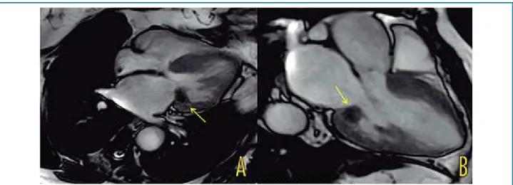
A magnetic resonance imaging of the heart (Figure 1) showed dilatation of the left atrium, mitral insufficiency, concentric and asymmetric left ventricular hypertrophy predominantly in the septum, measuring up to 23 mm in the middle and basal regions, and a cystic mass with well-defined borders adhered to the posterior leaflet of the mitral valve, with a heterogeneous content and a halo on late enhancement.
Coronary computed tomographic angiography (Figure 2) revealed no significant coronary obstructions and identified a well-delineated mass in the inferolateral portion of the mitral annulus, with regular borders and a fibrotic thin layer with high-intensity interior attenuation and focal coarse calcifications measuring 17 x 26 x 22 mm in the anteroposterior, transversal, and longitudinal axis, respectively.
178
Figure 1 – Cardiac magnetic resonance. A) Apical four-chamber view; B) Apical three-chamber view.
Figure 2 – Cardiac computed tomography. A) Two-chamber view; B) Four-chamber view; C) Calcium score.
Joaquim et al
Diagnosis of caseous necrosis of the mitral valve
Int J Cardiovasc Sci. 2017;30(2):177-180 Case Report
Since the patient was oligosymptomatic and had a mild mitral insufficiency, valvular intervention was not recommended at the time of diagnosis. The patient did not present any suggestive manifestations of previous embolism
179
1. Elgendy IY, Conti CR. Caseous calcification of the mitral annulus: a review. Clin Cardiol. 2013;36(10):E27-31.
2. Deluca G, Correale M, Ieva R, Del Salvatore B, Gramenzi S, Di Biase M. The incidence and clinical course of caseous calcification of the mitral annulus: a prospective echocardiographic study. J Am Soc Echocardiogr. 2008;21(7):828-33.
3. Pomerance A. Pathological and clinical study of calcification of the mitral valve ring. J Clin Pathol. 1970;23(4):354-61.
4. Stamou SC, Braverman AC, Kouchoukos NT. Caseous calcification of the anterior mitral valve annulus presenting as intracardiac mass. J Thorac Cardiovasc Surg. 2010;140(1):e9-10.
5. Harpaz D, Auerbach I, Vered Z, Motro M, Tobar A, Rosenblatt S. Caseous calcification of the mitral annulus: a neglected, unrecognized diagnosis. J Am Soc Echocardiogr. 2001;14(8):825-31.
6. Corre J, Leroux L, Coste P. Caseous necrosis of mitral annulus: a rare cause of stroke. Case Rep Cardiol. 2013;2013:748241.
References
Joaquim et al Diagnosis of caseous necrosis of the mitral valve Int J Cardiovasc Sci. 2017;30(2):177-180
Case Report
Discussion
The most common presentation of CNMV is as an intracardiac mass, and its diagnosis is incidental. When clinical manifestation occurs, it is usually with palpitations and dyspnea, and rarely with syncope due to atrioventricular blockade. These patients may present several other clinical findings related to the valvular lesion, with stenosis or insufficiency and, eventually, findings of systemic embolism.1,2,5
Harpaz et al.5 evaluated a series of 19 patients of whom
18 had isolated involvement of the posterior leaflet of the mitral annulus and one presented associated involvement of the anterior and posterior leaflets. In the same series, these authors observed that five patients (23%) presented some type of cerebrovascular event and only three patients underwent mitral valve replacement, while 84% of them were treated conservatively during a mean follow-up of 3.8 years.5 Several other cases of CNMV
presenting as a stroke have already been described in the literature, demonstrating the importance of such type of event in this population.6-8 The mechanisms for the
occurrence of stroke are unclear; it is unknown whether they occur due to their association with atherosclerosis, age and other risk factors related to the patients, or embolization of fragments of the mass.6
The differential diagnoses include intracardiac tumors, especially atrial myxoma, thrombosis of the coronary sinus, circumflex artery anomalies, and vegetations and abscesses of the mitral annulus.1,9,10 Multiple modalities
of cardiovascular imaging should be used to differentiate between the various lesions and, in these cases, are capable of establishing a definitive diagnosis without requiring pathological analysis of the mass, eliminating invasive surgical procedures.9,10 Follow-up after diagnostic
definition may be performed only with echocardiography at the discretion of the attending physician.10
CNMV is a dynamic process that, in some cases, may resolve spontaneously and even recur after surgically excised, depending on the valvular degeneration. Surgery is recommended upon dysfunction of the mitral valve with important stenosis or insufficiency, embolic events or when other types of tumors cannot be excluded regardless of the imaging method used.1 Anticoagulation
may be prescribed in cases of embolic events, but no consensus has been established in this regard.8
Even though CNMV is a rare condition, it is important for cardiologists performing imaging tests to be familiar with this diagnosis, in order to avoid confusion with other masses or abscesses, thus preventing unnecessary surgery.1
Author contributions
Conception and design of the research: Joaquim RM, Bezerra SL. Acquisition of data: Joaquim RM, Bezerra SL. Analysis and interpretation of the data: Pinto IMF, Santos TSG. Writing of the manuscript: Joaquim RM, Correia EB, Albrecht FC. Critical revision of the manuscript for intellectual content: : Joaquim RM, Correia EB, Albrecht FC.
Potential Conflict of Interest
No potential conflict of interest relevant to this article was reported.
Sources of Funding
There were no external funding sources for this study.
Study Association
180
7. Chevalier B, Reant P, Laffite S, Barandon L. Spontaneous fistulization of a caseous calcification of the mitral annulus: an exceptional cause of stroke. Eur J Cardiothorac Surg. 2011;39(6):e184-5.
8. Higashi H, Ohara T, Nakatani S, Hashimoto S, Torii T, Miyashita K, et al. A case of caseous calcification of the mitral annulus: a potential source of embolic stroke. J Cardiol Cases. 2010;2:e141-3.
9. Kydd AC, Gopalan D, Clarke SC, Rusk RA. Mitral annular caseous necrosis: insights from multimodality imaging. Eur Heart J. 2013;34(13):971.
10. Ribeiro S, Salgado A, Salomé N, Bettencourt N, Azevedo P, Costeira A, et al. Caseous calcification of the mitral annulus: A multi-modality imaging perspective. Rev Port Cardiol. 2012;31(4):313-6.
Joaquim et al
Diagnosis of caseous necrosis of the mitral valve