110
INTRODUCTION
The incidence of pulmonary tuberculosis (TB) is almost 500 times greater in acquired immu-nodeficiency syndrome (AIDS) patients com-pared with the general population.1 Moreover,
the incidence of central nervous system (CNS) TB is significantly high in AIDS patients.2
However, primary central nervous system lym-phoma (PCNSL) is also common in patients with AIDS.3 Although meningitis is the most
common presentation of CNS TB other forms including cerebritis, abscess, or tuberculoma are known to occur frequently in patients with AIDS.4
TB meningitis typically evolves over 1-2 weeks but may present acutely with headache, mental changes (confusion/lethargy), neck rigidity, low-grade fever, malaise, anorexia, irritability,
and paresis of cranial nerves. Involvement of cerebral arteries may produce focal ischaemia, coma with hydrocephalus and intracranial hy-pertension. Tuberculoma is uncommon and presents with seizures and focal signs. Although toxoplasma encephalitis and CNS lymphoma are more commonly encountered in HIV-in-fected patients, CNS TB is a significant cause of cerebral infection.
Lumbar puncture is the cornerstone for diag-nosis of TB meningitis. Computed tomogra-phy (CT) or magnetic resonance imaging (MRI) brain may show hydrocephalus and abnormal enhancement of basal cisterns or ependyma. Whereas, in CNS tuberculoma CT or MRI re-veals contrast-enhanced ring lesions. A biopsy is necessary to establish diagnosis.
Case Report:
Atypical presentation of a common opportunistic infection in advanced
AIDS
N. Chandra,1 N. Krishna,1 M.V.S. Subbalaxmi,1 M. Nageshwar Rao,1 Y.S.N. Raju,1 Y. Jyotsna Rani 2
Departments of 1General Medicine, 2Radiology & Imageology, Nizam's Institute of Medical Sciences, Hyderabad
ABSTRACT
Intracranial tuberculosis in immunocompromised patients can occasionally mimic central nervous system (CNS) neo-plasms radiologically and complicate the decisions regarding management. A 42-year-old male presented with a history of fever and vomitings of 5 days duration. On evaluation he was found to be reactive for human immunodeficiency virus 1 infection with a CD4+ count of 63 cells/mm3 and a viral load of 1,260,779 copies /mL. He was started on highly active
antiretroviral therapy with tenofovir, emtricitabine, efavirenz, Pneumocystis jiroveci prophylaxis and was discharged. After 5 months he developed aggressive behaviour, irrelevant talking and memory loss. On examination, he was irri-table with memory disturbances; no focal neurological signs were evident. Magnetic resonance imaging brain and magnetic resonance spectroscopy (MRS) showed a large heterogeneous enhancing ill-defined lesion in the left parieto-occipital lobe with a lipid lactate peak suggestive of infective aetiology. Cerebrospinal fluid (CSF) analysis showed glucose 33 mg/dL, protein 120 mg/dL, 40 cells/mm3 (all lymphocytes), adenosine deaminase 40U/L; Gram's stain was
negative, Ziehl-Neelsen stain did not reveal acid-fast bacilli, toxoplasma, cryptococcal antigen tests were negative. Polymerase chain reaction for Epstein-Barr virus was also negative. In view of the clinical setting, CSF analysis sup-ported by MRS findings he was started on antituberculosis treatment (ATT) and corticosteroids. Patient showed re-markable improvement clinically and radiologically with significant reduction in the size of the lesion. MRS is a useful non-invasive technique that can help in differentiating tuberculoma from lymphoma.
Key words:Tuberculoma, Lymphoma, Magnetic resonance spectroscopy
Chandra N, Krishna N, Subbalaxmi MVS, Nageshwar Rao M, Raju YSN, Jyotsna Rani Y. Atypical presentation of a common opportunistic infection in advanced AIDS. J Clin Sci Res 2013;2:110-3.
Corresponding author: Dr Naval Chandra, Associate Professor, Dept of General Medicine, Nizam's Institute of Medi-cal Sciences, Hyderabad. e-mail: [email protected]
Received: 08 November, 2012.
111 PCNSL is defined as lymphoma limited to the brain and spinal cord without systemic disease. As with other AIDS related lymphomas, these are also aggressive B-cell neoplasms, either dif-fuse large cell or difdif-fuse immunoblastic non-Hodgkin's lymphoma. However, unlike AIDS related systemic lymphomas where only 30%-50% of tumours are associated with Epstein-Barr virus (EBV) infection, AIDS related PCNSL has a 100% association with EBV.5
Clinical features range from focal seizures to rapidly growing mass lesion in the oral mucosa to persistent unexplained fever. It can present with B-grade symptoms of fever, night sweats, weight loss and focal neurological deficits, headache, seizures. MRI or CT may reveal a few 3 to 5 cm lesions. These lesions often show ring-enhancement on contrast administration and may occur in any location, the most com-mon locations being deep white matter.4
In clinical practice the presentation of CNS TB is not always classical. Very often there is a dilemma between CNS lymphoma and TB which have very similar clinical and radiologi-cal characteristics especially in the setting of AIDS. Often biopsy and histopathological, mi-crobiological examination is required to con-firm the diagnosis.
CASE REPORT
A 42-year-old male presented to our emergency medicine department with a history of fever and vomiting of 5 days duration. Physical exami-nation was unremarkable. On evaluation he was found to be reactive for human immunodefi-ciency virus1 (HIV1) infection with CD4+ count of 63 cells/mm3 and a viral load of
1,260,779 copies/mL. Upper gastrointestinal endoscopy, barium swallow and CT brain were normal. He was treated with supportive care and started on highly active antiretroviral therapy (HAART) with teno fovir, emtricitabine, efavirenz, Pneumocystis jiroveci
prophylaxis and was discharged in a stable
con-dition.
After 5 months he was again brought to emer-gency medicine department with complaints of aggressive behaviour, irrelevant talking and memory loss. On physical examination, he was irritable with memory disturbances; no focal neurological signs were evident.
Laboratory testing revealed an erythrocyte sedi-mentation rate of 48 mm at the end of first hour. Liver and renal function tests were normal. Chest radiograph showed no obvious radiologi-cal abnormality. Mantoux test was negative. Serological tests for hepatitis B surface anti-gen (HBsAg) and hepatitis C virus (HCV) were negative. MRI brain (Figure 1) showed a large heterogeneous peripherally enhancing ill-de-fined lesion in left parieto-occipital lobe. Cere-brospinal fluid (CSF) examination revealed glucose 33mg/dL, protein 120 mg/dL; 40 cells (all lymphocytes) and adenosine deaminase (ADA) 40 U/L. CSF cytology was negative; for malignant cells; Gram's stain was negative, Zeihl-Neelsen stain did not reveal acid fast bacilli. CSF serology for toxoplasma and her-pes simplex virus, as also the cryptococcal an-tigen test were negative. CT of chest revealed a fibrotic lesion in the right upper zone. EBV
Atypical presentation of tuberculoma in AIDS Chandra et al
112
Atypical presentation of tuberculoma in AIDS Chandra et al
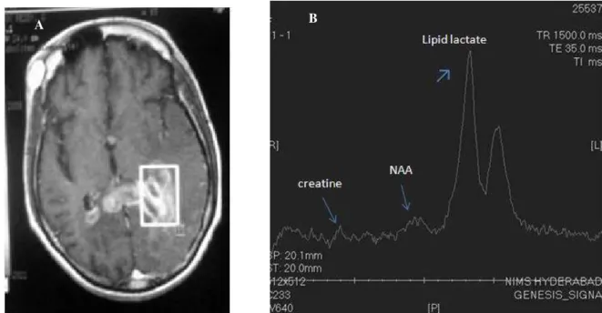
Figure 2: Singal voxel localizer (rectangle) placed on the lesion involving corpus callosum and periventricular white matter (A). Single voxel MRS reveals lipid-lactate peak (short arrow), reduced NAA and creatine (long arrows) and absence of choline typical for tuberculoma (B)
MRS= magnetic resonance spectroscopy; NAA=N-acetyl aspartate
A
polymerase chain reaction (PCR) was done to rule out PCNSL and was negative. Stereotac-tic biopsy of the lesion in the brain was con-sidered. Magnetic resonance spectroscopy (MRS) showed on elevated lipid lactate peak (Figures 2A and 2B) and a reduced N-acetylaspartate (NAA) and creatine suggestive of infective aetiology.
Given the above clinical scenario, CSF exami-nation findings, negative EBV PCR, MR Spec-troscopy suggesting infective aetiology and also given that tuberculosis is the most common op-portunistic infection in HIV/AIDS patient, CNS tuberculoma was considered. The patient was started on daily self-supervised antituberculo-sis treatment (ATT) and corticosteroids. Patient showed remarkable improvement clinically. MRI showed significant reduction in the size of the lesion at three months of follow up (Fig-ure 3).
DISCUSSION
In the background of immunocompromised host the most common differential diagnosis for a CNS mass lesion include primary CNS
lymphoma, toxoplasmosis and tuberculoma. While less common lesions are pyogenic ab-scess, cysticercosis, and syphilitic gummas. In our case we had a very large lesion in the brain parenchyma with a CD4+ count of 63 cells/µL. We initially suspected PCNSL, following which we have done EBV PCR which was negative. Then we considered second differen-tial diagnosis of tuberculoma. This patient had features common to PCNSL and tuberculoma, namely, altered sensorium, memory distur-bance, irrelevant talk, raised ESR, heteroge-neous peripherally enhancing ill defined lesion and lymphocytic predominance in CSF. Though sterotactic brain biopsy would confirm the tissue diagnosis, due to risks associated with doing the procedure we did MR spectroscopy to identify an infective lesion. MRS done at the site of lesions showed a increased lipid peak and a decreased NAA peak with increased cho-line/creatine ratio6. MRS helps to identify
lip-ids within the tuberculoma that are considered characteristic for TB. The characteristic MRS findings in tuberculomas also help in ruling out primary CNS lymphomas. However, the
113 cation of MRS in the diagnosis of tuberculo-mas in HIV-TB co-infected patients merits fur-ther detailed study. In vivo and in vitro MRS has shown elevated lipid peaks within the TB lesions.7 MRS study safely avoids the need of
brain biopsy to confirm the nature of lesion. Moreover CNS biopsy is invasive and in 5%-33% of cases no diagnosis can be reached due to sampling problems or tissue non-viability.8,9
MRS may prove a useful non-invasive investi-gational tool to differentiate tuberculoma and CNS lymphoma which are often confused.
REFERENCES
1. Berenguer J, Moreno S, Laguna F, Vicente T, Adrados M, Ortega A, et al. Tuberculous meningitis in patients infected with the human immunodefi-ciency virus. N Engl J Med 1992;326:668-72.
2. Sathe SS, Reichman LB. Mycobacterial disease in patients infected with the human immunodeficiency virus. Clin Chest Med 1989;10:445-63.
3. Ziegler JL, Beckstead JA, Volberding PA,Abrams DI, Levine AM, Lukes RJ, et al. Non-Hodgkin's lymphoma in 90 homosexual men. Relation to gen-eralized lymphadenopathy and the acquired immu-n odeficieimmu-n cy syimmu-n dr ome. N Eimmu-n gl J Med 1984;311:565-70.
4. Whiteman ML. Neuroimaging of central nervous system tuberculosis in HIV infected patients. Neuroimaging Clin North Am 1997;7:199-214.
5. MacMahon EM, Glass JD, Hayward SD, Mann RB, Becker PS, Charache P, et al. Epstein-Barr virus in AIDS related primary central nervous system lym-phoma. Lancet 1991;338:969-73.
6. Pretell EJ, Martinot C, Garcia HH, Alvarado M, Bustos JA, Martinot C. Differential diagnosis be-tween cerebral tuberculosis and neurocysticercosis by magnetic resonance spectroscopy. J Comput As-sist Tomogr 2005;29:112-4.
7. Santy K, Nan P, Chantana Y, Laurent D, Nadal D, Richner B. The diagnosis of brain tuberculoma by (1) H-magnetic resonance spectroscopy. Eur J Pediatr 2011;170:379-87.
8. Chappell ET, Guthrie BL, Orenstein J. The role of stereotactic biopsy in the management of HIV-r elated focal bHIV-r ain lesions. NeuHIV-r osuHIV-rgeHIV-r y 1992;30:825-9.
9. Karahalios D, Breit R, Dal Canto MC, Levy RM. Progressive multifocal leukoencephalopathy in pa-tients with HIV infection: lack of impact of early diagnosis by stereotactic brain biopsy. J Acquir Im-mune Defic Syndr 1992;5:1030-8.
Atypical presentation of tuberculoma in AIDS Chandra et al