depression in relation to cognition and white
matter integrity: follow up of two cases
Prognóstico de longo prazo da depressão geriátrica em relação à cognição
e à integridade da substância branca: acompanhamento de dois casos
Carlos Eduardo de Oliveira Alves
1,
Gilberto Sousa Alves
1, Felipe Kenji Sudo
1, Maria Elisa Lanna
2,
Letice Ericeira-Valente
1,
Denise Madeira Moreira
3, Jerson Laks
1, Eliasz Engelhardt
2ABSTRACT
Introduction: The geriatric depression (GD) represents one of the most frequent
psychia-tric disorders in outpatient services specialized in old-age treatment. Objective: The
cour-se of two illustrative cacour-ses of GD is discuscour-sed, highlighting its clinical picture after anti-depressant treatment and underlining variables related to disease prognosis, treatment efectiveness and conversion to major cognitive disorders such as vascular dementia (VD).
Methods: The cognitive performance, depressive symptoms, autonomy and brain
struc-tural measurements as white matter hyperintensities (WMH) and hippocampal size, and microstructural integrity of WM with difusion tensor imaging were followed during four
years. Results: Case 1, with a severe degree of WMH, was associated with worsening
nition and increasing functional disability. Case 2, with mild WMH, an improvement of
cog-nitive functioning could be seen. Conclusions: The existence of diferent subtypes of GD,
as presented in this report, points a pathophysiological heterogeneity of GD, and suggests
a possible continuum vascular depression (VaDp) and vascular cognitive impairment (VCI).
RESUMO
Introdução: A depressão geriátrica (DG) representa um dos mais frequentes transtornos
psiquiátricos em ambulatórios especializados em idosos. Objetivo: Discutir a evolução da
DG por meio de dois casos ilustrativos, destacando-se as variáveis relacionadas ao prog-nóstico da doença e à conversão para quadros cognitivos mais graves como demência
vas-cular (DV). Métodos: Os casos foram acompanhados por quatro anos com medidas do
desempenho cognitivo, funcional, sintomas depressivos, juntamente com as alterações de estruturas cerebrais, como hiperintensidades da substância branca (HSB), dimensões hipo-campais, e a integridade microestrutural da SB, por meio de imagens com tensor de difusão.
Resultados: Caso 1, com grave intensidade de HSB, evoluiu com piora cognitiva e
funcio-nal. Caso 2, com leve intensidade de HSB, evoluiu com melhora cognitiva após o tratamento
da depressão. Conclusões: A existência de diferentes subtipos de DG, como apresentado
neste relato, aponta para a heterogeneidade da isiopatologia da DG, sugerindo um possível
continuum entre depressão vascular (DpVa) e comprometimento cognitivo vascular (CCV).
1 Federal University of Rio de Janeiro (UFRJ), Psychiatry Institute (CDA/IPUB), Center for Alzheimer’s Disease and Related Disorders. 2 UFRJ, INDC-CDA/IPUB, Cognitive and Behavioral Unit.
3 Pró-Cardíaco Hospital, Radiology Service, Rio de Janeiro, RJ, Brazil.
Address for correspondence:Carlos Eduardo de Oliveira Alves Rua Professor Coelho e Souza, 48/201, Paineiras
36016-110 – Juiz de Fora, MG, Brazil E-mail: [email protected] Recebido em
30/3/2012 Aprovado em
11/5/2012
Keywords
Late life depression, geriatric depression, vascular depression, cognition, magnetic resonance imaging, difusion tensor.
Palavras-chave
INTRODUCTION
Late-life depression or geriatric depression (GD) represents one of the most frequent psychiatric disorders in
speciali-zed outpatient services1and afects nearly 9% to 18% of the
general elderly2,3. GD may afect from 3.2% to 15.4% of the
elderly Brazilian population4,5.
The course of GD has shown to be rather
heteroge-neous6,and it is assumed that the combination of white
matter lesions and cognitive symptoms related to frontal lobe dysfunction might be associated to GD with speci-ic features. This led to the concept of Vascular Depression
(VaDp) hypothesis7. This condition proposed by Alexopoulos
includes major depression occurring after 65 years of age, vascular risk factors and signiicant cerebrovascular disease, as main aspects, and cognitive impairment, represented by
executive dysfunction, as a secondary aspect7. In some
situa-tions a spectrum of cognitive and behavioral impairment of vascular etiology, comprising vascular depression (VaDp), mild vascular cognitive impairment (VMCI), and vascular
de-mentia (VaD) may be considered8,9.
The afective symptoms and executive dysfunction are both related to disconnection of prefrontal-subcortical cir-cuits10.Diferent frontal connections, such as the
cortico-lim-bic-striatum-pallidum-thalamic-cortical circuits, have been
shown to play an important role in afective modulation11,12,
contributing to the pathogenesis of some cases of
late--life depression13. Noteworthy, frontal vascular lesions have
shown to be associated with a poorer response to antide-pressant treatment14.
MR studies with difusion tensor (DTI) acquisitions and fractional anisotropy (DTI-FA) are used to evaluate white matter integrity in psychiatric illness, such as depression15.
This study aims to examine the heterogeneity of outco-me patterns of GD. Two cases of depression, with behavio-ral, neuropsychological, and neuroimaging assessments followed up for four years are described as examples. The
hypothesis of a continuum between VaDp and VCI (which
in-cludes VMCI and VaD) is analyzed, as well as the association between brain vascular lesions in speciic regions and poor response to antidepressant treatment.
METHODS
Two cases of GD were assessed over a 4-year-period at the specialized geriatric outpatient unit (Centre for Al zheimer Di-sease and Related Disorders) of the Federal University of Rio de Janeiro (CDA/IPUB-UFRJ-Brazil).
The cases fulilled Alexopoulous’ criteria for vascular de-pression7. Hamilton Depression Scale16 was applied to
inves-tigate depressive symptoms and the Neuropsychiatric Inven-tory (NPI) for a global evaluation of behavioral disturbances17.
Cognitive, functional, and behavioral performance and staging were assessed, as well as neuroimaging, according to the protocol of vascular dementia (VaD) recommendations of the DNCE-ABN which include the Mini-Mental State Exa-mination (MMSE), a validated Brazilian version of CAMCOG, executive function assessment (Trail-Making Test – TMT), Clock Drawing Executive Task – CLOX, and semantic verbal luency – VF [category animals]). TMT A can be considered as an indicator of visual-motor speed (processing speed), and TMT B can be considered as an indicator of cognitive
lexibility.CDR scale was used for staging of dementia
Func-tional Activities Questionnaire (FAQ) was applied to evaluate instrumental activities of daily-living (IADLs). The Hachinski Ischemic Score (EIH) for stroke risk assessment was used. The evaluation of the severity of white matter hyperintensities (WMH) was performed with a modiied Fazekas scale (mFa-zekas). De Leon score was applied to evaluate hippocampal atrophy18.
The neuroimaging evaluation was performed with MRI of the brain at baseline and at the end of the follow up (over three years later) with a GE Signa Horizon (1.5 T) equipment for standard acquisitions (including FLAIR sequence), and di-fusion tensor imaging (DTI), for fractional anisotropy measu-rements (DTI-FA). Post-processing of DTI was performed at a ADW 4.3 workstation with Functool 4.5.3 (GE Medical Systems) to obtain the quantitative values of DTI-FA.
RESULTS
The case 1 relates to a 73 years old, female with seven year
of schooling who sufered from hypertension and diabetes
mellitus. She reported that two years earlier she experien-ced her irst episode of depression, which caused a negative efect on her capacity to look after herself, pessimism, sad-ness, and increased dependency of her daily life activities. At that time, Sertraline 50 mg was introduced, with good clini-cal response.
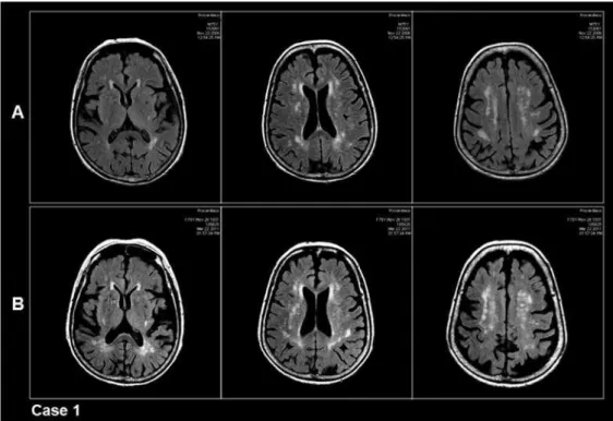
At the initial consultation at this clinic, she presented me-mory complaints. The neuropsychological assessment revea-led impairment of cognitive performance, with low global CAMCOG and attention and abstract thinking subscales, and the tests for executive function, TMT and CLOX, suggested impairment of executive function. CDR was 0.5 (Table 1). MRI showed moderate intensity of WMH (mFazekas score = 2) (Figure 1A). De Leon score for hippocampus was within nor-mal values (Table 2).
Over the four-year period the patient presented several episodes of depression, and progression of cognitive impair-ment, mainly of executive function. Sertraline was increased up to 200 mg/day for treatment of the relapse of depressive
symptoms in the irst six months. At the 6th month
together with diiculty in dealing with daily-life activities, such as controlling her own medication, preparing meals, keeping up to date with community life (FAQ = 13). Sertrali-ne was substituted for VenlafaxiSertrali-ne, 150 mg/day, and by the
end of the 1st year, the afective symptoms showed
rema-rkable improvement. The patient did not return to the clinic during the second year, continuing the treatment in a local health clinic. On returning to the clinic, she reported two new episodes of depression, partially controlled with Mirtazapine 45 mg in association with Bupropion 150 mg. One year after her last evaluation, the patient showed a relapse of the de-pressive symptoms (Table 3). In the last year of the follow-up the patient´s depressive symptoms showed improvement. However, global CAMCOG and memory subscale remained low, as well as executive function. CDR increased from 0.5 to 1 (Tables 1 and 3).
A new MRI showed an increase of white matter load (mFa-zekas score = 3) (Figure 1B), while there were no hippocam-pal changes in comparison to the initial indings (Table 2).
The comparative results of the DTI-FA are seen bellow. The case 2 is about a 67 years old, female, with three years of schooling, who was hypertensive but not diabetic, who-se irst interview revealed who-severe late-life depressive symp-toms, started one year before, when her son died. The global CAMCOG score was about average, with lower scores on at-tention and abstract thinking subscales, as well as low scores on TMT and CLOX, compatible with executive dysfunction. There was a mild functional decline. CDR was 0.5 (Table 1).
The initial MRI showed low level WMH (mFazekas score = 1) (Figure 2A), while the hippocampal were in the normal range.
After being treated for depression with Sertraline, increa-sed up to 100 mg, for one year, the depressive symptoms im-proved, accompanied by improvement in global CAMCOG and the subscales scores, as well as of executive function, and functional improvement. CDR was zero (Table 1). The-se improvements were sustained for the following 4 years (Table 1).
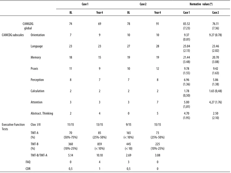
Table 1. Cognitive follow-up data
Case 1 Case 2 Normative values (*)
BL Year 4 BL Year 4 Case 1 Case 2
CAMGOG global
74 69 78 91 83.52
(7.23)
76.11 (7.36)
CAMCOG subscales Orientation 7 9 10 10 9.37
(0.81)
9.27 (0.78)
Language 23 23 27 28 25.04
(2.13)
23.46 (2.02)
Memory 18 15 19 19 21.44
(3.48)
20.70 (3.08)
Praxis 11 9 10 12 9.78
(1.55)
9.42 (1.63)
Perception 8 7 7 8 6.96
(1.36)
5.86 (1.38)
Calculation 2 2 2 2 1.78
(0,50)
1.65 (0,48)
Attention 3 3 3 7 5.00
(1,81)
4,27 (1.76)
Abstract. Thinking 2 4 0 5 4.70
(1.95)
2.50 (2.10)
Executive Function Tests
Clox I/II 11/15 13/15 9/15 15/15
TMT-A (%)
70 (50%-75%)
85 (25%-50%)
165 (< 10%)
73 (25%-50%)
TMT-B (%)
360 (10%-25%)
859 (< 10%)
445 (< 10)
225 (10%-25%)
TMT-B/TMT-A 5.14 10.10 2.69 3.08
FAQ 0 4 3 0
CDR 0,5 1 0,5 0
*Normative value by Bueno 200919.
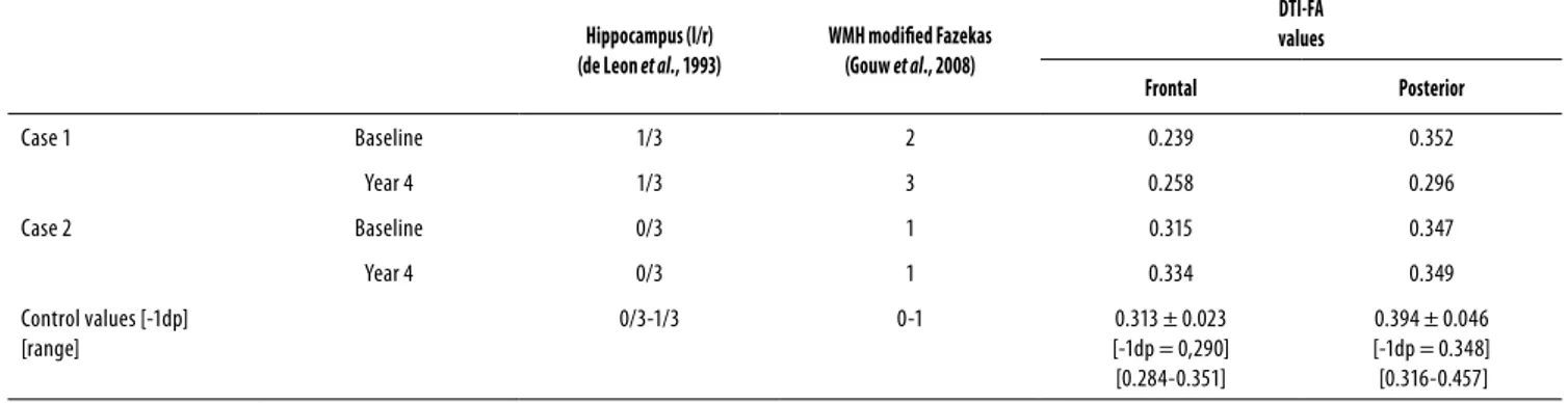
Table 2. Neuroimage data
Hippocampus (l/r) (de Leon et al., 1993)
WMH modiied Fazekas (Gouw et al., 2008)
DTI-FA values
Frontal Posterior
Case 1 Baseline 1/3 2 0.239 0.352
Year 4 1/3 3 0.258 0.296
Case 2 Baseline 0/3 1 0.315 0.347
Year 4 0/3 1 0.334 0.349
Control values [-1dp] [range]
0/3-1/3 0-1 0.313 ± 0.023
[-1dp = 0,290] [0.284-0.351]
0.394 ± 0.046 [-1dp = 0.348] [0.316-0.457]
Table 3. Behavioral follow-up data
Year Case 1 Case 2
HAM-D NPI HAM-D NPI
BL 5 8 20 14
0.5 15 38 1 3
1 2 9 5 6
3 18 16 2 0
4 7 28 1 0
HAM-D: Hamilton Depression Scale; NPI: Neuropsychiatric Inventory; BL: baseline.
At the end of this study, the patient was again submitted to MRI, which revealed a very slight increase of WMH, main-taining the same score (mFazekas score = 1) (Figure 2B), and no hippocampal changes were found (Table 2).
The comparative results of the DTI-FA are as follows.
Results of DTI-FA
The DTI data of both cases were submitted to post-proces-sing to obtain DTI-FA values of the subcortical white matter, which were compared with a control sample of normal el-derly individuals22. At the beginning of the study, case 1
pre-sented frontal DTI-FA values signiicantly inferior compared to those of case 2 and to the control group. The evaluation 4 years latter showed a slight loss of frontal DTI-FA values in both case 1 and 2, in comparison to the control sample. A loss in the posterior white matter in case 2 was also obser-ved (Table 2).
DISCUSSION
The evolution of GD may vary signiicantly6, as shown by the
illustrative cases.It is believed that in some situations the
afective symptoms might be a response to psychological
stress23. In other cases, however, it can be considered within
a spectrum of cognitive and behavioral impairment of
vas-cular etiology, comprising vasvas-cular depression and VMCI9.
Some studies suggest that these behavioural symptoms may be the earliest signs of VCI in progress8.
It may be diicult to distinguish between cases of de-pressed patients demonstrating initial signs of VCI and those whose cognition will improve to treatment with antidepres-sants. Executive function appears to be the most relevant element in patients with GD, and is outstanding manifesta-tion in vascular depression. However, these afective symp-toms may represent an important, confounding element in the interpretation of cognitive deicits in non-demented and
demented patients24. Considering transversal studies, there
is agreement about the frequent presence of executive
dys-function in DG25. However, results showed variability when
considering the speciicity of executive deicits associated
with depression26-29. Thus, long-term follow-up of cases is
fundamental in determining diagnosis.
Long-term follow-up permitted the assessment of the cases in the absence and presence of depressive symptoms. This made possible to clarify the role of afective symptoms in each case. The initial cognitive evaluation of case 1, in the absence of depressive symptoms, revealed executive dysfunction (TMT and CLOX, and CAMCOG subscales atten-tion and abstract thinking). The reappearance of depressi-ve symptoms during the third year was accompanied by a global cognitive worsening, as shown by global CAMCOG, and executive function lower scores (attention and abstract thinking, TMT and CLOX). In the last year the patient’s de-pressive symptoms showed improvement. However, global CAMCOG and memory subscale remained low, as well as executive function.
The initial evaluation of case 2 showed high scores regar-ding depressive symptoms (Table 3) and executive dysfunction (TMT and CLOX) (Table 1), and impairment of attention and abs-tract thinking. These cognitive parameters improved with the improvement of the depressive symptoms in the long-term.
evol-Figure 2. Case 2. MRI-FLAIR acquisition of three representative axial sections that show WMH burden. A: mFazekas score = 1;
B: mFazekas score = 1.
Figure 1. Case 1. MRI-FLAIR acquisition of three representative axial sections that show WMH burden. A: mFazekas score = 2;
B: mFazekas score = 3.
ved to normalization of cognition (CDR 0). Patients with GD frequently demonstrate executive dysfunction, associated with diiculties in performing instrumental daily-life activi-ties, which may persist even after the successful treatment of mood disorder. These particular patients, may, over time, develop VD6.
studies demonstrating this relation30-32
. Stefens et al.
33
descri-bed a patient with recurring GD over a 4-year period, which evolved to cognitive decline and VaD. This patient was also screened for the presence of WMH at the end of the
follow--up, which showed worsening of the lesions33. Another study
showed the same pattern with a two-year follow-up34.
The comparison between the baseline and inal neuroi-maging studies were able to explain diferent patterns of GD. The lesions may contribute not only to the etiology of the afective symptoms and cognitive impairment in some case of GD13,35, but also to a negative prognosis of the cases36. The
DTI-FA values were reduced in the frontal white matter of case 1, and were in normal range in case 2, as compared to a normal group, which seems to be the determining factor for the diferent prognosis in each case. The posterior white matter showed reduced values in case 1, and remained in the normal range in case 2 (Table 2). Reduced DTI-FA values are related to loss of axonal integrity, and taking into account the frontal white mater. It is compatible with disconnection of prefrontal-subcortical circuits, considered the neuroana-tomical substrate underlying afective and cognitive
ma-nifestations10, and appear to have prognostic relevance in
GD14. Other studies corroborate these indings, despite some
variations37,38. The reduction of posterior DTI-FA values in
case 1 is possibly related to the interruption of other circuits, not identiied in the present study.
The two cases difered with regard to the time needed to show a response to treatment with anti-depressants, which is an important factor in deining the sub-types of elderly
de-pression. Dew et al.39 described four groups of patients with
GD, each with diferent responses to therapy. The irst group showed rapid improvement with the use of antidepressants (the best prognosis), the second, showed improvement, but required a longer period of antidepressant use, the third group demonstrated an oscillating course with improvement, and the fourth group proved to be resistant to treatment (the worst prognosis)39. In a recent study Alexopoulos et al.14
eva-luated 48 elderly depressed individuals (23 with refractory depression and 25 who responded to treatment with antide-pressant medication). Just as described by Dew et al., the case 2 of this study had a rapid response to treatment and evolved better, while case 1 showed a poorer response to antidepres-sant treatment with subsequent cognitive impairment.
CONCLUSION
The association of vascular factors (including vascular risk factors and WMH load) and GD has guided the development of the VaDp hypotheses and the mechanisms which predis-pose, initiate or perpetuate this subtype of depression. Neu-roimaging studies, especially those with WMH assessment and quantitative DTI-FA have revealed the disruption of
pre-frontal-subcortical circuits. These changes might represent a neuroanatomical substrate associated with VaDp and the accompanying cognitive impairment, as well as inluencing the kind of response to antidepressant treatment.
The existence of diferent subtypes of GD, as presented in this report, one with cognitive improvement and the other with cognitive decline, points out the pathophysiological
heterogeneity of DG, and suggests a possible continuum
VaDp and VCI (including VMCI and VaD), with signiicant im-pact on case management.
ACKNOWLEDGEMENTS
To Luzinete Alvarenga, librarian, for organizing the referen-ces. My wife Clarisse and my son Felipe for their inspiration and encouragement.
REFERENCES
1. Panza F, Frisardi V, Capurso C, D’Introno A, Colacicco AM, Imbimbo BP, et al. Late-life
depression, mild cognitive impairment, and dementia: possible continuum? Am J Geriatr Psychiatry. 2010;18:98-116.
2. Copeland JR, Beekman AT, Braam AW, Dewey ME, Delespaul P, Fuhrer R, et al. Depression
among older people in Europe: the EURODEP studies. World Psychiatry. 2004;3:45-9.
3. Mulsant BH, Ganguli M. Epidemiology and diagnosis of depression in late life. J Clin
Psychiatry. 1999;60(Suppl 20):9-15.
4. Andrade LH, Walters EE, Gentil V, Laurenti R. Prevalence of ICD – 10 mental disorder
in a catchment area in the city of São Paulo, Brazil. Soc Psychiatry Psychiatr Epidemiol. 2002;37:316-25.
5. Costa E, Barreto SM, Uchoa E, Firmo JO, Lima Costa MF, Prince M. Prevalence of
Inter-national Classiication of diseases, 10th Revision Common Mental Disorders in the elder-ly in a Brazilian community: the Bambui Health Ageing study. Am J Geriatr Psychiatry. 2007;15:17-27.
6. Bhalla RK, Butters MA, Becker JT, Houck PR, Snitz BE, Lopez OL, et al. Patterns of mild
cognitive impairment after treatment of depression in the elderly. Am J Geriatr Psychiatry. 2009;17:308-16.
7. Alexopoulos GS, Meyers BS, Young RC, Campbell S, Silbersweig D, Charlson M. The vascular
depression hypothesis. Arch Gen Psychiatry. 1997;54:915-22.
8. Baernes E, Alexopoulos G, Lopez O, Wilhanson J, Yafe K. Depressive symptoms, vascular
disease, and mild cognitive impairment. Arch Gen Psychiatry. 2006;63:273-80.
9. Zimmerman JA, Mast BT, Miles T, Markides KS. Vascular risk and depression in the Hispanic
Established Population for the Epidemiologic Study of the Elderly (EPESE). Int J Geriatr Psychiatry. 2009;24:409-16.
10. Tekin S, Cummings JL. Frontal-subcortical neuronal circuits and clinical neuropsychiatry: an update. J Psychosom Res. 2002;53:647-54.
11. Davidson RJ, Lewis DA, Alloy LB, Amaral DG, Bush G, Cohen JD, et al. Neural and behavioral substrates of mood and mood regulation. Biol Psychiatry. 2002;52:478-502.
12. Drevets WC. Geriatric depression: brain imaging correlates and pharmacologic considera-tions. J Clin Psychiatry. 1994;55(Suppl A):71-81.
13. Taylor WD, MacFall JR, Payne ME, McQuoid DR, Stefens DC, Provenzale JM, et al. Greater MRI lesion volumes in elderly depressed subjects than in control subjects. Psychiatry Res. 2005;139:1-7.
14. Alexopoulos GS, Murphy CF, Gunning-Dixon FM, Latoussakis V, Kanellopoulos D, Klimstra S, et al. Microstructural white matter abnormalities and remission of geriatric depression. Am J Psychiatry. 2008;165:238-44.
16. Moreno RA, Moreno DH. Escalas de depressão de Montgomery & Asberg (MADRS) e de Hamilton (HAM-D). Rev Psiq Clín. 1998;25:262-72.
17. Camozzato AL, Kochhann R, Simeoni C, Konrath CA, Pedro Franz A, arvalho A, et al. Reabili-ty of Brazilian Portuguese version of the neuropsychiatric inventory (NPI) for patients with Alzheimer’s disease and their caregivers. Int Psychogeriatr. 2008;20:383-93.
18. Engelhardt E, Tocquer C, Charles A, Moreira DM, Okamoto IH, Cavalcanti JLS. Demên-cia vascular: critérios diagnósticos e exames complementares Dement Neuropsychol. 2011;5(Suppl1):49-77.
19. Bueno D. Perfil de idosos com demência e depressão: status cognitivo medido pelo CA-MCOG, escolaridade e histórico das habilidades sociocognitivas Campinas, SP. 2009 [s.n.]: Dissertação (Mestrado) Universidade Estadual de Campinas. Faculdade de Ciências Mé-dicas.
20. Gouw AA, van der Flier WM, van Straaten EC, Pantoni L, Bastos-Leite AJ, Inzitari D, et al. LADIS study group. Reliability and sensitivity of visual scales versus volumetry for evalua-ting white matter hyperintensity progression. Cerebrovasc Dis. 2008:25(3):247-53.
21. de Leon MJ, Golomb J, George AE, Convit A, Tarshish CY, McRae T, et al. The radiologic prediction of Alzheimer disease: the atrophic hippocampal formation. AJNR. Am J Neuro-radiol. 1993;14(4):897-906.
22. Engelhardt E, Moreira DM, Laks J. The brain subcortical white matter and aging. A quanti-tative fractional anisotropy analysis. Dement Neuropsychol. 2009;3:228-33.
23. Anstey KJ, Burns R, Butterworth P, Windsor TD, Christensen H, Sachdev P. Cardiovascular risk factors and life events as antecedents of depressive symptoms in middle and early-old age: PATH Through Life Study. Psychosom Med. 2009;71:937-43.
24. Wright SL, Persad C. Distinguishing between depression and dementia in older persons: neu-ropsychological and neuropathological correlates. J Geriatr Psychiatry Neurol. 2007;20:189-98.
25. Roman G. Vascular depression: an archetypal neuropsychiatric disorder. Biol Psychiatry. 2006;60:1306-8.
26. Kramer-Ginsberg E, Greenwald B, Krishnan R, Christiansen B, Hu J, Ashtari M, et al. Neu-ropsychological functioning and MRI signal hyperintensities in geriatric depression. Am J Psychiatry. 1999;156:438-44.
27. Hart RP, Kwentus JA. Psychomotor slowing and subcortical-type dysfunction in depres-sion. J Neurol Neurosurg Psychiatry. 1987;50:1263-6.
28. Boone KB, Lesser IM, Miller BL, Bruce L, Wohl M, Berman N, et al. Cognitive functioning in older depressed outpatients: relationship of presence and severity of depression to neu-ropsychological tests cores. Neuropsychology. 1995;9:390-8.
29. Butters M, Whyte E, Nebes R, Begley AE, Dew MA, Mulsant BH, et al. The nature and de-terminants of neuropsychological functioning in late-life depression. Arch Gen Psychiatry. 2004;61:587-95.
30. Stefens DC, Potter GG, McQuoid DR, MacFall JR, Payne ME, Burke JR, et al. Longitudinal resonance imaging vascular changes, apolipoprotein E genotype, and development of de-mentia in the neurocognitive autcomes of depression in the elderly study. Am J Geriatr Psychiatry. 2007;15:839-49.
31. Taylor WD, Stefens DC, Macfall JR, McQuoid DR, Payne ME, Provenzale JM, et al. White matter hyperintensity progression and late-life depression outcomes. Arch Gen Psyshiatry. 2003;60:1090-6.
32. Nebes RD, Reynolds CF, Boada F, Meltzer CC, Fukui MB, Saxton J, et al. Loongitudinal increa-se in the volume of white matter hyperintensities in late-onincrea-set depression. Int J Getriatr Psychiatry. 2002;17:526-30.
33. Stefens DC, Taylor WD, Krishnan R. Progression of subcortical ischemic disease from vas-cular depression to vasvas-cular dementia. Am J Psychiatry. 2003;160:1751-6.
34. Loganathan S, Phutane VH, Prakash O, Varghese M. Progression of vascular depression to possible vascular dementia. J Neuropsychiatry Clin Neurosci. 2010;22:451.
35. Thomas AJ, O’Brien JT, Davis S, Ballard C, Barber R, Kalaria RN, et al. Ischemic basis for deep white matter hyperintensities in major depression: a neuropathological study. Arch Gen Psychiatry. 2002;59:785-92.
36. Sheline YI, Pieper CF, Barch DM, Welsh-Bohmer K, McKinstry RC, MacFall JR, et al. Support for the vascular depression hypothesis in late-life depression: results of a 2-site, prospecti-ve, antidepressant treatment trial. Arch Gen Psychiatry. 2010;67:277-85.
37. Nobuhara K, Okugawa G, Sugimoto T, Minami T, Tamagaki C, Takase K, et al. Frontal white matter anisotropy and symptom severity of late-life depression: a magnetic resonance di-fusion tensor imaging study. J Neurol Neurosurg Psychiatry. 2006;77:120-2.
38. Yang Q, Huang X, Hong N, Yu X. White matter microstructural abnormalities in late-life depression. Int Psychogeriatr. 2007;19:757-66.