Treatment of pelvic congestion associated with varicose veins
of the lower limbs: report of a small number of cases
Tratamento da congestão pélvica associada a varizes dos membros inferiores: relato de uma
pequena série de casos
Fábio Augusto Cypreste Oliveira1, Carlos Eduardo de Sousa Amorelli1, Fábio Lemos Campedelli1, Juliana Caetano Barreto2,
Mariana Caetano Barreto3, Philippe Moreira da Silva4, Fernanda Lauar Sampaio Meirelles5
Abstract
he pelvic congestion syndrome is deined by anatomical criteria, hemodynamic and clinical result of hypertension and venocapillary stasis in the pelvic area and can be extended to the lower limbs. he presence of varicose veins of the lower limbs may or may not correlate relux of the saphenous system and, in cases of absence of saphenous relux, the relux pelvic presents itself as an important etiological factor responsible for recurrence of venous disease in lower limbs. he authors report a small series of three cases of young patients sufering from pelvic congestion syndrome associated with varicose veins of the lower limbs treated by pelvic embolization and surgical treatment of varicose veins of the lower limbs during the surgical procedure with good initial results and no embolic complications.
Keywords: pelvic pain; embolization, therapeutic; varicose veins.
Resumo
A síndrome de congestão pélvica é deinida por critérios anatômicos, hemodinâmicos e clínicos, decorrentes da hipertensão e estase venocapilar no território pélvico, podendo ser estendido aos membros inferiores. A presença de varizes dos membros inferiores pode estar ou não relacionada ao reluxo do sistema safeno e, nos casos de ausência do reluxo safeno, o reluxo pélvico apresenta-se como importante fator etiológico responsável pelo aparecimento e recidiva da doença venosa nos membros inferiores. Os autores relatam uma pequena série de três casos de pacientes jovens portadoras de síndrome de congestão pélvica associada à varizes dos membros inferiores tratadas por embolização pélvica e tratamento cirúrgico das varizes de membros inferiores, no mesmo tempo cirúrgico, com bons resultados iniciais e ausência de complicações embólicas.
Palavras-chave: dor pélvica; embolização terapêutica; varizes.
Study carried out at the Service of Angiology, Vascular Surgery, Endovascular and Laser herapy of Hospital São Francisco de Assis – Goiânia (GO), Brazil.
1 Specialist in Vascular Surgery with experience in Endovascular Surgery and Angiography by the Brazilian Society of Angiology and Vascular Surgery (SBACV), of Colégio Brasileiro de
Radiolo-gia (CBR) and the Brazilian Medical Association (AMB) – Goiânia (GO), Brazil.
2 General practitioner at Angiogyn – Goiânia (GO), Brazil.
3 General Surgeon at Hospital de Urgências de Goiânia – Goiânia (GO), Brazil. 4 Nursing and Instrumentation Technician at Angiogyn – Goiânia (GO), Brazil.
5 Medical Student (6th year) at Pontifícia Universidade Católica of Goiás (PUC-Goiás) – Goiânia (GO), Brazil.
Financing source: none.
Conlict of interest: nothing to declare. Submitted on: 08.24.11. Accepted on: 10.28.11. J Vasc Bras. 2012;11(1):62-66.
Introduction
he pelvic congestion syndrome (PCS) has been de-scribed since the past century1. Its main clinical manifes-tations are: non-cyclical chronic pelvic pain (>6 months), dyspareunia or post-coital discomfort, pre-menstrual pain, sensation of pelvic/perineal “weight” and menstrual changes associated with the presence of pelvic, perineal and vulvar
at endovascular therapy. Beard et al.11 describe phlebo-graphic criteria: contrast retention >20 seconds, gonadal vein >6mm and contrast retention in varicose tributaries in the vulva and the perineum. Intravascular ultrasound (USIVU) is mostly used for the diferential diagnosis of the “nutcracker” and the May-hurner syndrome, or when the previously described methods fail.
Once the diagnosis of PCS and chronic venous insuf-iciency of the lower limbs is conirmed, medical treatment is established with pharmacotherapy, elastic stocking com-pression, physiotherapy and postural treatment. In refrac-tory cases, invasive treatment is indicated.
Case report
Case 1
he patient is a 26-year old multiparous female patient (GIVPIIIAI), with PCS due to let gonadal vein relux mani-fested as non-cyclical chronic pelvic pain, dyspareunia and metrorrhagia. At physical examination, she presented with vulvar and perineal varicose veins associated with symptom-atic lower limb varicose veins (CEAP classiication III) with no saphenous relux at vascular echography of the lower limbs. Endovaginal ultrasound suggested pelvic varicose veins, es-pecially on the let side, which were conirmed by angiores-onance. No sign of external compression or thrombosis was identiied. Under spinal anesthesia and sedation, lower limb varicose vein operation was performed, with segmental phle-bectomies and insuicient Cockett’s perforating vein ligation, followed by embolization of pelvic varicose veins and let go-nadal vein with Histoacryl glue and lipiodol 3:1, via right fem-oral vein, all done in an one-stage procedure (Figures 1 and 2). he procedure was uneventful and the patient was discharged from the hospital ater 24 hours, with no changes in the kid-ney function or signs of pulmonary embolism and no bleeding from the surgical incisions. She has been on outpatient follow-up for 21 months since the operation, presenting signiicant improvement in pelvic symptoms. She has been asymptomatic and has a satisfactory cosmetic result, with no recurrence of vulvar or lower limb varicose veins.
Case 2
he patient is a 41-year old multiparous female pa-tient (GIVPIVA0), with PCS manifested by non-cyclical chronic pelvic pain, post-coital discomfort and sensation of weight in the lower abomen. At physical examination, she had symptomatic vulvar, gluteal and lower limb varicose hus, the pelvic renal relux can be transmitted to the lower
limbs, presenting as recurrent varicose veins. Labropoulos et al.6 studied 834 limbs and found 84 limbs (10%) of non-sa-phenous relux. In that series, the relation with pelvic venous relux was 34%, and 68% originated in vulvar veins medial to the femoral-saphenous junction, and 32% of gluteal veins. Lower limb varicose veins with pelvic origin can pres-ent in four ways:
1. Sapheno-femoral junction relux;
2. Relux to the saphena magna and saphena parva; 3. Relux parallel to the saphenous axis (medial side of the
thigh);
4. Sciatic relux (posterior side of the thigh and gluteal).
Recurrence of lower limb varicose veins of abdominal/ pelvic origin may happen in up to 17% of the cases7,8.
he diagnosis of PCS is deined by a detailed clinical history and physical examination, including gynecologi-cal and perianal exams. Non-invasive vascular testing in-cludes vascular ultrasound with transparietal abdominal/ pelvic color Doppler ultrasonography (to analyze the iliac axis, cava and renal) and endovaginal ultrasound (pelvic evaluation) associated with the ultrasonographic study of the lower limbs. Some echographic criteria can deine syndromes (May-hurner and “nutcracker”), according to Engelhorn et al.9 However, the presence of tributary dilated and tortuous veins in the pelvic adnexal area with diam-eter ≥7mm associated with bidirectional low during the Valsalva Maneuver and endovaginal ultrasound is diagnos-tic of pelvic varicose veins9. he diagnosis of pelvic venous thrombosis could also be performed with this method.
Pelvic angioresonance has been used as the non-inva-sive diagnostic method of choice for the anatomical dei-nition of the pelvic congestion, its relation with the lower limb venous system and the etiological deinition of PCS. Coakley et al.10 describe criteria for the angioresonance/ angiotomography of pelvic relux, which include tributary to uterine veins >4 mm, and gonadal vein >8 mm. Pelvic phlebography is not a routine test, used exceptionally and
Table 1. Relation between physiopathology and etiology of pelvic ve-nous congestion
Physiopathology Etiology
Relux Ovarian veins
Internal iliac veins Pelvic vascular malformations
Post-thrombotic syndrome
External compression “Nutcracker” syndrome
May-hurner syndrome Pelvic tumors
veins (CEAP classiication III), (Figure 3). Doppler ultraso-nography showed let saphena magna relux of both lower limbs. She underwent endovaginal ultrasound that was suggestive of pelvic varicose veins. he diagnosis was was conirmed by angioresonance which showed relux in the internal right iliac vein with contrast retention in tributary varicose veins due to probable relux along the sciatic nerve. here were no signs of external compression or thrombosis. Under spinal anesthesia and sedation, surgery of varicose veins in the lower limbs with segmental phlebectomy and thermal ablation of the let saphena magna with semicon-ductor laser (980 mm of wavelength and 30 watts of power with 600 micron iber) was performed, followed by emboli-zation of tributary pelvic varicose veins in the internal right iliac vein and its trunk with histoacryl and Gianturco coils, via right femoral vein. he procedure was uneventful and the patient was discharged from the hospital ater 24 hours, with no changes in the kidney function or signs of pulmo-nary embolism and no bleeding from the surgical inci-sions. She has been on outpatient follow-up for 18 months since the operation, with signiicant improvement of pelvic symptoms, and asymptomatic in the lower limbs with satis-factory cosmetic results, with no varicose veins recurrence.
Case 3
he patient is a 28-year-old multiparous female pa-tient (GIIIPIIIA0), with PCS due to let gonadal vein mani-fested by non-cyclical chronic pelvic pain, dyspareunia and metrorrhagia. Physical examination showed vulvar and perineal varicose veins associated with symptomatic lower limb varicose veins (CEAP classiication III). No saphe-nous vein relux was seen at Doppler ultrasonography of the lower limbs. An endovaginal ultrasound was suggestive of pelvic varicose veins. Angioresonance conirmed the echo-graphic indings and also indentiied an incompetent let ovarian vein. here were no signs of external compression
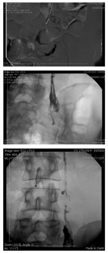
Figure 1. Selective venography of left ovarian vein with relux and pel-vic varicose vein illing with contrast retention.
Figure 2. Superselective catheterization with microcatheter and injec-tion of lipiodol and histoacryl 3:1, with distal to proximal embolizainjec-tion.
he procedure was uneventful and the patient was discharged from the hospital ater 24 hours, with no changes in the kid-ney function,or signs of pulmonary embolism and no bleed-ing from the surgical incisions. She has been on outpatient follow-up for two months since the operation, with signiicant improvement of pelvic symptoms, and asymptomatic in the lower limbs with satisfactory cosmetic results, with no varicose veins recurrence
Discussion
Several techniques have been described to treat the symptoms of PCS that are refractory to the initial therapy. The classical treatment consists of the surgical ligation by retroperitoneal access of gonadal and/or in-ternal iliac veins12. Laparoscopic ligation of pelvic veins is feasible, with improvement of abdominal symp-toms, and it is also an important method in the dif-ferential diagnosis of causes for chronic pelvic pain13. Embolization of tributary varicose veins and venous trunks has been showing satisfactory results, compa-rable to surgical ligation in the clinical control of PCS, with lower morbidity rates and length of hospital stay14. In cases of external compression or chronic iliac vein and inferior vena cava thrombosis, simple angioplasty, or with a stent, has been performed with satisfactory initial results15,16. Hysterectomy, with or without oopho-rectomy, will only be indicated when the previously de-scribed therapeutic options have failed.
We could not ind any contraindications or factors that increase the morbidity rates of surgical treatment of lower limb varicose veins associated with the endovascular treat-ment of PCS. hus, we chose to treat PCS and chronic ve-nous insuiciency of the lower limbs at the same time, with the goal of performing the deinitive treatment under a sin-gle anesthetic session and hospital admission, thus lowering the patients´ surgical risk.
Conclusion
he synchronous treatment of the pelvic congestion syndrome by percutaneous embolization, and lower limb varicose veins by surgery is feasible, with good initial re-sults and low complication rates, however, we need more cases and long term follow-up.
References
1. Taylor Jr HC. Vascular congestion and hyperhemia: the efect on function in the female reproductive organs. Part I. Physiological ba-sis and history of the concept. Am J Obstet Gynecol. 1949;57:211-30.
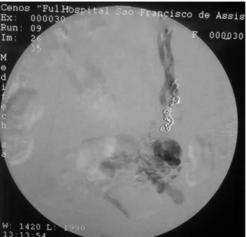
Figure 5. Superselective catheterization with mixed embolization, in the varicose plexus with glue and in the proximal gonadal stem with Gi-anturco coils. Control venography without illing of the varicose plexus.
Figure 3. Demonstration of relux to the lower limbs. It is important to observe the prevalence in the posterior side of the thigh and buttocks due to relux originated in the tributaries of internal iliac vein.
2. Greiner M, Gillin-Smith GL. Leg varices originating from the pelvis: diagnosis and treatment. Vascular. 2007;15(2):70-8.
3. Creton D, Hennequin L, Kohler F, et al. Embolisation of symp-tomatic pelvic veins in women presenting with non-saphenous varicose veins of pelvic origin - three-year follow-up. Eur J Vasc Endovasc Surg. 2007;34(1):112-7.
4. Engelhorn CA, Engelhorn AL,Cassou MF, et al. Classiicação an-otomofuncional da insuiciência das veias safenas baseada no eco-Doppler colorido dirigida para o planejamento cirúrgico das varizes. J Vasc Br. 2004;3(1):13-9.
5. Leal Monedero J, Zubicoa Ezpeleta S, Castro Castro J et al. Embolization treatment of recurrent varices of pelvic origin. Phlebology. 2006;1(1):3-11.
6. Labropoulos N. Nonsaphenous supericial vein relux. J Vasc Surg. 2001:34(5):872-7.
7. Van Rij AM, Jiang P, Solomon C, et al. Recurrence after varicose vein surgery (a prospective long-term clinical study with du-plex ultrasound scanning and air plethysmography). J Vasc Surg. 2003;38:935-43.
8. Perrin MR, Labropoulos N, Leon LR Jr. Presentation of the pa-tient with recurrent varices after surgery (REVAS). J Vasc Surg. 2006;43(2):327-34.
9. Engelhorn CA, Morais-Filho DM, Barros FS, et al. Investigação da síndrome de quebra-nozes e Síndrome compressiva da veia ilíaca comum esquerda pela artéria ilíaca comum direita. Engelhorn CA, Morais-Filho DM, Barros FS, et al. Guia prático de ultrassonograia vascular. 2. ed. Rio Janeiro: Di Livro, 2011. p. 213-26.
10. Coakley FV, Varghese SL, Hricak H. CT and MRI of pelvic vari-ces in women. Journal of Computer Assisted Tomography. 1999;23(3):429-34.
11. Beard RW, Highman JH, Pearce S, et al. he diagnosys of pelvic vari-cosities in women with chronic pelvic pain. Lancet. 1984;2:946-9.
12. Edwards RD, Robertson AB, MacLean AB, et al. Case report pelvic pain syndrome - successful treatment of a case by ovarian vein embolization. Clin Radiol. 1993;47:429-31.
13. Mathis BV, Miller JS, Lukens ML, et al. Pelvic congestion syn-drome: a new approach to an unusual problem. Am Surg. 1995;61: 1016-8.
14. Cordts PR, Eclavea A, Buckley PJ, et al. Pelvic congestion syndrome: early clinical results after transcatheter ovarian vein embolization. J Vasc Surg. 1998;28:862-8.
15. Ferreira M, Lanziotti L, Abuhadba G, et al . Dor pélvica crônica: o papel da síndrome do quebra-nozes. Chronic pelvic pain: the role of the nutcracker syndrome. J Vasc Bras. 2008;7(1):76-9.
16. Cunha Júnior JR, Neves DQ, Fontes FA, et al. Tratamento endo-vascular da síndrome de compressão da veia ilíaca (May-hurner) – relato de caso. J Vasc Bras. 2011;10(1):1-5.
Correspondence
Fabio Augusto Cypreste Oliveira Avenida Alphaville Flamboyant, 3.900, casa 283 CEP 74884-527 – Goiânia (GO), Brazil
E-mail: [email protected]
Author’s contribution