1027
Early peritoneal-scrotal leakage in a patient submitted to
peritoneal dialysis demonstrated by dynamic peritoneal
99mTc-Phytate scintigraphy
_______________________________________________
Andrés Martínez-Esteve
1, Francisco Javier García-Gómez
1, Juan Ignacio Cuenca-Cuenca
1, Juan Luis
Tirado-Hospital
11 Departamento de Medicina Nuclear, Hospital Virgen del Rocío Universitario, Sevilla, España
1027
RADIOLOGY PAGE
A 77 year old male with history of renal lithiasis leading to right nefrectomy and end stage renal disease (ESRD) secondary to possible vas-cular nephropathy and focal segmental glomeru-losclerosis diagnosed in 2009, started ambulatory peritoneal dialysis (APD) due to clinical decline. Insufficient drainage of the peritoneal dialysis solution with progressive bilateral testicular ede-ma (more severe in right side) was observed from the first APD session. As a consequence, the
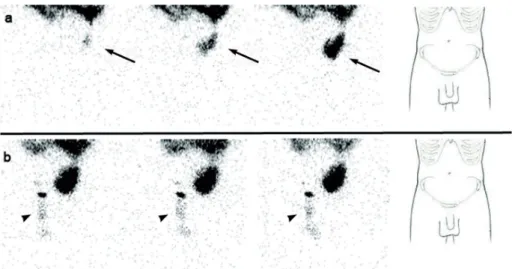
pa-tient needed temporarily hemodialysis. In order to diagnose a patent peritoneal-vaginal duct, a dynamic 99mTc-Phytate scintigraphy was perfor-med after introduction of peritoneal dialysis so-lution (600, 1200 and 2000 mL) labelled with 74 MBq of 99mTc-Phytate in the abdominal cavity. A total of 60 images (30 seconds per image) were acquired and a hypogastric uptake of radiotracer was observed (Figure 1 - Panel a). Right inguinal and scrotal uptake was observed only after
per-Figure 1 - Dynamic peritoneal 99mTc-Phytate scintigraphy. Panel a: Images acquired during the filling of peritoneal dialysis solution (600, 1200 and 2000 ml) labeled with 74 MBq of 99mTc Phytate, revealing a hypogastric uptake of radiotracer (arrow), not reaching the inguinal canal or the scrotal area. On the right side, anatomical reference contour. Panel b: Inguinal and right scrotal uptake of radiotracer (head arrow) after Valsalva’s maneuver. On the right side, anatomical reference contour.
IBJU| RADIOLOGY PAGE
1028
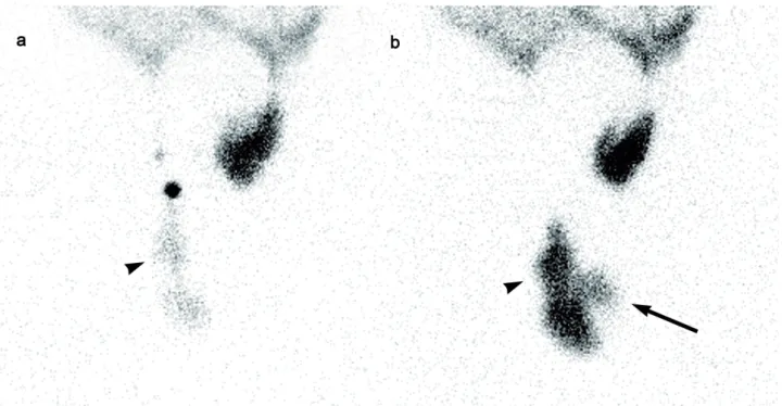
forming the Valsalva’s maneuver (Figure 1 - Pa-nel b). Delayed sectorial images (Figures 2 and 3) were acquired following the peritoneal drainage period (250 seconds per image), revealing the presence of peritoneal dialysis solution in both inguinal canal and right scrotal area (Arrow and head arrow). Due to the peritoneal-vaginal le-akage an inguinal hernioplasty with raquideal anesthesia was performed.
The peritoneal-scrotal dialysate leaka-ge is the major non-infectious catheter-related complication in patients receiving APD, caused by increased intraperitoneal pressure and the loss of integrity of the peritoneal membrane, al-though the most common cause is fluid
extra-Figure 2 - Sectorial peritoneal 99mTc-Phytate scintigraphy. Panel a: Post-filling period image revealing the right inguinal and scrotal uptake of radiotracer (head arrow). Panel b: Post peritoneal lavage image, showing the appearance of left inguinal canal uptake of radiotracer (arrow).
vasation from an indirect hernial sac or patent peritoneal-vaginal duct (1). Pleural, abdominal or genital dialysate leaks tend to develop during the first year of APD, while early leaks after ca-theter insertion are usually observed in the first 30 days of APD (2).
IBJU| RADIOLOGY PAGE
1029
ARTICLE INFO
Int Braz J Urol. 2015; 41: 1027-9
_____________________
Submitted for publication: December 11, 2014 _____________________
Accepted after revision: May 12, 2015
REFERENCES
1. Engeset J, Youngson GG. Ambulatory peritoneal dialysis and hernial complications. Surg Clin North Am. 1984;64:385-92. 2. Leblanc M, Ouimet D, Pichette V. Dialysate leaks in peritoneal
dialysis. Semin Dial. 2001 Jan-Feb;14:50-4.
3. Tokmak H, Mudun A, Türkmen C, Sanli Y, Cantez S, Bozfakioğlu S. The role of peritoneal scintigraphy in the detection of continuous ambulatory peritoneal dialysis complications. Ren Fail. 2006;28:709-13.
_______________________ Correspondence address:
Andrés Martínez Esteve, MD Departamento de Medicina Nuclear Hospital Virgen del Rocío Universitario Sevilla, 41013, España Telephone: + 34 63 545-0488. E-mail: [email protected]