SURGICAL TECHNIQUE
168
Single-port retroperitoneal renal biopsy using standard
urological instruments
_______________________________________________
Rodrigo Guerra1, Flávio Vasconcelos Ordones1, Hamilto Akihissa Yamamoto1, Paulo Roberto Kawano1, João Luiz Amaro1
1Department of Urology, Medical School, São Paulo State University (UNESP), Botucatu, SP, Brazil
ABSTRACT ARTICLE INFO
______________________________________________________________ ______________________
Objective: To describe the surgical technique and initial experience with a single-port retroperitoneal renal biopsy (SPRRB).
Materials and Methods: Between January and April 2013, five children underwent SPRRB in our hospital. A single 1.5 cm incision was performed under the 12th rib at mid-axillary line, and an 11 mm trocar was inserted. A nephroscope was used to iden-tify the kidney and dissect the perirenal fat. After lower pole exposure, a laparoscopic biopsy forceps was introduced through the nephroscope working channel to collect a renal tissue sample.
Results: SPRRB was successfully performed in five children. The mean operative time was 32 minutes, and mean estimated blood loss was less than 10 mL. The hospital stay of all patients was two days because they were discharged in the second postoperative day, after remaining at strict bed rest for 24 hours after the procedure. The average number of glomeruli present in the specimen was 31.
Conclusion: SPRRB is a simple, safe and reliable alternative to open and videolaparos-copic approaches to surgical renal biopsy.
Key words:
Kidney; Biopsy; Retroperito-neal Space; Surgical Proce-dures, Operative; Minimally Invasive Surgical Procedures; Laparoscopy
Int Braz J Urol. 2015; 41: 168-71
_____________________
Submitted for publication: February 09, 2014
_____________________
Accepted after revision: May 13, 2014
INTRODUCTION
Image-guided percutaneous renal biopsy is the most widely used method to sample renal parenchyma for the evaluation of malignancy or diffuse renal disease. The risks of this procedure are minimal and the overall success rate of all re-nal biopsies varies from 70 to 100% (1). Its major indications rest on diagnosis and follow-up of se-veral systemic and nephrological conditions that lead to glomerular damage and renal function im-pairment, providing useful data for treatment and prognosis. It may also be used for evaluation of solid renal masses and cystic renal lesions (1).
However, this method has absolute and re-lative contraindications that may hamper or pre-clude it, such as the presence of a solitary kidney, uncontrolled arterial hypertension, coagulation disorders, renal artery aneurysm, previous percu-taneous needle biopsy failure and obesity. Blee-ding and inadequate amounts of renal tissue for diagnosis are not infrequent, and constitute po-tential disadvantages of the procedure. In addi-tion, children may be unable to cooperate, requi-ring general anesthesia.
In these settings, open and laparoscopic approaches are well-established alternatives and should be considered, although with a higher level
IBJU| SINGLE-PORT RENAL BIOPSY
169
of invasiveness and complexity. In search for an alternative that could minimize surgical aggres-siveness of these procedures and hence spread its use, we outlined a renal biopsy technique throu-gh a single retroperitoneal laparoscopic access using standard urological instruments. The aim of this paper is to describe the technique now standardized in our institution and our initial experience with the Single Port Retroperitoneal Renal Biopsy (SPRRB).
SURGICAL TECHNIQUE
After receiving general anaesthesia, oro-gastric and bladder catheterization, the patient is usually positioned in the left flank position, as the kidney is more easily accessible at the right side due to its lower position. Standard preparation of the surgical field is performed. A 1.5 cm incision is carried out just below the tip of the 12th rib, at the mid-axillary line, and is followed by blunt access to the retroperitoneum space. An initial digital dissection is done aim-ing to identify the lower renal pole, while also displacing the peritoneum anteriorly. During this step, care must be taken in order to avoid peritoneal tearing, as the pneumoperitoneum re-sulting from gas insufflation would hamper the maintenance of adequate retroperitoneal work-ing space. Next, a rubber balloon is positioned between the kidney and the posterior abdomi-nal wall, and is filled with 300-400 cc of saline, creating a virtual cavity. The saline is drained after a few minutes, to achieve hemostasis, and the balloon is then removed. An 11 mm trocar is inserted and carbon dioxide is used to main-tain pneumoretroperitoneum at 12 to 15mmHg. Retroperitoneal inspection and identification of the psoas muscle and the lower pole of the kidney are now performed with a standard 26 French nephroscope, as shown in Figure-1. It is frequently possible to expose the renal sur-face bluntly, by using gentle movements of the tip of the scope to drag the perirenal fat away from the intended site of biopsy. Alterna-tively, standard laparoscopic surgical aspirator, scissors or hooks can be inserted through the working channel of the nephroscope, and then
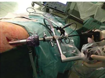
Figure 1 - Positioning of trocar and use of nephroscope for retroperitoneoscopy.
Figure 2 - Sampling renal parenchyma with a toothed biopsy forceps, through the nephroscope working channel.
IBJU| SINGLE-PORT RENAL BIOPSY
170
COMMENTS
At our institution, laparoscopic retroperito-neal renal biopsy is currently often performed for pediatric patients with nephrological conditions (2). As the surgical team’s experience progressed and the procedure was standardized, however, we felt that it should be even less invasive, especially for this very young population. Additionally, in order to spread and popularize its execution, we devised how to use instruments that are already present in a regular urological operating room, such as the nephroscope and laparoscopic scissors and forceps, in a different fashion. A similar ap-proach has been described previously, in pediatric surgery, for appendectomies and varicocelecto-mies, but with only one case of renal biopsy (3).
Between January and April/2013, five children underwent SPRRB in our hospital, refer-red from the Nephrology Clinic for renal biopsy. Informed consent was previously obtained from parents, respecting our institution’s Ethics Com-mittee recommendations and approval. Clinical characteristics of these patients are summarized in Table-1. In all cases, the procedure was succes-sfully performed with the technique above
des-cribed, by a supervised resident in-training. The overall mean operative time was 32 minutes, and mean estimated blood loss was less than 10 mL. No open conversion was needed. The hospital stay of all patients was two days because they were kept in absolute bed rest for 24 hours after the procedure, before being discharged home. Pain and analgesics use were low, and there were no significant detected complications. Regarding the obtained samples, the average number of glo-meruli present in the specimens was 31, and the histopathological findings showed focal prolife-rative lupus glomerulonephritis in two cases, di-ffuse mesangial proliferative glomerulonephritis in another two, and nephritis related to Henoch--Schönlein purpura in one child. These results are comparable to those previously shown by us, with laparoscopic renal biopsy in children, regarding operative time, blood loss, hospital stay and suc-cess in obtaining adequate samples (2).
Although it is likely that the same appro-ach could be used in adult patients as well. Our experience with this very initial group was com-posed entirely of children, and SPRRB has been shown to be a very simple, safe and reliable al-ternative to other laparoscopic approaches. The use of a nephroscope, instead of a regular lapa-roscope, obviates the need to place an additional trocar for using an auxiliary instrument to dissect the perirenal fat, as is the standard practice (4, 5). Its working channel finely substitutes that, sparing one incision, the cost of another trocar and also surgical time to place it. Because a second trocar traditionally would be only 5 mm wide, it may seem that the benefit here is not strongly relevant in terms of postoperative pain or cosmetic results, but it is our understanding that no technical di-fficulty was added whatsoever, by using only one access. Moreover, especially children could benefit the most even of a small effect, and coincidently they constitute the majority of patients requiring a surgical renal biopsy in our hospital. Mini-perc nephroscopes are not available at our institution at this time, but its use could be a step forward, in this regard, and further decrease the required size of the access port incision. Additionally, the ease for urologists in using regular urological equipment, and the possibility that the surgeon simultaneously
IBJU| SINGLE-PORT RENAL BIOPSY
171
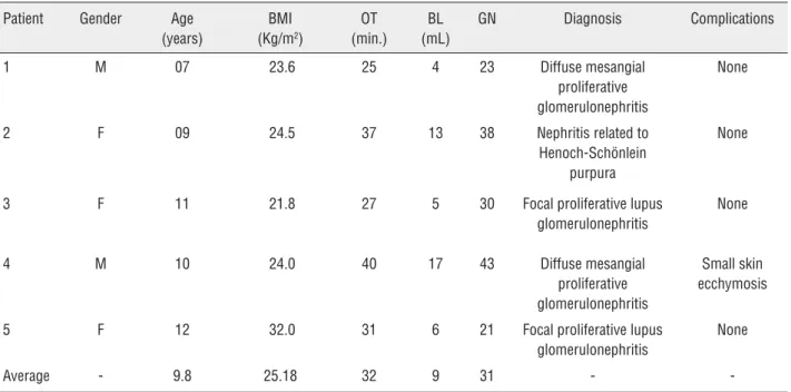
Table 1 - Clinical features of patients submitted to Single-port retroperitoneal renal biopsy.
Patient Gender Age (years)
BMI (Kg/m2)
OT (min.)
BL (mL)
GN Diagnosis Complications
1 M 07 23.6 25 4 23 Diffuse mesangial
proliferative glomerulonephritis
None
2 F 09 24.5 37 13 38 Nephritis related to
Henoch-Schönlein purpura
None
3 F 11 21.8 27 5 30 Focal proliferative lupus
glomerulonephritis
None
4 M 10 24.0 40 17 43 Diffuse mesangial
proliferative glomerulonephritis
Small skin ecchymosis
5 F 12 32.0 31 6 21 Focal proliferative lupus
glomerulonephritis
None
Average - 9.8 25.18 32 9 31 -
-BMI = Body mass index (kg/m2); OT = Operative time (minutes); BL = Blood loss (milliliters); GN = Number of glomeruli per biopsy.
controls both the camera and laparoscopic scissor/ biopsy forceps, are other advantages of this alter-native method.
In our hospital, retroperitoneal laparosco-py is the procedure of choice for renal biopsy in children and the SPRRB is an even less invasi-ve option for these patients, performed through a single incision and with very satisfactory results and only minor pain.
ABBREVIATIONS
SPRRB = Single Port Retroperitoneal Renal Biopsy
CONFLICT OF INTEREST
None declared.
REFERENCES
1. Uppot RN, Harisinghani MG, Gervais DA. Imaging-guided percutaneous renal biopsy: rationale and approach. AJR Am J Roentgenol. 2010; 194: 1443-9.
2. Jesus CM, Yamamoto H, Kawano PR, Otsuka R, Fugita OE. Retroperitoneoscopic renal biopsy in children. Int Braz J Urol. 2007; 33: 536-41; discussion 541-3
3. Martino A, Zamparelli M, Cobellis G, Mastroianni L, Amici G. One-trocar surgery: a less invasive videosurgical approach in childhood. J Pediatr Surg. 2001; 36: 811-4.
4. Luque Mialdea R, Martín-Crespo Izquierdo R, Díaz L, Fernández A, Morales D, Cebrían J. Renal biopsy through a retroperitoneoscopic approach: our experience in 53 pediatric patients. Arch Esp Urol. 2006; 59: 799-803. 5. Takeda M, Watanabe R, Kurumada S, Saito K, Tsutsui T,
Takahashi K, et al. Endoscopic renal biopsy in pediatric patients: comparison of retroperitoneoscopy-assisted and retroperitoneoscopic methods. Nephron. 2000; 84: 199-200.