Risk groups in bladder cancer patients treated with radical
cystectomy
_______________________________________________
Eva Mallen1, Pedro Gil2, Maria Jesus Gil2
1Department of Urology, Hospital Royo Villanova, Zaragoza, Spain; 2Deparment of Urology Hospital
Miguel Servet, Zaragoza, Spain
ABSTRACT ARTICLE INFO
______________________________________________________________ ______________________
Objective: To stratify patients with bladder cancer into homogeneous risk groups ac-cording to statistically significant differences found in PFS (progression-free survival). To identify those patients at increased risk of progression and to provide oncological follow-up according to patient risk group.
Materials and Methods: A retrospective study of 563 patients treated with radical cystectomy (RC). In order to determine which factors might predict bladder tumour progression and death, uni- and multivariate analyses were performed. The risk groups were identified according to “inter-category” differences found in PFS and lack of differences, thus revealing intra-category homogeneity.
Results: Median follow up time was 37.8 months. Recurrence occurred in a total of 219 patients (38, 9%). In 63% of cases this was distant recurrence.
Only two variables retained independent prognostic value in the multivariate analysis for PFS: pathological organ confinement and lymph node involvement. By combining these two variables, we created a new “risk group” variable. In this second model it was found that the new variable behaved as an independent predictor associated with PFS. Four risk groups were identified: very low, low, intermediate and high risk:
s Very low risk: pT0 N0
Conclusions: We retrospectively identified 4 risk groups with an independent prognos-tic value for progression-free survival following RC.
Differences in recurrence patterns after RC between risk groups have led us to set di-fferent intervals in monitoring for cancer.
Key words:
Urinary Bladder Neoplasms; Cystectomy; Pathology
Int Braz J Urol. 2015; 41: 30-9
_____________________
Submitted for publication: January 26, 2014
_____________________
Accepted after revision: February 02, 2014
INTRODUCTION
Bladder cancer is major health problem in Spain, with high incidence and elevated mortality (1-3). Radical cystectomy is the standard treatment for patients with muscle-invasive bladder cancer, ho-wever this is generally insufficient. In fact, it is very important to identify those at high risk of progression
from disease progression. These patients are therefore less likely to require adjuvant treatment, and follow--up intervals may also be more distanced. We propo-se a follow-up strategy for RC-treated bladder cancer patients that identifies most cases of recurrence while at the same time avoids monitoring patients too clo-sely. This implies in a reduction of costs related to tests and number of visits.
While it is true that our study is not a multi-disciplinary project, we believe that our single-center study includes a sample sufficiently large to draw conclusions similar to those drawn from studies un-dertaken by multidisciplinary groups (6). The aim of the present study was establish risk groups and tailor follow-up accordingly with a schedule appropriate to the likelihood of progression.
MATERIALS AND METHODS
Patient Population
We retrospectively reviewed all patients who underwent radical cystoprostatectomy and pelvic lymphadenectomy for bladder cancer with curative intent at Miguel Servet University Hospital betwe-en 1975 and 2007. The study population consisted of 599 patients who underwent radical cystectomy. Thirty-six patients were eliminated from the study due to missing data. Therefore, analysis was perfor-med on the 563 remaining patients.
Radical cystoprostatectomy and lymphade-nectomy were always performed according to stan-dard protocol; indications for cystectomy did not change during the time period studied. Cystectomy is indicated in patients with invasive bladder car-cinoma, endoscopically uncontrollable superficial bladder cancer and high risk bladder tumors and for those with BCG-resistance bladder tumors. Radical cystectomy and limited pelvic lymph node dissection were performed in 84% of the patients. In only 10 cases it was performed an extended pelvic lympha-denectomy. It is true that currently, this procedure is routinely performed in most cases.
All cystectomy specimens were subjected to routine pathological examination. In the last two decades the same pathologist examined the specimens microscopically. Primary tumors and lymphadenectomy were restaged based on the 2002 UICC TNM system.
Statistical analysis
“FileMaker Pro 11.0, version 11.0 v2” (Fi-leMaker Inc©) was used as database software and
“PASW Statistics 18, version 18.0.0” (IBM©) was
used as statistics software.
Paper-based patient records were reviewed and data were analyzed for possible predictive factors. Univariante analysis was performed using the Kaplan-Meier (or Mantel-Haenszel) test. Sig-nificant variables (p<0.05) and those close to sig-nificance (p<0.1) from univariate analysis were analyzed using backwards multivariate analysis with the Cox proportional hazards regression model. To search for a clinical application and to check the strength of the model, a new model was set up, with the combination of the statistically most powerful variable taken from multivariate analysis. We created a new variable called “risk groups”, to try to identify any diffe-rences in PFS and CSS between the different cate-gories of this variable. The risk groups identified were compared using the Kaplan-Meier method and log-rank test.
RESULTS
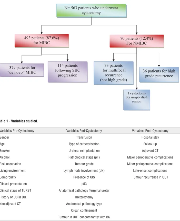
Cystectomy was indicated for muscle-inva-sive bladder cancer (MIBC) in 493 patients. In the remaining 70 patients, the indication for cystectomy was superficial bladder cancer. In these 70 cases, 33 underwent cystectomy for multifocal recurrence of NMBC (non muscle-invasive bladder cancer) such as TaG1-2, T1G1-2, 36 patients for recurrent high-grade tumours (TaG3, T1G3, and CIS) and in one case the reason for cystectomy was not specified. These data are summarized in the chart below (Figure-1).
The variables investigated in patients treated with cystectomy in the period of study were grouped into: pre-, peri- and post-cystectomy variables, as shown in Table-1.
At the time of cystectomy, median patient age was 65.3 years (IQR 13.1). In the analysis by de-cades it can be seen that patients were progressively older, with statistically significant differences.
1 SDWLHQWVZKRXQGHUZHQW F\VWHFWRP\
SDWLHQWV
IRU0,%& SDWLHQWV)RU10,%&
SDWLHQWV IROORZLQJ6%&
SURJUHVVLRQ SDWLHQWVIRU
³GHQRYR´0,%&
SDWLHQWV IRUPXOWLIRFDO
UHFXUUHQFH QRWKLJKJUDGH
SDWLHQWVIRUKLJK JUDGHUHFXUUHQFH
F\VWHFWRP\ IRUXQVSHFLILHG
UHDVRQ
Figure 1 - Indication of radical cystectomy.
Table 1 - Variables studied.
Variables Pre-Cystectomy Variables Peri-Cystectomy Variables Post-Cystectomy
Gender Transfusion Hospital stay
Age Type of catheterisation Follow-up
Smoker Ureteral reimplantation Adjuvant CT
Alcohol Pathological stage (pT) Major perioperative complications
Risk occupation Tumour grade Minor perioperative complications
Living environment Lymph node involvement (pN) Late-onset complications
Comorbidity Presence of CIS Tumour recurrence in UUT
Clinical presentation p53
Clinical stage of TURBT Anatomical pathology Terminal ureter
History of UC in UUT Ureterectomy
Neoadjuvant CT Anatomical pathology type
Organ confinement
Tumour in UUT concomitantly with BC
studied, such as for example the number of units transfused (p<0,00), length of hospital stays and the number of complications (p<0.03). All these variables decreased in number/duration. Conver-sely, continent urinary derivations (p<0.00) incre-ased during the study period. In terms of studied tumor characteristics no clear differences were observed over the four decades of the study. In the percentage of patients with organ-confined tumors there were no statistically significant di-fferences (p=0.714). In the case of lymph node in-volvement prevalence increased over the decades, but not significantly (p=0.250).
Progressive disease occurred in 219 pa-tients of the total series (38.9%). We classified re-currence as: local, regional lymph node and dis-tant. Local recurrence: we considered this to be the appearance of local recurrence in the pelvis urothelial tumor, surgical site and/or urinary tract without distant involvement. Locoregional nodal recurrence: this included pathological lymph node involvement and distant metastasis.
In patients who progressed, the predomi-nant type of progression was distant metastasis in 63% of cases. Mean PFS (progression-free sur-vival) in the 563 patients was 157 (144.2-169.9) months (the median could not be calculated).
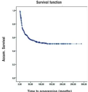
It can be observed that almost all events (recurrence from bladder cancer) occurred in the first two years of follow up. In fact, by the end of the second year of follow-up, 77% of the events had already occurred. There was a 73% probabi-lity of survival in the first year, falling to 55% at the end of the fifth year, with little further change to the end of the tenth year (51%) (Figure-2 and Table-2).
In patients who progressed, n=219 (38.9%),
median progression-free survival was 9.7 months (CI 95% 8.3-11.1); there were no differences in PFS by type of progression. Progression appea-red to occur shortly before lymph node recurrence (median PFS=8.6 months [6.3-11]), but significant differences were found in the univariate analysis.
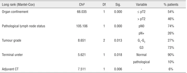
Univariante analysis demonstrated that pathological organ confinement, lymph node in-volvement, tumour grade, terminal ureter invol-vement and the administration of adjuvant che-motherapy were variables that were significantly
associated with lower progression-free survival (Table-3).
In the multivariate analysis (Table-4) we entered the significant variables detected in the univariate analysis and found that pathological organ confinement and lymph node involvement were independent variables that were significantly associated with PFS.
In order to identify patients at increased risk of urothelial disease progression or recurrence following cystectomy, we developed a classifica-tion system based on the multivariate predictive variables for PFS: pathological organ confinement and lymph node involvement. We created a new variable called “risk groups”, using a combination of the two previous variables (pT and pN), in order
Table 2 - Mean PFS in the whole series.
T (months) Cumulative survival probability
12m 83.1%
36m 65.5%
60m 61.5%
120m 55.2%
Survival function
Accum. Sur
vival
Time to progression (months)
Survival function Censured
Table 3 - Univariante analysis of predictive factors for PFS.
Long rank (Mantel-Cox) Chi² Df Sig. Variable % patients
Organ confinement 66.035 1 0.000 d pT2 54%
> pT2 46%
Pathological lymph node status 105.106 1 0.000 pN0 74%
pN+ 26%
Tumour grade 8.651 2 0.013 G1-G2 27%
G3 73%
Terminal ureter 5.621 1 0.018 Normal 90%
pathological 10%
Adjuvant CT 7.511 1 0.006 - 6%
PFS = progression-free survival; CT = chemotherapy
Table 4 - Multivariate analysis. Predictive model for PFS.
Variable B SE Wald Gl p O.R. 95% CI for EXP(B)
Lower Upper
Pathological organ confinement 1.077 0.283 14.445 1 0.000 2.936 1.685 5.117
Lymph node involvement 0.861 0.214 16.219 1 0.000 2.365 1.556 3.595
PFS = progression-free survival
to identify any differences in PFS between the di-fferent categories of this variable.
We analysed the survival for each pT va-riables (pT0 vs. pTa-Tis-T1 vs. pT2 vs. pT3-4) and pN variables (pN0 vs pN1-3). We observed clear differences between the patients with pT0 and the group stages PTa, pTis, pT1, pT2, and of course among the rest of groups, which led us to differen-tiate between a group of very low risk, separating it from the rest. According to this analysis, four independent groups were identified (Table-5).
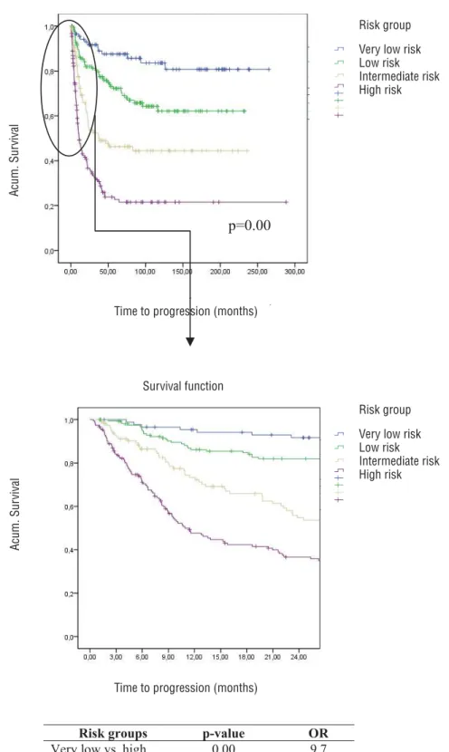
With respect to the survival graph (Fi-gure-3), firstly it can be seen that there are sta-tistically significant inter-category differences (p<0.001), which appear to confirm the homoge-nous nature of the composition of the four ca-tegories. Of note are the findings in PFS in the
Table 5 - Risk groups.
“Risk group” variable
Categories Description
Very low risk pT0 and pN0
Low risk pTa, pT1, pTis or pT2, and pN0
Intermediate risk pT3 and pN0
High risk pT4N0 or pN1-3
5LVNJURXSV SYDOXH 25
9HU\ORZYVKLJK
/RZYVKLJK
,QWHUPHGLDWHYVKLJK 7LPHWRSURJUHVLRyQPRQWKV
S
9 Figure 3 - PFS by risk group.
Time to progression (months)
Survival function
Very low risk Low risk Intermediate risk High risk Risk group
Acum. Sur
vival
Acum. Sur
vival
Time to progression (months)
9
The survival graphs show that the risk of progression stabilizes. This is similarly reflected in the survival table (Table-6). This shows the pro-bability of reaching a certain time point, in the post-cystectomy follow-up, without progression.
In this second model it was found that the new “risk group” variable behaved as an indepen-dent predictor associated with PFS.
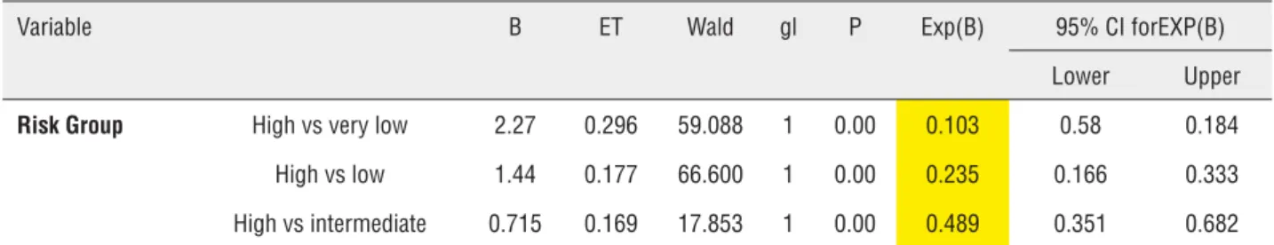
From the data of Table-7 it can be deduced that a patient in the high risk group has an OR of progression that is 9.7 times (1/0.103) higher than patients with a very low risk. The same high risk patient has an OR that is 4.25 times (1/0.235) hi-gher for progression than low-risk patients.
DISCUSSION
This study was undertaken primarily due to the high rates of incidence and mortality of bla-dder cancer in Spain and in our region, Aragon. Rates are above the mean for Europe (7, 8), and this, together with the marked ageing of popula-tion, suggests that the upward trend will continue.
In the literature there are several studies on the prognostic value of different variables such as stage, lymph node and lymphovascular invol-vement, type of urinary diversion, margin status, etc. on invasive bladder cancer progression and survival. However, organ confinement and lymph node involvement are repeatedly found to be in-dependent risk factors for PFS and cancer-specific mortality in most series (4, 9, 10).
In a study at Gregorio Marañón Hospital (4), both pathological stage and lymph node in-volvement were found to be independent predic-tors for cancer-specific survival. In that study the authors, like us, also constructed a second model by grouping local stage (pT) and lymph node in-volvement (pN) into risk groups in order to study CSS. Thus, the association by risk group allowed them to predict the risk of death from bladder can-cer more reliably, and to identify patients in whom cystectomy is insufficient and who could benefit from adjuvant treatment.
Solsona (5) also stratified patients by risk. In this study, the records of 298 patients who
un-Table 6 - Survival table (PFS) by "Risk group".
PFS by “Risk group”. Survival table
Categories End 1st year
End 2nd year
End 3rd year
End 4th year
End 5th year
End 10th year
Very low pT0 and pN0 0.954 0.917 0.903 0.875 0.875 0.836
Low pTis, pTa-1, pT2 and pN0 0.861 0.82 0.797 0.758 0.722 0.621
Intermediate pT3 and pN0 0.734 0.547 0.525 0.476 0.463 0.444
High pT4 or pN1-3 0.485 0.367 0.315 0.238 0.227 0.215
Table 7 - Subvariables that retain independent statistical significance for PFS.
Variable B ET Wald gl P Exp(B) 95% CI forEXP(B)
Lower Upper
Risk Group High vs very low 2.27 0.296 59.088 1 0.00 0.103 0.58 0.184 High vs low 1.44 0.177 66.600 1 0.00 0.235 0.166 0.333
derwent cystectomy were retrospectively analysed and risk groups were established based on lymph node involvement, pathological stage and pros-tatic stromal involvement as predictors of morta-lity in the multivariate analysis. Their figures for 5-year CSS, by risk group, were 86.4% for the low risk group (P1-2N0St-), 64.4% for the intermedia-te risk group (60.9%-65.3%) (P1-2N1St-, P3N0St-, HR=2.7) and 28.1% (0%-47.7%) for the high risk group (N2-3, P4, St+, N1P3, HR=8.7). These figures were not far off our 5-year CSS of 89% for the very low risk group, 75% for low risk, 54% for interme-diate and 30% for low grade (data not shown).
Sonpavde (11) investigated bladder cancer risk following cystectomy using pathological fac-tors to facilitate an indication for adjuvant tre-atment. This series was more similar to ours in terms of sample size and study time (although the follow-up period was longer in their study), and it was also found that pathological stage was a predictor. Like us, these authors constructed a mo-del with prognostic groups and found that stage, lymphovascular involvement and poorly-differen-tiated cells were predictors of PFS. They distingui-shed three groups according to the presence of the three variables in the multivariate analysis, assig-ning a score of 0-4, according to the presence of these variables, and depending on the cumulative score, they divided patients into low, intermediate and high risk groups.
In the case of the Abol-Enein and Ghoneim group (12), which involved a large series because of the high incidence of bladder cancer in Egypt, these authors concluded that PFS predictors are lymph node involvement, stage, lymphovascular invasion and type of urinary diversion. Using the-se four variables, patients were stratified into four risk groups ranging from low risk (T1,N0, LV-, or-thotopic diversion) to maximum risk (T4,N+, LV+, rectal diversion), comparing survival curves and likelihood of progression among these groups. They found a 5-year PFS of 64.5%, which does not differ greatly from our figure of 55.6%. Like these authors, we divided patients into categories or risk groups, with two objectives. Firstly, in re-ference to follow-up we aimed to identify risk of progression and detect this as early as possible. The second objective was to identify candidates
for adjuvant treatment, with the final objective of increasing CSS.
In our study, differences in progression patterns after radical cystectomy suggest the need for varied follow-up protocols for each group. We proposed a stage-based protocol for monitoring of patients with bladder cancer treated with radical surgery that captures most recurrences while li-miting over-investigation. Multicenter studies are consistent with our study, showing that in most patients the tumor recurs in the first two years after radical cystectomy and over half of these re-currences are distant (6).
Table-6 shows the probabilities of progres-sion-free survival at a certain time point according to risk group. Looking at how to apply these data in clinical practice, we can draw up guidelines and recommendations for monitoring these patients. To date, there has been no consensus on how to follow up these patients after surgical intervention. Avai-lable guidelines include the National Comprehensi-ve Cancer Network and the European Association of Urology recommendations, which acknowledge the need for risk-stratified surveillance but do not clearly delineate any protocols (13, 14) .The ESMO (European Society for Medical Oncology) guidelines, by contrast, do not address any stage-specific sur-veillance regimen (15).
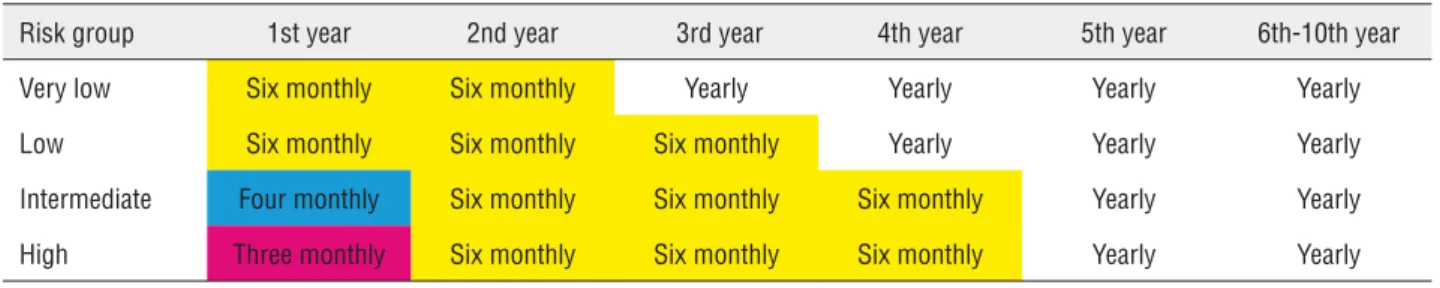
Table 8 - Follow-up recommendations.
Risk group 1st year 2nd year 3rd year 4th year 5th year 6th-10th year
Very low Six monthly Six monthly Yearly Yearly Yearly Yearly
Low Six monthly Six monthly Six monthly Yearly Yearly Yearly
Intermediate Four monthly Six monthly Six monthly Six monthly Yearly Yearly
High Three monthly Six monthly Six monthly Six monthly Yearly Yearly
so follow-up can be performed at six-monthly in-tervals until the probability of progression stabili-ses (in the 4th year). In the low risk group, patients could be followed up less frequently from the third year onwards, when the probability of progression stabilises. Faysal et al. (6) proposed stage-based protocols for surveillance of patients with bladder cancer based on recurrence patterns that coincide with our group in terms of the frequency of visits especially in the first year of follow-up in patients with very high risk of recurrence.
Since there is such a high probability of progression in the intermediate and high risk groups, intensive follow-up of these patients is clearly essential. However, the aim must be to pre-vent progression from occurring or to delay it as much as possible. Therefore, an assessment should be made of whether to administer adjuvant che-motherapy in these two groups of patients.
It is true that there are no randomised trials that demonstrate the superiority of adjuvant chemotherapy in MIBC treatment, but until such studies are available, we would recommend that chemotherapy - preferably within the framework of a clinical trial - as it is one of the few tools at our disposal that lengthens CSS.
Our study has several potential limi-tations, including those inherent to any ret-rospective study. For example, the variable margin status; this was not studied in the ear-ly years of the study period and therefore could not be included in the study. Extent of surgery, such as the upper limit of lymph node dissec-tion, changed slightly and was not consistent during the study period. Our median follow-up time may seem short (37.8 months) however these are patients with an ominous prognosis.
Further-more, this is a single institution study. Find-ings must be evaluated externally in a larger patient cohort and validated prospectively for practical use.
CONCLUSIONS
Non-organ confinement and lymph node involvement in radical cystectomy specimens are factors that retain independent prognostic value in progression-free survival in the multivariate analysis. We retrospectively identified four risk groups (very low, low, intermediate and high) with an independent prognostic value for progression--free survival following radical cystectomy. This would be useful in order to provide information to patients and physicians and to improve stratifica-tion for future clinical trials. This would serve to optimize indications for treatment, avoiding ex-cessive monitoring and reducing costs.
ABBREVIATIONS
PFS = progression-free survival
RC = radical cystectomy
CSS = cancer-specific survival
MIBC = muscle-invasive bladder cancer
NMBC = non muscle-invasive bladder cancer
CONFLICT OF INTEREST
None declared.
REFERENCES
2. National Epidemiology Center. Area Environmental Epidemiology and Cancer. Cancer mortality in Spain. Consulted 2011. Available at: http://cne.isciii.es
3. World Health Organization. Available at: http://www.who.int 4. Monzó Gardiner JI, Herranz Amo F, Díez Cordero JM, Cabello Benavente R, Silmi Moyano A, Hernández Fernández C. Prognostic factors for survival in patients with transitional bladder cancer treated with radical cystectomy. Actas Urol Esp. 2009;33:249-57.
5. Solsona E, Iborra I, Dumont R, Rubio J, Casanova JL, Almenar S. Risk groups in patients with bladder cancer treated with radical cystectomy: statistical and clinical model improving homogeneity. J Urol. 2005;174:1226-30. 6. Yafi FA, Aprikian AG, Fradet Y, Chin JL, Izawa J, Rendon R,
et al. Surveillance guidelines based on recurrence patterns after radical cystectomy for bladder cancer: the Canadian Bladder Cancer Network experience. BJUI. 2012;110:1317-1324.
7. Serrano P. Study of bladder cancer in the health area II of the province of Zaragoza: Epidemiology, anatomo and clinical outcome prediction model for the period 1994-2003 [MS thesis]. University of Zaragoza, 2007.
8. Romero FJ, Bernal M. Evolución en el carcinoma vesical en el área III de Zaragoza en los últimos 30 años. Póster XXVII Reunión del Grupo de Urología Oncológica, Sevilla, 2010.
9. Bassi P, Ferrante GD, Piazza N, Spinadin R, Carando R, Pappagallo G, et al. Prognostic factors of outcome after radical cystectomy for bladder cancer: a retrospective study of a homogeneous patient cohort. J Urol. 1999;161:1494-7. 10. Frazier HA, Robertson JE, Dodge RK, Paulson DF. The value of
pathologic factors in predicting cancer-specific survival among patients treated with radical cystectomy for transitional cell carcinoma of the bladder and prostate. Cancer. 1993;71:3993-4001.
11. Sonpavde G, Khan MM, Svatek RS, Lee R, Novara G, Tilki D, et al. Prognostic risk stratification of pathological stage T2N0 bladder cancer after radical cystectomy. BJU Int. 2011;108:687-92.
12. Baco E, Rud E, Vlatkovic L, Svindland A, Eggesbø HB, Hung AJ, et al. Predictive value of magnetic resonance imaging determined tumor contact length for extracapsular extension of prostate cancer. J Urol. 2015;193:466-72.
13. Stenzl A, Witjes JA, Comperat E, et al.Guidelines on Bladder Cancer 2012: Muscle-invasive and Metastatic. Available at: http://www.uroweb.org. Accessed November 2012.
14. NCCN Clinical Practice Guidelines in Oncology (NCCN Guidelines TM). Bladder Cancer. 2012. Available at: http:// www.nccn.com. Accessed November 2012.
15. Bellmunt J, Albiol S, Kataja V; ESMO Guidelines Working Group. Invasive bladder cancer: ESMO clinical recommendations for diagnosis, treatment and follow-up. Ann Oncol. 2008;19 Suppl 2:ii47-8.
_______________________ Correspondence address:
Eva Mallen, MD Department of urology Hospital Royo Villanova Avenida San Gregorio, 30,