*Correspondência: Genética Clínica UFCSPA Rua Sarmento Leite, 245 - sala 403
Centro
CEP: 90050-170 - Porto Alegre – RS, Brazil Tel: (51) 3303-8771 - Fax: (51) 3303-8810
AbstrACt
Objective. To verify the frequency and types of congenital heart defects in a sample of patients with oculo-auriculo-vertebral spectrum (OAVS), in order to correlate the presence of these defects with other clinical characteristics and evolution.
MethOds. The sample comprised 33 subjects, all attended in the same center, between January 1975 and December 2007. Twenty two of them were male and eleven female with ages ranging from 1 day to 17 years old. All presented normal karyotype by GTG-Banding. A data collection related to their clinical history, physical examination and result of complementary evaluations was performed.
Results. Cardiac abnormalities were observed in 13 patients (39.4%). Of these, 5 (38.5%) were conotruncal, tetralogy of Fallot being the main malformation (n=2). Unusual anomalies identiied included cor triatriatum and double inlet left ventricle. Signiicant differences among the clinical characteristics of the group with and without heart defect were only veriied in relation to age at irst
evaluation that was lower in subjects with cardiac malformations. Five patients died, four of them carriers of congenital heart defects.
cOnclusiOn. Cardiac malformations, mainly of the conotruncal and septal types, are frequent among patients with OAVS. The frequency found in our study was statistically similar to the one found in the majority of works described in literature, which ranges from 18 to 58%. Congenital heart defects also represent the main cause of death of these subjects. Thus, a cardiac evaluation should always be performed in these patients, especially at an early age.
Keywords:Goldenhar syndrome. Facial asymmetry. Congenital heart defects.
OculO
-
auRiculO
-
veRtebRal
spectRuM
and
caRdiac
MalfORMatiOns
Rafael fabianO MachadO ROsa1, lisiane dall’agnOl2, paulO RicaRdO gazzOla zen3*, veRa lúcia beRenstein peReiRa4, caRla gRaziadiO5, giORgiO adRianO paskulin3 Study conducted at Universidade Federal de Ciências da Saúde de Porto Alegre –UFCSPA, Porto Alegre, RS, Brazil
1. Geneticista Clínico e Doutorando pelo Programa de Pós-Graduação em Patologia da Universidade Federal de Ciências da Saúde de Porto Alegre -UFCSPA e Complexo Hospitalar Santa Casa de Porto Alegre –CHSCPA, Porto Alegre, RS
2. Aluna de Graduação do Curso de Medicina da Universidade Federal de Ciências da Saúde de Porto Alegre – UFCSPA, Porto Alegre, RS
3. Professores Doutores - Geneticista Clínico da Universidade Federal de Ciências da Saúde de Porto Alegre - UFCSPA e Complexo Hospitalar Santa Casa de Porto Alegre – CHSCPA; Professores da Disciplina de Genética Clínica e do Programa de Pós-Graduação em Patologia da Universidade Federal de Ciências Médicas da Saúde de Porto Alegre, UFCSPA, Porto Alegre, RS
4. Graduação em Farmácia - Citogeneticista do Laboratório de Citogenética da Universidade Federal de Ciências Médicas da Saúde de Porto Alegre UFCSPA, Porto Alegre, RS 5. Professora com mestrado - Geneticista Clínica da UFCSPA e CHSCPA, Professora da Disciplina de Genética Clínica da UFCSPA
i
ntROductiOnOculo-auriculo-vertebral spectrum (OAVS), also known as Goldenhar syndrome and hemifacial microsomia (OMIM 164210),1 is an etiologically heterogeneous and phenotypi-cally quite variable condition. Most cases occur in a sporadic manner; however, familial cases, suggesting a pattern of inheritance, both autosomal dominant and recessive, have also been described.2-4 OAVS presents an estimated preva-lence that ranges from 1 to 5,600 in 45,000 of the live-born, and is considered the result of a blastogenesis defect that involves particularly the structures originated from the first branchial arches.2-7 Thus, its main findings are anomalies, generally asymmetric, involving ears (especially microtia and
preauricular skin tags ), face (hemifacial microsomia), eyes (epibulbar dermoid), and spine (vertebra l alterations with fused vertebrae or hemivertebrae). However, its phenotypical spectrum is very broad, and involvement of other organs and systems is frequent.4,8-10 Cardiac malformations, on their turn, are common in OAVS individuals. However, its frequency has been shown to be very variable, oscillating between 5% and 58%.4-14
M
ethOdsThe sample was composed of individuals attended by the Clinical Genetics Service of the Universidade Federal de Ciências da Saúde de Porto Alegre/Complexo Hospitalar Santa Casa de Porto Alegre, Rio Grande do Sul, Brazil, diagnosed with OAVS from January 1975 to December 2007. This study was approved by the Ethics Committee of the Institution. This study included only patients submitted to chromosome evaluation through karyotype exam by GTG banding and who presented phenotypical abnormalities in at least two or the following regions: 1) orocraniofacial, 2) ocular, 3) auricular and 4) vertebral. This approach was in accordance with the one adopted by Strömland et al. (2007).4 Individuals carrying chromosome alterations or having incomplete medical files were excluded from the study.
A retrospective analysis with collection of data related to sex, age, reason for the referring, as well as clinical history, physical examination, and complementary evaluations result was performed. For evaluating anthropometrical measures, standard growth curves were used,18 and values two standard deviations higher or lower than the average, according to the age (with due correction for length/height), were considered abnormal. A delay in neuropsychomotor development was considered when the patient presented the description of starting walking alone only after 18 months old.9 The side affected presented by the syndrome (right, left, or bilateral) was determined according to microtia’s and facial microsomia’s localization, similarly to the method adopted by Rollnick et al. (1987).8
Thus, during this period, 42 patients fulfilled the clinical criteria for inclusion. However, six of them were excluded due to not having a karyotype evaluation. Out of the other 36, one did not have complete medical files and two had chromosome abnormalities. Thus, the final sample was constituted of 33 patients. Twenty two were male and 11, female, their ages at the first evaluation ranged from 1 day to 17 years old (69.7% of them presented less than 1 year). The majority was referred by pediatrics (n=21); out of the rest, four came referred by plastic surgery, two by pediatric surgery, two by cardiology, and four by other specialties. As for clinical characteristics presented by the patients, two had phenotypic alterations involving the four regions belonging to the study’s inclusion criteria, 14 had 3 regions, and 17 had 2.
For statistic analysis, PEPI program was used, more specifically Fisher’s exact test for comparing the frequencies found between the subgroups with and without cardiopathy in our sample and for comparing with other studies described in literature. Values of p <0.05 were considered significant.
R
esultsAll the 33 patients included in the study were submitted to cardiac evaluation through clinical exam and complementary tests, such as thorax radiography and electrocardiogram. In 23 it was complemented through echocardiography. Cardiac abnormalities were observed in 13 patients (39.4%). Defects presented by them are exposed in Table 1. Conotruncal defects
were present in 5 patients (38.5%), the most relevant of them being tetralogy of Fallot (n=2). One of the patients without cardiopathy had the description of an innocent murmur. Out of the 13 individuals with congenital heart defect, 4 died within the first two years of life, while in the group without cardio-pathy only one presented a similar description (see Table 1). The other clinical characteristics observed in the 33 patients of the total sample are found in Table 2. A significant difference between the group with and without cardiopathy was seen only in relation to the age at first evaluation, lower among cardiac malformation carriers.
d
iscussiOnCongenital heart defects are frequently observed among OAVS individuals. The prevalence of these conditions seen in our study (39%) was statistically similar to that of some reports (varying between 18% and 58%),4-7,10-14 and different from others with values between 5% and 15%8,9 (see Table 3). This variability is very probably due to the different inclusion criteria adopted and the varying sample sizes of the studies.6 For example, Rollnick et al. (1987),8 despite their big sample (n=294), included among their OAVS patients many subjects who presented only microtia and few with more severe abnor-malities, as pointed by Kumar et al (1993),6 which may justify the low frequency of congenital cardiopathies found (5%). On
table 1 – type and frequency of congenital cardiopathies observed in the patients with oculo-auriculo-vertebral spectrum (OAVs)
Cardiologic inding N Heart
surgery
Death
Normal 20 NA 1
Abnormal 13 3 4
- Conotruncal / outlet defects 5 2 2
Fallot’s tetralogy (2) -
-Intraventricular communication with pulmonary atresia (IVC + PA)
(1) (1) (1)
Great arteries transposition (GAT) (1) -
-Double inlet left ventricle (1) (1) (1)
- Septal defects 3 -
-Interatrial communication ostium secundum (OS)
(2) -
-Intraventricular communication (1) -
-- Others 5 1 2
patent ductus arterious (2) -
-Atrioventricular septal defect (1) (1) (1)
Pulmonary artery stenosis (1) -
-Cor triatriatum (1) - (1)
tOtAL 33 3 5
Table 2 – Clinical indings presented by patients with OAVS, divided in accordance with the presence or not of congenital cardiopathy
Clinical indings
Congenital cardiopathy total Yes
N= 13
No N=20
sex M10 / F 3 M12 / F 8 M22 / F11
Age at irst evaluation 1d – 5y 1d – 17y 1d – 17y
< 6 months + 11 8 19
Affected side
Right 7 8 15
Left 6 9 15
Bilateral - 4 4
Low stature 7 6 13
Craniorofacial abnormalities
Microcephaly - 2 2
Facial assimetry 7 13 20
Atresia / choanal stenosis 2 - 2
Labiopalatine cleft 6 6 12
Microretrognathia 4 9 13
Tongue hypoplasia 1 1 2
Machrostomia 2 5 7
Ophtalmologic anomalies
Upper palpebral coloboma 2 - 2
Microftalmia / anoftalmia 2 1 3
Epibulbar dermoid 3 3 6
Dacryostenosis - 1 1
Nystagmus 1 - 1
Ears anomalies
Preauricular fossette 2 1 3
Preauricular appendixes 6 14 20
Agenesis / auditory canal stenosis 3 6 9
Microtia 12 15 27
Esophagic / pulmonary abnormalities
Esophagus atresia 2 2 4
Tracheoesophageal istula 2 1 3
Laryngotracheomalacia 2 - 2
Broncogenic cyst 1 - 1
skeleton alterations
Anormalidades radiais de Upper limbs radial abnormalities 2 1 3
Vertebral abnormalities 6 7 13
Ribs alterations 4 3 7
Lower limbs reduction defects - 2 2
Congenital clubfoot 2 2 4
Abdominal anomalies
Pyloric hypertrophic stenosis 1 - 1
Accessory spleen 1 - 1
Renal anomalies 2 2 4
Umbilical / inguinal hernia 4 1 5
Anogenital alterations
Genital abnormalities - 3 3
Anal imperforation 1 - 1
Neurological abnormalities
Neuropsychomotor delay * 7 / 12 13 / 18 20 / 30
Hypotonia 5 3 8
CNS abnormalities 4 5 9
M: male; F: female; d: days; y: years;
CNS: central nervous system; + Clinical inding with statistically signiicant difference between the groups with and without cardiopathy
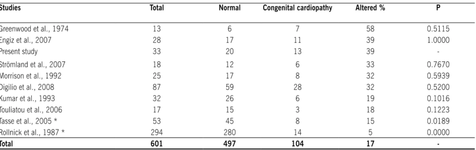
the other hand, Greenwood et al. (1974)12 attributed the high level of cardiac malformations observed in their study (58%) to a probable selection bias, as the researchers were more interested in patients with these defects and in the use of bigger medical centers as a source. Taking into conside-ration all the studies together (n=601), we observed that congenital heart defects frequency found was 17% (Table 3). Nevertheless, we believe that the true index of these defects may be higher, due to total value dilution provoked by the work of Rollnick et al. (1987).8 Apart from this study, the frequency of cardiac defects would go to 29%. Besides that, as in our study, the most works described in literature evaluated OAVS patients from tertiary centers, where indi-viduals are frequently hospitalized for complications related to major abnormalities, such as congenital cardiopathies. Thus, we believe that this may also have some influence on the real frequency of cardiac malformations in these patients.
Despite the variability of defects described in the syndrome, there seems to be a predominance of those of the conotruncal (involving the heart’s outlets) and septal types among these patients.4-12,16 Digilio et al. (2008)7 believe that this heterogeneity is related with the different pathogenic causes associated to the syndrome.
In our study, conotruncal and septal defects were also predominant, being observed in 39% and 23% of the patients, respectively. Among them, tetralogy of Fallot is highlighted,6,7,11,12,16,19 the main cardiac defect discovered in our sample (15%), and interventricular communica-tion,5-7,9,11,19 identified in one patient (8%). Defects concer-ning abnormal expected growth (such as total anomalous venous pulmonary return) and laterality (such as heterotaxy) have also been very common;6,7,10,15,16,19 however, none of our patients presented them. On the other hand, cor triatriatum and the double inlet of left ventricle, defects observed in our sample, are malformations usually not observed in individuals with OAVS. It is believed that the high frequency of defects of conotruncal type described
among the patients with the syndrome may be related to the hypothesis that its etiology is related to an abnormality in neural crest cells, which would justify both cardiac pheno-type and the other abnormalities related to ear, mandible, and neck presented by the patients.2,4,6,20
As for the other clinical characteristics, we noticed that, in the whole sample, there was a predominance of male over female patients, with a proportion of 2:1. A similar finding has been described in literature, but with a slightly lower proportion, 3:2.2,9 In relation to age at first evaluation, we believe it to be lower in cardiopathy carriers due to the fact that these malformations, because of their own severity, frequently require a special attention in an early age. In addition to that, almost all our patients were not born in our hospital, having been referred for evaluation to this center mainly due to its major findings (as congenital cardiopathy). In Bustamante et al.’s cases series (1989),16 for example, congenital cardiopathy’s clinical repercussion was the main motive of hospital admission in the five evaluated patients. The side affected by the disease (right or left) was similar in both groups, with or without cardiopathy. Interestingly, we did not observe a significant difference in relation to the presence of neuropsychomotor delay, which is a common finding in individuals with congenital cardiopathy. In addi-tion to that, hypotonia, a neurologic finding closely related with the acquisition of developmental marks, was present in a similar way in patients of both groups.
Congenital heart defects were the main cause of death in our patients (five of them died; four of these had cardiac defects, and three died due to clinical complications directly related to this malformation), which did agree with reports in literature.3,5,12 It is known that conotruncal cardiac defects present a higher severity and are associated to a high mortality index, especially in face of the lack of early clinical and surgical care,21 happening in the first years of life.3,5,12 Thus, congenital cardiopathies are very important in these individuals’ clinical evolution and prognostics. table 3 – Comparison of congenital cardiopathies frequency observed in our sample of patients with OAVs with that observed in studies
described in the literature.
studies total Normal Congenital cardiopathy Altered % P
Greenwood et al., 1974 13 6 7 58 0.5115
Engiz et al., 2007 28 17 11 39 1.0000
Present study 33 20 13 39
-Strömland et al., 2007 18 12 6 33 0.7670
Morrison et al., 1992 25 17 8 32 0.5939
Digilio et al., 2008 87 59 28 32 0.5200
Kumar et al., 1993 32 26 6 19 0.1016
Touliatou et al., 2006 17 15 3 18 0.1223
Tasse et al., 2005 * 53 45 8 15 0.0189
Rollnick et al., 1987 * 294 280 14 5 0.0000
total 601 497 104 17
Artigo recebido: 09/06/09 Aceito para publicação: 20/10/09
c
OnclusiOnCongenital cardiac defects are frequent in OAVS patients, especially of the conotruncal and septal type. They also represent the main cause of death in these individuals, which occurs early in their lives.3,5,12 Therefore, all the patients with OAVS diagnosis should be submitted early to cardiologic evaluation, aiming at identifying the presence of these defects and, consequently, try to lessen the possible complications resulting from them.
No conlicts of interest declared concerning the publication of
this article.
R
efeRences1. Online Mendelian Inheritance in Man, OMIM (TM). Bethesda: BeMcKusick-Nathans Institute for Genetic Medicine, Johns Hopkins University and National Center for Biotechnology Information, National Library of Medicine; 2000. [cited 2009 may 20]. Available from: http://www.ncbi.nlm.nih.gov/omim/. 2. Cohen MM Jr, Rollinck BR, Kaye CI. Oculoauriculovertebral spectrum: an
updated critique. Cleft Palate J. 1989;26:276-86.
3. Castori M, Brancati F, Rinaldi R, Adami L, Mingarelli R, Grammatico P, et al. Antenatal presentation of the oculo-auriculo-vertebral spectrum (OAVS). Am J Med Genet. 2006;140A:1573-9.
4. Strömland K, Miller M, Sjögreen, Johansson M, Joelsson B-ME, Bills-tedt E. Oculo-auriculo-vertebral spectrum: associated anomalies,
func-tional deicits and possible developmental risk factors. Am J Med Genet.
2007;143A:1317-25.
5. Morrison PJ, Mulholland HC, Craig BG, Nevin NC. Cardiovascular abnormalities in the oculo-auriculo-vertebral spectrum (Goldenhar syndrome). Am J Med Genet. 1992;44:425-8.
6. Kumar A, Friedman JM, Taylor GP, Patterson MWH. Pattern of cardiac malfor-mation in oculoauriculovertebral spectrum. Am J Med Genet. 1993;46:423-6. 7. Digilio MC, Calzolari F, Capolino R, Toscano A, Sarkozy A, Zorzi A, et al.
Congenital heart defects in patients with oculo-auriculo-vertebral spectrum (Goldenhar syndrome). Am J Med Genet. 2008;146A:1815-9.
8. Rollnick BR, Kaye CI, Nagatoshi K, Hauck W, Martin AO. Oculo auriculovertebral dysplasia and variants: phenotypic characteristic of 294 patients. Am J Med Genet. 1987;26:361-75.
9. Tasse C, Böhringer S, Fischer S, Lüdecke H-J, Albrecht B, Horn D, et al. Oculo-auriculo-vertebral spectrum (OAVS): clinical evaluation and severity
scoring of 53 patints and proposal for a new classiication. Eur J Med Genet.
2005;48:397-411.
10. Engyz O, Balel S, Unsal M, Ozer S, Oguz KK, Aktas D. 31 cases with oculo-auriculovertebral dysplasia (Goldenhar syndrome): clinical, neuroradiologic,
audiologic and cytogenetic indings. Genet Couns. 2007;18:277-88.
11. Friedman S, Saraclar M. The high frequency of congenital heart disease in oculo-auriculo-vertebral dysplasia (Goldenhar’s syndrome). J Pediatr. 1974;85:873-4.
12. Greenwood RD, Rosenthal A, Sommer A, Wolff G, Craenen J. Cardiovascular malformations in oculoauriculovertebral dysplasia (Goldenhar syndrome). J Pediatr. 1974;85:816-8.
13. Werler MM, Sheehan JE, Hayes C, Padwa BL, Mitchell AA, Mulliken JB. Cleft Palate Craniofac J. 2004;41:494-500.
14. Touliatou V, Fryssira H, Mavrou A, Kanavakis E, Kitsiou-Tzeli S. Clinical manifestations in 17 Greek patients with Goldenhar syndrome. Genet Couns. 2006;17:359-70.
15. Lisbôa RC, Mendez HMM, Paskulin GA. Síndrome de Goldenhar e variantes: relato de sete pacientes. Rev AMRIGS. 1987;31:265-9.
16. Bustamante LN, Guerra IV, Iwahashi ER, Ebaid M. Síndrome de Goldenhar. Relato de cinco casos em associação com malformações cardíacas. Arq Bras Cardiol. 1989;53:287-90.
17. Verona LL, Damian NGC, Pavarina LP, Ferreira CHF, Melo DG. Monozygotic twins discordant for Goldenhar syndrome. J Pediatr. 2006;82:75-8. 18. Jones KL. Smith’s recognizable patterns of human malformation. Philadelphia:
Elsevier Saunders; 2006.
19. Pierpont MEM, Moller JH, Gorlin RJ, Edwards JE. Congenital cardiac, pulmo-nary, and vascular malformations in oculoauriculovertebral dysplasia. Ped Cardiol 1982;2:297-302.
20. Lammer EJ, Chen DT, Hoar RM, Agnish ND, Benke PJ, Braun JT, et al. Retinoic acid embriopathy. N Engl J Med. 1985;313:837-41.