Sao Paulo Med J. 2012; 130(1):53-6 53
CASE REPORT
Type 1 diabetes in a patient with Ellis-van Creveld syndrome
Diabetes tipo 1 em um paciente com a síndrome de Ellis-van Creveld
Carla Graziadio
I, Pricila Bernardi
II, Rafael Fabiano Machado Rosa
II, Paulo Ricardo Gazzola Zen
III, Giorgio Adriano Paskulin
IVUniversidade Federal de Ciências da Saúde de Porto Alegre (UFCSPA) and Complexo Hospitalar Santa Casa de Porto Alegre (CHSCPA),
Porto Alegre, Rio Grande do Sul, Brazil
ABSTRACT
CONTEXT: Ellis-van Creveld (EVC) syndrome is a rare autosomal recessive disease characterized by dispro-portionate short stature, narrow thorax, postaxial polydactyly, nail and tooth abnormalities and congenital heart disease.
CASE REPORT: The patient was a 22-year-old Caucasian man, the third child of consanguineous parents. He received the diagnosis of insulin-dependent diabetes mellitus (DM) at 16 years of age, and around one year later, he underwent surgery to correct a partial atrioventricular septal defect. Upon physical examina-tion, at 22 years of age, he presented stature of 145.5 cm (P3), weight of 49 kg (P3), head circumference of 54 cm (P2-50), high palate, absence of one of the lower lateral incisor teeth, narrow shoulders, narrowing of the upper thorax, scoliosis, rhizomelic shortening of the upper limbs, brachydactyly, postaxial poly-dactyly and clinopoly-dactyly of the second and third ingers. The lower limbs showed rhizomelic shortening with signiicant genu valgum (knock-knee deformity), small feet with postaxial polydactyly, syndactyly between the second and third toes and hallux valgus. Multiple melanocytic nevi were evident on the face, thorax and limbs. At that time, he was using neutral protamine Hagedorn (NPH) insulin, with poorly controlled DM. The clinical indings presented led to the diagnosis of EVC syndrome. Only one case of this syndrome has been described with DM so far. Attention is drawn to the fact that the genes associated with this syndrome are located close to those of the Wolfram syndrome, a condition that leads to early-onset diabetes.
RESUMO
CONTEXTO: A síndrome de Ellis-van Creveld (EVC) é uma doença autossômica recessiva rara, caracteriza-da por baixa estatura desproporcionacaracteriza-da, tórax estreito, policaracteriza-dactilia pós-axial, anormalicaracteriza-dades em unhas e dentes e cardiopatia congênita.
RELATO DO CASO: O paciente é um rapaz caucasiano de 22 anos, o terceiro ilho de pais consanguíneos. Recebeu diagnóstico de diabetes melito (DM) insulino-dependente aos 16 anos, sendo que, cerca de um ano depois, foi submetido a cirurgia cardíaca de correção de defeito de septo atrioventricular parcial. Ao exame físico, aos 22 anos, ele apresentava estatura de 145,5 cm (P3), peso de 49 kg (P3), perímetro cefálico de 54 cm (P2-50), palato alto, ausência de um dos dentes incisivos inferiores laterais, ombros estreitos, estreitamento do tórax superior, escoliose, encurtamento rizomélico dos membros superiores, braquidactilia, polidactilia pós-axial e clinodactilia dos segundo e terceiro dedos. Nos membros inferiores, observava-se encurtamento rizomélico com importante geno valgo (deformidade dos joelhos-batidos), pés pequenos com polidactlia pós-axial, sindactilia entre segundo e terceiro dedos, e háluces valgos. Múl-tiplos nevos melanocíticos eram evidentes na face, tórax e membros. Neste momento ele está em uso de insulina NPH (neutral protamine Hagedorn), com um controle inadequado do DM. Seus achados clínicos levaram ao diagnóstico de síndrome de EVC. Apenas um caso desta síndrome foi descrito com DM até o momento, sendo que chama a atenção o fato de que os genes associados à síndrome se localizam próxi-mo ao da síndrome de Wolfram, uma condição que cursa com diabetes de início precoce.
IMD. Assistant Professor and Clinical Geneticist,
Universidade Federal de Ciências da Saúde de Porto Alegre (UFCSPA), and Complexo Hospitalar Santa Casa de Porto Alegre (CHSCPA), Porto Alegre, Rio Grande do Sul, Brazil.
IIMD. Clinical Geneticist, Universidade Federal
de Ciências da Saúde de Porto Alegre (UFCSPA), and Complexo Hospitalar Santa Casa de Porto Alegre (CHSCPA), Porto Alegre, Rio Grande do Sul, Brazil.
IIIPhD. Adjunct Professor of Clinical Genetics and Professor of the Postgraduate Program on Pathology and Clinical Genetics, Universidade Federal de Ciências da Saúde de Porto Alegre (UFCSPA), and Complexo Hospitalar Santa Casa de Porto Alegre (CHSCPA), Porto Alegre, Rio Grande do Sul, Brazil.
IVPhD. Associate Professor of Clinical Genetics and professor of the Postgraduate Program on Pathology, Clinical Genetics and Cytogenetics, Universidade Federal de Ciências da Saúde de Porto Alegre (UFCSPA), and Complexo Hospitalar Santa Casa de Porto Alegre (CHSCPA), Porto Alegre, Rio Grande do Sul, Brazil.
KEY WORDS:
Diabetes mellitus, type 1. Ellis-Van Creveld syndrome. Polydactyly.
Dwarism. Consanguinity.
PALAVRAS-CHAVE:
Diabetes mellitus tipo 1. Síndrome de Ellis-van Creveld. Polidactilia.
Nanismo. Consanguinidade.
INTRODUCTION
CASE REPORT | Graziadio C, Bernardi P, Rosa RFM, Zen PRG, Paskulin GA
54 Sao Paulo Med J. 2012; 130(1):53-6
EVC syndrome is oneof the short-rib polydactyly syndromes characterized by disproportionately short stature, small chest, postaxial polydactyly, abnormalities in nails and teeth and con-genital heart defect. However, inter and intra-familial variabil-ity has been reported. Mortalvariabil-ity and morbidvariabil-ity in cases of this condition are frequently determined by pulmonary and cardiac complications and, additionally, some disability may result from knock-knee deformity. No other health problems are common in this syndrome.2-4
We report here a man presenting a very rare association between EVC syndrome and type 1 diabetes mellitus (DM).
CASE REPORT
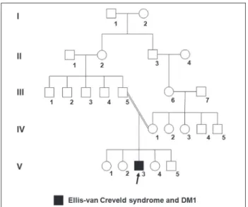
he patient was a 22-year-old Caucasian man, the third child of a young, healthy consanguineous couple (the parents were irst-de-gree cousins) (Figure 1), without similar cases in the family. He was born by means of vaginal delivery, at term, weighing 3,350 g (P25-50) and measuring 38 cm (P < 3). He did not present any intercurrence at birth, but limb abnormalities were identiied. he pregnancy had been uneventful.
he child evolved with a normal neuropsychomotor devel-opment: he could hold up his head at three months of age, sat up without support at ive months, walked alone at 11 months and pronounced his irst words at around one year of age. At two years of age, he was evaluated because of growth retarda-tion, and at this stage, an endocrinologist indicated treatment with calcium and vitamin D. At three years of age, he began to develop genu valgum. At 12 years of age he underwent fem-oral varization osteotomy. He began school at seven years of age, continued as far as the ith year, which he did twice, and then dropped out of school. Insulin-dependent diabetes was
diagnosed at 16 years of age at a primary healthcare center, and around one year later, he underwent correction of a partial atrioventricular septal defect (AVSD).
On physical examination, at 22 years of age, he presented height of 145.5 cm (P3), weight of 49 kg (P3), head circumfer-ence of 54 cm (P2-50), bulbous nasal point, high arched palate, absence of one of the lower lateral incisors, narrow shoulders, narrowing of the upper thorax, scoliosis, rhizomelic shortening of the upper limbs with cubitus valgus, brachydactyly (most pro-nounced in the third ingers, which also showed nails that were smaller than those of the other ingers), postaxial polydactyly, clinodactyly (ulnar deviation) of the second and third ingers and several hypoplastic nails. he lower limbs showed rhizomelic shortening with signiicant genu valgum (knock-knee deformity), small feet with postaxial polydactyly, small toes (especially the third toes) with several hypoplastic nails, syndactyly between the second and third toes, bilateral enlargement of the space between the irst and second toes, and hallux valgus. Multiple melanocytic nevi were evident on his face, thorax and limbs (Figure 2). Mul-tiple labial-gingival frenulae were not observed.
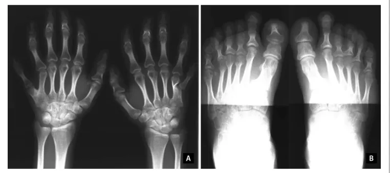
Radiographic evaluation revealed dextroconvex thoracic sco-liosis; rectiication with inversion of the physiological kyphosis; brachyphalangia with shortening of the metacarpal and metatar-sal bones of both the hands and the feet; postaxial polydactyly of the hands with duplication of the ith metacarpal bones; coni-cal epiphyses; signiicant bilateral genu valgum; curving of the tubular bones (most notably in the lower limbs) with deformity of the epiphyses; foot deformity with enlargement of the metatar-sal bones and phalanges; postaxial polydactyly with six metatarmetatar-sal bones in both feet; and presence of osteochondromas and osteo-porosis (Figure 3).
Figure 1. Pedigree of the patient’s family (subject V-3), showing the consanguinity observed between his parents (III-5 and IV-1) (DM1: type 1 diabetes mellitus).
Type 1 diabetes in a patient with Ellis-van Creveld syndrome | CASE REPORT
Sao Paulo Med J. 2012; 130(1):53-6 55 He had symptoms of dysuria and mictional urgency, and a
urodynamic evaluation revealed detrusor hyperactivity, a blad-der with diminished capacity and complacency, diminished uri-nary low and increased sensitivity suggestive of irritative and infectious factors, including diabetes. Abdominal ultrasound did not identify any presence of structural abnormalities. he patient also had a history of knee and back pain that had increased over the last few months. He was being followed up at the orthopedics department of the hospital because of the signiicant genu val-gum. Furthermore, he was receiving care in the endocrinology department and was using NPH (neutral protamine Hagedorn) insulin, but with poor diabetes control. He presented glucose levels ranging between 180 and 314 mg/dl in the morning, and monitoring was being conducted at a primary healthcare center.
He started treatment in the hospital at the age of 22 years. NPH insulin was initially prescribed, consisting of 25 units before breakfast and 10 units before dinner. However, his lack of diabetes control persisted, and he showed symptoms of polyuria and nocturia. His blood glucose levels before breakfast ranged from 194 to 333 mg/dl; before lunch from 179 to 27 mg/dl and before dinner from 99 to 222 mg/dl. His glycated hemoglobin level was 8.7% (normal values range from 3.9 to 6.1%). Subse-quently, the NPH insulin dose was changed to 26 units before breakfast and 14 units before dinner. Ater this, the patient devel-oped episodes of hypoglycemia. His blood glucose levels before breakfast ranged from 153 to 225 mg/dl, before lunch from 51 to 282 mg/dl and before dinner from 54 to 229 mg/dl. he new gly-cated hemoglobin level was 9.5%, while the qualitative examina-tion of urine showed a cross (+) for the presence of glucose. In
Table 1. Review of medical databases using the descriptors corresponding to the main features presented by the patient, conducted on December 13, 2010
Data base Strategy of search Results*
PubMed “Ellis Van Creveld Syndrome” AND “Diabetes Mellitus”
1 case report 1 article 0 reviews Scirus “Ellis Van Creveld Syndrome”
AND “Diabetes Mellitus” 60 articles Embase “Ellis Van Creveld Syndrome”
AND “Diabetes Mellitus”
0 case reports 2 reviews
*There were no results from using the same search strategies in the Cochrane Library, SciELO and Lilacs databases.
Figure 3. Radiographs on the patient’s hands and feet. Note especially the post-axial polydactyly, brachydactyly with short metacarpal and metatarsal bones and phalanges, and conical epiphyses (A and B).
the latest assessment, the scheme was changed to an NPH dose of 28 units before breakfast and 18 units before dinner.
An ophthalmological evaluation showed the presence of cat-aracts, with an indication for surgery. His blood pressure was normal (mean of 120 x 80 mmHg). hyroid, liver and kidney function tests were all normal. he patient was also reevaluated in the cardiology department and underwent an electrocardio-gram, which was normal.
DISCUSSION
CASE REPORT | Graziadio C, Bernardi P, Rosa RFM, Zen PRG, Paskulin GA
56 Sao Paulo Med J. 2012; 130(1):53-6
problems, especially during the neonatal period. Congenital heart defects have been described in up to 60% of the patients with EVC syndrome, and atrioventricular canal defects are the most frequently observed malformations,3,4 as seen in our patient. Other congenital heart malformations that have been described include a single atrium, defects of the mitral and tricuspid valves, patent ductus, ventricular septal defect, atrial septal defect and hypoplastic let heart syndrome.3,4
Diabetes mellitus is not considered to be a component of EVC syndrome, and, in our literature review, using the PubMed, Sci-rus, Embase, Cochrane Library, Lilacs and SciElo databases, we saw only one report describing this association5 (Table 1). About half of the cases of EVC syndrome are determined by EVC and EVC2 gene mutations, and these genes are located in the 4p16
region.4 We draw attention to the fact that the WFS1 gene is also located in this region and determines the Wolfram syndrome (WS, OMIM 2223000),1 which is an autosomal recessive condi-tion also referred to as DIDMOAD (diabetes insipidus, diabetes mellitus, optic atrophy and deafness).6,7 Occurrences of diabetes mellitus and optic atrophy together is suicient for diagnosing WS.6,7 However, until the latest assessment of our patient, at the age of 22 years, only the DM was observed, which would be not expected for a patient with WS (usually, patients with WS present optic atrophy before the age of 19 years).7
Autoimmune conditions are uncommon among patients with EVC syndrome.2-4 Type 1 DM is responsible for about 5-10% of all cases of diabetes, and susceptibility to this is considered to be largely inherited, residing especially in the HLA genotypes DR and DQ.8 However, several diferent candidate genes have been proposed,8 and a relationship between type 1 DM and the con-sanguinity observed in our patient cannot be ruled out.
hus, more reports will be necessary in order to determine whether the type 1 DM observed in our patient may be a true fea-ture of the clinical spectrum of the syndrome. We cannot rule out the possibility that the association between EVC syndrome and DM observed in our patient may have been merely coincidental.
REFERENCES
1. Online Mendelian Inheritance in Man. OMIM® - Online Mendelian
Inheritance in Man. Available from: http://www.ncbi.nlm.nih.gov/ omim. Accessed in 2011 (Apr 6).
2. Al-Khenaizan S, Al-Sannaa N, Teebi AS. What syndrome is this? Chondroectodermal dysplasia--the Ellis-van Creveld syndrome. Pediatr Dermatol. 2001;18(1):68-70.
3. Baujat G, Le Merrer M. Ellis-van Creveld syndrome. Orphanet J Rare Dis. 2007;2:27.
4. Ruiz-Perez VL, Goodship JA. Ellis-van Creveld syndrome and Weyers acrodental dysostosis are caused by cilia-mediated diminished response to hedgehog ligands. Am J Med Genet C Semin Med Genet. 2009;151C(4):341-51.
5. Postek G. Przypadek zespolu Ellisa-van Crevelda [A case of Ellis-van Creveld syndrome]. Pol Tyg Lek. 1990;45(38-39):787-9.
6. Zen PRG, Pinto LLC, Schwartz IVD, Paskulin G. Relato de um paciente brasileiro com síndrome de Wolfram [Report of a Brazilian patient with Wolfram Syndrome]. J Pediatr (Rio J). 2002;78(6):529-32. 7. Rigoli L, Lombardo F, Di Bella C. Wolfram syndrome and WFS1 gene.
Clin Genet. 2011;79(2):103-17.
8. Daneman D. Type 1 diabetes. Lancet. 2006;367(9513):847-58.
Sources of funding: None
Conlict of interest: None
Date of irst submission: October 13, 2010
Last received: March 3, 2011
Accepted: April 18, 2011
Address for correspondence:
Giorgio Adriano Paskulin
Genética Clínica, Universidade Federal de Ciências da Saúde de Porto Alegre (UFCSPA)
Rua Sarmento Leite, 245/403 Centro — Porto Alegre (RS) – Brasil CEP 90050-170