Clinical description of 41 Brazilian patients with
oculo-auriculo--vertebral dysplasia
JOSÉ ROBERTO MENDES PEGLER¹, DIOGO CORDEIRODE QUEIROZ SOARES²*, CAIO ROBLEDO D’ANGIOLI COSTA QUAIO³,
NATALIA FERNANDES4, LUIZ ANTONIO NUNESDE OLIVEIRA5, RACHEL SAYURI HONJO6, DEBORA ROMEO BERTOLA7, CHONG AE KIM8
1MD – Pediatric Resident Physician, Instituto da Criança do Hospital das Clínicas da Faculdade de Medicina da Universidade de São Paulo (ICr-HC-FMUSP), São Paulo, SP, Brazil 2MD – Specialist in Medical Genetics and PhD candidate from FMUSP. Preceptor, Medical Genetics Unit, ICr-HC-FMUSP, São Paulo, SP, Brazil
3Specialist in Medical Genetics – Collaborating Physician at the Medical Genetics Unit, ICr-HC-FMUSP, São Paulo, SP, Brazil 4Biomedical Student – Intern in the Medical Genetics Unit, ICr-HC-FMUSP, São Paulo, SP, Brazil
5Radiologist – Assistent Physician, Radiology Service, ICr-HC-FMUSP, São Paulo, SP, Brazil
6PhD in Medicine from FMUSP – Assistant Physician, Medical Genetics Unit, ICr-HC-FMUSP, São Paulo, SP, Brazil 7PhD in Medicine from FMUSP – Head of the Medical Genetics Unit, ICr-HC-FMUSP, São Paulo, SP, Brazil 8PhD in Medicine from FMUSP. Associate Professor, Department of Pediatrics, FMUSP, São Paulo, SP, Brazil
S
UMMARYStudy conducted at Unidade de Genética Médica, Instituto da Criança do Hospital das Clínicas da Faculdade de Medicina da Universidade de São Paulo (ICr-HC-FMUSP), São Paulo, SP, Brazil
Article received: 8/6/2014 Accepted for publication: 8/24/2014 *Correspondence: Address: Av. Enéas de Carvalho Aguiar, 647, 7º andar Cerqueira César São Paulo, SP – Brazil Postal code: 05403-000 [email protected]
http://dx.doi.org/10.1590/1806-9282.62.03.202
Objective: To describe the most prominent clinical features of a cohort of pa-tients with oculo-auriculo-vertebral (OAV) dysplasia in Brazil.
Method: A review of medical records of patients with diagnosis of OAV from 1990 to 2010 was performed in a medical genetics center.
Results: 41 patients were included in the study. Their average age at diagnosis was 2y 10mo (34,4±48,8 months) and the female proportion was 53.7%. Mean mater-nal age at patient’s birth was 28.5y (min: 17, max: 46y) for mothers and 31.4y (min: 21, max: 51y) for fathers. Most patients (97.5%) had auricular involvement, with facial manifestation in 90.2%, spinal in 65.9%, ocular in 53.7%, 36.6% with cardio-vascular involvement, 29.3% urogenital, and 17% of the cases with central nervous system (CNS) involvement. The classic OAV triad was present in only 34%. All pa-tients except one had concomitant problems in other organs or systems.
Conclusion: Since the diagnosis of OAV dysplasia relies only on a comprehen-sive medical evaluation, it is imperative that clinicians be aware of the most com-mon presentation of the syndrome. Once suspected, every patient should under-go a complete medical evaluation of multiple systems including complementary exams. Treatment of these patients is based on surgical correction of malforma-tions and rehabilitation.
Keywords: Goldenhar syndrome, facial asymmetry, craniofacial abnormalities.
I
NTRODUCTIONThe Goldenhar syndrome was described in 1845 by Carl Ferdinand von Arlt and recognized as a clinical entity in 1952 by Maurice Goldenhar who described it in a child,
as reported Salvitti et al.1 It has been known as irst
bran-chial arch syndrome, Gorlin’s syndrome and hemifacial microsomia (OMIM 164210), but currently it is best known as oculo-auriculo-vertebral (OAV) dysplasia, a no-menclature given by Gorlin et al.2 and Sugar.3
Its prevalence has been estimated at about one case per 5,600 to 26,550 births,4-6 with greater involvement of males
than females (in a ratio of about 3:2).7 The cases are
most-ly sporadic, but families with autosomal recessive or
auto-somal dominant inheritance have been described;8,9 thus,
the hypothesis that there is no kind of genetic factor in-volved that would inluence susceptibility to the disease has been ruled out.10 In this regard, reports of
monozygot-ic twins,11 both dichorionic and monochorionic,12
discor-dant for the disease have been made, suggesting a correla-tion with a multifactorial inheritance pattern.
the abuse of alcohol during pregnancy as well.13-15
Mou-noud et al. reported a case of OAV dysplasia in a child whose mother had a history of hypervitaminosis A. It is known that daily doses of vitamin A higher than 25,000 IU have teratogenic effects. This teratogen has harmful effects in the formation of neural crest cells, which are essential for the formation of the pharyngeal arches.16,17
Affected individuals may present: malar and/or man-dibular hypoplasia, hypoplasia of the facial muscles, mi-crotia, preauricular tags and outer ear dysplasia, hemi-vertebrae and hypoplasia of cervical thoracic or lumbar vertebrae, epibulbar dermoids, microphthalmia, cleft pal-ate and/or lip, cardiac, kidney or central nervous system (CNS) anomalies.2 In addition, there are reports on the
association of OAV dysplasia with other conditions, such as genitourinary,18,19 cardiovascular,14,20 or psychiatric21,22
changes, and obstructive sleep apnea.23 However, due to
clinical variability, some patients have minimal manifes-tations, predominantly facial asymmetry and dysplasia of the auricular pavilion.2
Based on the above, and considering the importance of the topic due to prevalence, wide spectrum of clinical manifestations and the lack of studies that describe a sig-niicant number of patients suffering from OAV dyspla-sia in our midst, we describe a case series of patients di-agnosed with OAV dysplasia followed in our service over the past 20 years.
M
ETHODThe sample consisted of individuals followed in the Med-ical Genetics Unit at Instituto da Criança, Hospital das Clínicas, Faculdade de Medicina da Universidade de São Paulo (ICr-HC-FMUSP), diagnosed with OAV dysplasia from 1990 to 2010. This study was approved by the Eth-ics Committee for Research Project Analysis – CAPPesq of HC-FMUSP (No. 0667-07).
Patients included in the study were those with nor-mal G-banding karyotype and involvement of at least two of the following sites: 1) mouth, skull and face, 2) eyes, 3) ears and 4) vertebrae. This approach was consistent with that adopted by Strömland et al. (2007).12 Individuals
with chromosomal abnormalities or incomplete medical records were excluded from the study.
We conducted a retrospective analysis, and collected data on clinical manifestations (ear, face, spine, eyes and more), demographics (gender, date of birth, age at irst consultation, and age of parents) and results of addition-al tests. The affected side (right, left or bilateraddition-al) was that where microtia or facial microsomia was located, similar-ly to the method adopted by Rollnick et al. (1987).24
R
ESULTSAmong the 41 patients studied, 19 (46.3%) were male and 22 (53.7%) female. The mean age at irst consultation was 2 years and 10 months (34.4±48.8 months). The mean age of parents at the birth of the child in cases where in-formation was available (39/41 patients) was 28.5±6.9 years for the mothers and 31.4±7.4 years for the fathers.
With regard to clinical manifestations, 97.5% of the pa-tients had some degree of ear involvement, 90.2% facial, 65.9% vertebral, and 53.7% ocular. 89% of children had in-volvement of other organs. The classic OAV triad was pres-ent in only 34% (15 children). All patipres-ents except one had concomitant problems in other organs or systems.
Facial manifestations
Facial abnormalities were observed in 90.2% (37/41) of patients. Of these, 83.8% (31/37) had some degree of hemi-facial microsomia. In 46% (17/37) of the cases, change in facial expression was observed, suggesting some degree of facial paralysis. We also found that 14% (6/37) of the cases had cleft palate and/or lip and 7% (3/37) had ocu-lar hypertelorism.
Ocular manifestations
From the classic triad of changes described as OAV dys-plasia, ocular changes were less frequent in our series, with about 53.7% (22/41) of patients presenting some in-volvement. Epibulbar dermoids or dermoid cysts were seen in 45.4% (10/22), representing the vast majority in the group of eye diseases, followed by the inding of epi-canthus, present in 22.7% (5/22) and other epibulbar tu-mors, present in 13.6% (3/22). Other ocular manifesta-tions found in our series at a lower prevalence (1 or 2 cases) are: coloboma, changes in the lacrimal gland, an-ophthalmia and amaurosis.
Auricular manifestations
Vertebral manifestations
65.9% (27/41) of the patients had vertebral abnormali-ties. Of these, spinal axis changes were found most of-ten including marked kyphosis and/or scoliosis in 48.2% (13/27) of cases. Localized vertebral involvements were also prevalent, including the inding of hemivertebrae in 37% (10/27) cases, most often in the thoracic segment with 25% (7/27) patients affected. Block or fused verte-brae were diagnosed in 33% (9/27) of cases, with greater involvement of the cervical spine, affected in 77.7% (7/9) of patients, as well as the presence of incomplete fusion of vertebral arches in 14.8% (4/27) of the patients. Oth-er reports included: sacral dimples in 22% (6/27) of pa-tients, spina biida in 14.8% (4/27), and transitional ver-tebrae in 14.8% (4/27).
Other systemic manifestations Cardiovascular system
Cardiovascular involvement was found in 36.6% (15/41) of the patients. The spectrum is quite heterogeneous, but the most frequent changes were communications between heart chambers, which were present in 36.6% of the pa-tients with cardiac involvement. Interatrial communica-tion was responsible for 40% (6/15) of the cases, and ven-tricular septal defects for 33.3% (5/15) of the cases. Complex congenital heart disease were seen in 26.7% (4/15) patients, the most common being the tetralogy of Fallot, present in 50% (2/4) of cases. Persistent arterial duct was found in 20% (3/15) of the patients.
Urogenital system
In our sample, 29.3% (12/41) of patients showed abnor-malities in the urinary tract, of which 41.7% (5/12) had concomitant change in the cardiovascular system. Among the abnormalities found, pelvic kidneys and unilateral renal agenesis were the most prevalent occurrences, both present in 41.7% (5/12) patients each. Of note, among patients diagnosed with pelvic kidney, 80% (4/5) had ip-silateral ear involvement. Other changes less frequently observed (1 or 2 cases) were pyelocaliceal ectasia, pyelo-ureteral duplication, vesicopyelo-ureteral relux and hypospa-dias.
Central nervous system
17% (7/41) of the patients had CNS changes, especially expansion of the cerebral ventricles, found in 43% (03/07) of cases. 28.6% (2/7) of these cases had dysgenesis of the
corpus callosum associated with ventriculomegaly, and
14.3% (1/7) showed absence of the septum pellucidum as-sociated with ventriculomegaly. Occipital encephalocele
associated with posterior parietal meningoencephalocele was less frequent alteration found in one patient.
D
ISCUSSIONOAV dysplasia is a well-deined entity, characterized by unilateral or bilateral craniofacial anomalies, to a variable degree, involving the irst and second branchial arches, and vertebral and eye abnormalities. Clinically, it varies from an isolated microtia, with or without mandibular hypoplasia, to a more complex phenotype involving skel-etal, cardiac, renal, lung and CNS disorders.24,25
As previously mentioned, much has been speculated about the etiological and pathogenic mechanisms that lead to the development of OAV dysplasia. In this sense, several studies have been performed to identify genetic changes that may be related to the phenotype displayed by patients with OAV, but so far such correlation could not be established.26
Our results reveal that 21 (48%) patients were male and 23 (52%) were female, a proportion similar to that found in a study conducted in the city of Bauru, state of São Pau-lo, 27 and different from that observed in other studies that
found male predominance with a ratio of 3:2.7
Regarding the therapeutic approach, in less complex cases interventions vary according to the age and system-ic involvement and are mainly intended to improve esthet-ics. In patients with mandibular hypoplasia, reconstruc-tive surgery can be performed using bone grafts taken from the ribs and, in some instances, bone stretching. In cases where there is cleft lip and/or palate, surgical correction is usually performed followed by the use of orthodontic de-vices after correction of mandibular defects.
Reconstructive surgery to correct auricular malfor-mation is usually performed at the age of 6 to 8 years. In patients with milder involvement, mandibular reconstruc-tion surgery can be performed in early adolescence. Epi-bulbar dermoids must be removed surgically. Structural ocular anomalies and those of the outer ear should be corrected by plastic surgeons.
C
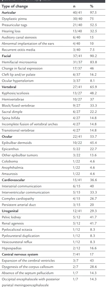
ONCLUSIONTABLE 1 Type and frequency of changes observed in patients with OAV dysplasia.
Type of change n %
Auricular 40/41 97.5
Dysplastic pinna 30/40 75
Preauricular tags 21/40 52.5
Hearing loss 13/40 32.5
Auditory canal stenosis 6/40 15
Abnormal implantation of the ears 4/40 10
Recurrent otitis media 3/40 7.5
Facial 37/41 90.2
Hemifacial microsomia 31/37 83.8
Change in facial expression 17/37 46
Cleft lip and/or palate 6/37 16.2
Ocular hypertelorism 3/37 8.1
Vertebral 27/41 65.9
Kyphosis/scoliosis 13/27 48.2
Hemivertebrae 10/27 37
Block/fused vertebrae 9/27 33.3
Sacral dimple 6/27 22.2
Spina biida 4/27 14.8
Incomplete fusion of vertebral arches 4/27 14.8
Transitional vertebrae 4/27 14.8
Ocular 22/41 53.7
Epibulbar dermoids 10/22 45.4
Epicanthus 5/22 22.7
Other epibulbar tumors 3/22 13.6
Coloboma 1/22 4.6
Anophthalmia 1/22 4.6
Amaurosis 1/22 4.6
Cardiovascular 15/41 36.6
Interatrial communication 6/15 40
Interventricular communication 5/15 33.3
Complex cardiopathy 4/15 26.7
Persistent arterial duct 3/15 20
Urogenital 12/41 29.3
Pelvic kidney 5/12 41.7
Renal agenesis 5/12 41.7
Pyelocaliceal ectasia 1/12 8.3
Pyeloureteral duplication 1/12 8.3
Vesicoureteral relux 1/12 8.3
Hypospadias 2/12 16.6
Central nervous system 7/41 17
Expansion of the cerebral ventricles 3/7 43 Dysgenesis of the corpus callosum 2/7 28.6
Absence of the septum pellucidum 1/7 14.3 Occipital encephalocele with posterior
parietal meningoencephalocele
1/7 14.3
R
ESUMODisplasia óculo-aurículo-vertebral: aspectos clínicos de 41 pacientes brasileiros
Objetivo: descrever os principais achados clínicos de uma coorte de pacientes com a displasia óculo-aurícu-lo-vertebral (OAV).
Método: revisão de prontuários médicos dos pacientes com diagnóstico de OAV no período de 1990 a 2010, acom-panhados em um centro de genética médica.
Resultados: foram incluídos no estudo 41 pacientes. A média de idade ao diagnóstico foi de 2 anos e 10 meses (34,4±48,8 meses) e a proporção de pacientes do sexo fe-minino foi de 53,7%. A média de idade dos pais ao nasci-mento do paciente foi de 28,5±6,9 anos para as mães e 31,4±7,4 anos para os pais. A maioria dos indivíduos (97,5%) possuía acometimento auricular, 90,2% tinham manifes-tações faciais, 65,9%, vertebrais, 53,7%, oculares, 36,6%, car-diovasculares, 29,3%, urogenitais e 17%, no sistema nervo-so central. Além disnervo-so, 34% dos pacientes apresentavam a tríade clássica óculo-aurículo-vertebral, e todos os pacien-tes exceto um apresentavam concomitantemente proble-mas em outros órgãos ou sisteproble-mas.
Conclusão: já que o diagnóstico desta entidade é emi-nentemente clínico, é imprescindível que os médicos das mais diversas especialidades conheçam os achados mais frequentes na OAV. Diante de um paciente com suspeita diagnóstica, deve ser realizada avaliação detalhada de ou-tros órgãos, tanto clínica como por meio de exames com-plementares. O tratamento é baseado na correção cirúr-gica das malformações e na reabilitação.
Palavras-chave: síndrome de Goldenhar, assimetria fa-cial, anormalidades craniofaciais.
R
EFERENCES1. Salvitti C, Azulay RD, Heringer ML, Almeida de Faria LA. [Oculo-auriculo-vertebral dysplasia: presentation of a case and attempt at organizing the symptomatology]. Rev Ass Med Bras. 1978; 24(5):160-2.
2. Gorlin RL. Branchial arch and oro-acral disorders. In: Gorlin JJ, Cohen Jr MM, Hennekam RC (eds.). Syndromes of the head and neck. London: Oxford University Press, 2001. p.790-97.
3. Sugar HS. The oculoauriculovertebral dysplasia syndrome of Goldenhar. Am J Ophthalmol. 1966; 62(4):678.
4. Grabb WC. The irst and second branchial arch syndrome. Plast Reconstr Surg. 1965; 36(5):485-508.
5. Stoll C, Roth MP, Dott B, Bigel T. Discordance for skeletal and cardiac defect in monozygotic twins. Acta Genet Med Gemellol. 1984; 33(3):501-4. 6. Melnick M. The etiology of external ear malformations and its relation to
abnormalities of the middle ear, inner ear and other organ systems. Birth Defects Orig Artic Ser. 1980; 16(4):303-31.
8. Pearson A. Developmental anatomy of the ear. In: English M (ed.). Otolaryngology. New York: Harper and Row, 1978. p.1-68.
9. Scholtz AW, Fish III JH, Kammen-Jolly K, Ichiki H, Hussl B, Kreczy A, et al. Goldenhar’s syndrome: congenital hearing deficit of conductive or sensorineural origin? Temporal bone histopathologic study. Otol Neurotol. 2001; 22(4):501-5.
10. Phelps PD, Lloyd GA, Poswillo DE. The ear deformities in craniofacial microsomia and oculo-auriculo-vertebral dysplasia. J Laryngol Otol. 1983; 97(11):995-1005.
11. Bisdas S, Lenarz M, Lenarz T, Becker H. Inner ear abnormalities in patients with Goldenhar syndrome. Otol Neurotol. 2005; 26(3):398-404. 12. Strömland K, Miller M, Sjögreen L, Johansson M, Joelsson B-ME, Billstedt
E, et al. Oculo-auriculo-vertebral spectrum: associated anomalies, functional deicits and possible development risk factors. Am J Med Genet. 2007; 143A(12):1317-25.
13. Mehta B, Nayak C, Savant S, Amladi S. Goldenhar syndrome with unusal features. Indian J Venerol Dermatol Leprol. 2008; 74(3):254-6.
14. Nakajima H, Goto G, Tanaka N, Ashiya H, Ibukiyama C. Goldenhar syndrome associated with various cardiovascular malformations. Jpn Circ J. 1998; 62(8):617-20.
15. Das A, Ray B, Das D. A case of Goldenhar-Gorlin syndrome with unusual association of hypoplastic thumb. Indian J Ophthamol. 2008; 56(2):150-2. 16. Mounoud RL, Klein D, Weber F. [A case of Goldenhar syndrome: acute vitamin A intoxication in the mother during pregnancy]. J Genet Hum. 1975; 23(2):135-54.
17. Maj V, Col R. Golden-har syndrome. Mjai. 2000; 56:231-2.
18. Ishitoya S, Arai Y, Okubo K, Suzuki Y. Left retrocaval ureter associated with the Goldenhar syndrome (branchial arch syndrome). J Urol. 1997; 158(2):572-3. 19. Ritchey ML, Norbeck J, Huang C, Keating MA, Bloom DA. Urologic
manifestations of Goldenhar syndrome. Urology. 1994; 43(1):88-91. 20. Kumar A, Friedman JM, Taylor GP, Patterson MWH. Pattern of cardiac
malformation in oculoauriculovertebral spectrum. Am J Med Genet. 1993; 46(3):423-6.
21. Brieger P, Bartel-Friederich S, Haring A, Marneros A. Oculo-auriculo-vertebral spectrum disorder (Goldenhar “syndrome”) coexisting with schizophreniform disorder. J Neurol Neurosurg Psychiatry. 1998; 65(1):135-6.
22. Lleonart M, Sarduy A, Gil M, Lois L. Displasia oculoauriculovertebral o síndrome de Goldenhar: estudio multidisciplinario de un caso clínico. Rev Cubana Oftalmol. 2001; 14(1):42-6.
23. Hoch B, Hochban W. Four-year-old girl with Goldenhar-sequence and severe obstructive sleep apnea, symptoms, diagnosis and therapy. Int J Pediatr Otorhinolaryngol. 1998; 43(3):277-81.
24. Rollnick BR, Kaye CI, Nagatoshi K, Hauck W, Martin AO. Oculo auriculovertebral dysplasia and variants: phenotypic characteristic of 294 patients. Am J Med Genet. 1987; 26(2):361-75.
25. Kelberman D, Tyson J, Chandler DC, McInerney AM, Slee J, Albert D, et al. Hemifacial microsomia: progress in understanding the genetic basis of a complex malformation syndrome. Hum Genet. 2001; 109(6):638-45. 26. Brosco KC, Zorzetto NL, Costa AR. [Audiology proile in patients with
Goldenhar’s syndrome]. Rev Bras Otorrinolaringol. 2004; 70(5):645-9. 27. Smith DW. Síndromes de malformações congênitas: aspectos genéticos,