Inluence of prone positioning on premature
newborn infant stress assessed by means of
salivary cortisol measurement: pilot study
INTRODUCTION
he ability of preterm infants to perceive pain and stress is well established.(1-3) In particular, the actions of stressors may disorganize various systems in newborn preterm infants, including the autonomic, motor, and information systems, resulting in physiological and behavioral expressions of stress.(4) In regard to behavioral characteristics, preterm infants exhibit observable signs in response to stress.(5,6) With the aim of systematically assessing newborn infants’ behavior, Als et al. formulated the Behavioral Assessment Scale (Brazelton Maria Fernanda Cândia1, Erica Fernanda Osaku1,
Marcela Aparecida Leite1, Beatriz Toccolini1,
Nicolle Lamberti Costa1, Sandy Nogueira
Teixeira1, Claudia Rejane Lima de Macedo Costa1,
Pitágoras Augusto Piana2, Marcos Antonio da
Silva Cristovam3, Nelson Ossamu Osaku1,3,4
1. Physical Therapy in Intensive Care Residency Program, Hospital Universitário do Oeste do Paraná - Cascavel (PR), Brazil.
2. Universidade Estadual do Oeste do Paraná - Campus Toledo (PR), Brazil.
3. Undergraduate Medice Course, Universidade Estadual do Oeste do Paraná - Cascavel (PR), Brazil. 4. Neonatal Intermediate Care Unit,
Hospital Universitário do Oeste do Paraná - Cascavel (PR), Brazil.
Objective: his study sought to assess the inluence of prone positioning on the stress of newborn premature infants through the measurement of the salivary cortisol concentration and the evaluation of physiological and behavioral responses before and after changes in body positioning.
Methods: Saliva samples were collected from newborn infants at two diferent times: the irst (corresponding to the baseline) after a period of 40 minutes during which the infants were not subjected to any manipulation and were placed in the lateral or supine position, and the second 30 minutes after placement in the prone position. Variables including heart rate, respiratory rate, peripheral oxygen saturation, and the Brazelton sleep score were recorded before, during, and at the end of the period in the prone position.
Results: he sample comprised 16 newborn premature infants (56.3% male) with a gestational age between 26 and
Conflicts of interest: None. Submitted on November 20, 2013 Accepted on April 6, 2014
Corresponding author:
Maria Fernanda Cândia Rua Otto Edmundo Riihmann, 840 Zip code: 86.073-675 - Londrina (PR), Brazil E-mail: [email protected]
Inluência do posicionamento em prona sobre o estresse no
recém-nascido prematuro avaliada pela dosagem de cortisol
salivar: um estudo piloto
ABSTRACT
Keywords: Adrenal córtex/metabolismo; Hydrocortisone/analysis; Infant newborn/ metabolism; Infant premature/metabolism; Prone position; Saliva/analysis; Stress, physiological; Intensive care units, neonatal
36 weeks, postnatal age between 1 and 33 days, birth weight of 935 to 3,050g, and weight at the time of intervention of 870 to 2,890g. During the intervention, six participants breathed room air, while the remainder received oxygen therapy. he median salivary cortisol concentration was lower in the prone position compared to baseline (0.13 versus 0.20; p=0.003), as was the median Brazelton sleep score (p=0.02). he average respiratory rate was lower after the intervention (54.88±7.15 versus 60±7.59; p=0.0004). he remainder of the investigated variables did not exhibit signiicant variation.
Conclusion: Prone positioning signiicantly reduced the salivary cortisol level, respiratory rate, and Brazelton sleep score, suggesting a correlation between prone positioning and reduction of stress in preterm infants.
Neonatal Behavioral Assessment Scale - BNBAS) in 1973. his research tool was adapted to the physical and neurological responses and emotional behaviors of newborn infants while taking individual diferences into account.(6) In regard to the physiological changes caused by stress, cortisol is one of the stress-related hormones commonly measured in newborn infants.(7) After the correlation between plasma and salivary cortisol levels was demonstrated,(8,9) subsequent studies on neonatal pain and stress were conducted using the noninvasive method of saliva collection for the measurement of cortisol concentration.(10-17)
As newborn infants exhibit physiological and behavioral signs of pain and stress that can be recognized by healthcare professionals, pharmacological and non-pharmacological resources may be employed in the neonatal intensive care unit (NICU) to manage such conditions.(4,12,13) In this regard, appropriate positioning is considered an important non-pharmacological intervention in premature newborns admitted to NICUs,(17-19) and according to some studies, the frequency of stress behaviors is lower when infants are placed in the prone position.(20,21)
he main objective of the present study was to establish whether prone positioning inluences stress in premature newborn infants, as indicated by the salivary cortisol concentration. In addition, we also investigated the correlation between salivary cortisol concentration and physiological parameters, including heart rate (HR), respiratory rate (RR), and oxygen saturation (SatO2), and behavioral signs according to an adaptation of the BNBAS sleep score.
METHODS
he present study was a pilot intervention study conducted at the NICU of the Hospital Universitário do
Oeste do Paraná from August to September 2013. he
study was approved by the research ethics committee of the Universidade Estadual do Oeste do Paraná, under protocol no. 322,268. All of the participants’ parents or guardians signed an informed consent form.
he study population comprised newborn infants with a gestational age of 25 to 36 weeks who were clinically stable and exhibited normal physiological parameters before sample collection. he samples were collected at least one hour after the last feeding and 40 minutes after any manipulation of the infants. Premature infants in unstable hemodynamic conditions with grade III or IV intraventricular hemorrhage or subsequent leukomalacia, congenital defects of the nervous system, neurological
malformations or impairments, or kidney disease; using opiates, corticosteroids, or other drugs liable to interfere with the response to nociception; and requiring invasive procedures at the time of sample collection were excluded.
Study protocol
he study design took into account the fact that the circadian cortisol cycle is not yet established in newborn infants.(10) hus, each individual was considered as his or her own control, whereby the baseline and response measurements were paired. To minimize the presence of other potential stressors, sample collection was performed between 6:00 and 7:30 in the morning, a time when newborn infants are less subjected to manipulation and activities are minimal at the NICU, thus resulting in low levels of noise and exposure to light. he newborn infants were monitored by means of conventional pulse oximetry, digital skin thermometers, and observation of the respiratory pattern and behavioral responses by the principal investigator. he variables were recorded in an ad hoc form at two diferent times: before baseline sample collection (before oral hygiene) and after prone positioning of the infants.
eyes, occasional smooth movements; state 3 - drowsy, occasional opening of the eyes; state 4 - awake, eyes open, few bodily movements; state 5 - fully awake, continuous bodily movements; state 6 - crying.
One hour after the last feeding and immediately before the irst saliva collection, the infant’s oral cavity was gently but thoroughly cleansed using sterile cotton swabs and water, and all care was taken to avoid contamination of the samples by milk remnants. Next, 0.5 to 0.7mL of saliva was collected through careful aspiration of the loor of the mouth using a 10mL syringe coupled to a needleless lexible 18G intravenous catheter. Two saliva samples were collected. he irst sample, corresponding to the baseline level, was collected after a period of at least 40 minutes during which the infants were subjected to no manipulation whatsoever before the oral cleansing. his sample was collected immediately after the oral cleansing, with no stabilization interval allowed between the cleansing procedure and saliva collection; this sample represented the pre-manipulation state, as the stress associated with such a procedure could have induced an increase in salivary cortisol secretion, the peak of which usually occurs after 20 to 40 minutes. After the baseline sample was collected, the newborn infants were placed in the prone position and inclined forward, with the head to one side and all four limbs in lexion. No restraints were removed or added beyond those already in use by the newborn infant at the time of the baseline sample collection.
Ten minutes were allowed for the infants to achieve stabilization in the prone position, and the second saliva sample was collected 20 minutes later. he samples were stored and transported to the Laboratório de Análises ClínicasAlvaro located in the city of Cascavel, Paraná state, Brazil, where the measurement of cortisol was performed.
he samples were processed using electrochemiluminescence (Roche Diagnostics Elecsys®
2010 Immunoassay System) and stored at 2 to 8 ºC for 5 days and then discarded. he following parameters were recorded in an ad hoc form before the irst sample collection, while the infants were placed in the prone position, and at the end of the procedure: HR, RR, SatO2, body temperature (T), and the Brazelton sleep score. Intercurrent events and other observations were also recorded in the abovementioned form.
Statistical analysis
As this was a pilot study, the sample size was gradually increased to calculate the standard error and compensate for the 0.015-unit margin of error in the cortisol
measurement (value right below the precision level of the measurement) and 95% conidence interval relative to the sample mean. Parametric data were analyzed by means of the paired t-test, and non-parametric data were analyzed by means of the Wilcoxon test. he signiicance level was set at 5%. Although the paired t-test controls for variations that occur among newborn infants, to assess the variations that occur as a result of treatment only (i.e., before and after treatment), regression analysis was performed to assess the possible inluence of the variables gestational and postnatal age. he statistical analyses were performed using the software Statistica 10®
(Stat Soft, 2011). he clinical characteristics of the participants are described as frequencies, means, and standard deviations. he paired t-test and the Wilcoxon test were applied to matched pairs, and the signiicance level was set as p<0.05.
RESULTS
A total of 21 newborn infants were recruited. Of these, 16 were included in the analysis, and 5 were excluded because their saliva samples were too viscous for laboratory processing.
All 16 participants were preterm infants, with an average gestational age of 31.2 weeks, ranging from 26 to 36 weeks. he mean birth weight of the infants was 1,813.7g, ranging from 935 to 3,050g. On the day of the intervention, the mean body weight was 1,705g, ranging from 870 to 2,890g, and the mean age of the infants was 9.4 days old, ranging from 1 to 33 days. Nine participants were male, and nine participants had been delivered by cesarean section. he mean 5-minute Apgar score was 7.6, ranging from 4 to 10. Two participants were placed in oxygen hoods, eight were given oxygen masks, and six were not given oxygen therapy. Most participants (11) preferentially adopted the right lateral position; in addition, four adopted the left lateral position and one the supine position (Table 1).
From the 42 collected saliva samples, 32 (76.19%) were appropriately processed and included for analysis. he salivary cortisol level after prone positioning decreased in 13 newborn infants (81.25%), increased in one (6.25%), and did not change in two (12.5%). he median salivary cortisol level after prone positioning was signiicantly lower compared to the baseline measurement (0.13 (0.1125-0.465) versus 0.20 (0.100-0.250); p=0.003) (Figure 1).
Table 1 - Clinical data of premature newborn infants
Variables Results
Cesarean birth 9
Gestational age 31.3 (26-36)
Birth weight (g) 1,813.8 (935-3,050)
Weight on intervention day (g) 1,705 (870-2,890)
Five-minute Apgar score 7.6 (4-10)
Age (days) 9.4 (1-33)
Male gender 9
Oxygen therapy
Hood 2
Mask 8
None 6
Baseline body position
Right lateral 11
Left lateral 4
Supine 1
Values expressed as weeks and means.
Figure 1 - Variation in salivary cortisol concentration before and after prone positioning. * p value=0.003/Wilcoxon test.
at baseline to 54.88±7.15 after prone positioning (Figure 2). he mean HR was 150.8±18.00 (minimum/maximum: 119/186) at baseline versus 146.5±17.12 (114/175) after prone positioning, with no signiicant diference between measurements (p=0.17). Neither the SatO2 (94.69±3% versus 95.56±2.22%; p=0.33) nor T measurements (36.72 versus 36.73; p=0.75) exhibited signiicant diferences before and after the intervention.
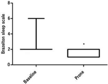
he Brazelton sleep score did not increase in any participant after prone positioning, decreased in seven (43.75%) and did not change in nine (56.25%) in comparison to the baseline score (p=0.02) (Figure 3).
Figure 2 - Comparison of the variation in respiratory rate before and after prone positioning of infants. p value=0.0004/paired t-test.
Figure 3 - Variation in Brazelton sleep score before and after prone positioning.
* p value=0.02/Wilcoxon test.
DISCUSSION
Technological advances in neonatal intensive care and research have contributed to an increase in the survival of newborn infants, including extremely premature ones.(22) At the same time, approximately 15% of surviving premature infants exhibit signiicant sequelae,(23) which may be related to the routine interventions applied by healthcare providers at NICUs that, while on the one hand provide life support, on the other cause pain, stress, and discomfort.(23,24) hus, life support in premature infants should prioritize care measures centered on their development, among which body positioning is important.(5,6,18,19)
correlation between the salivary cortisol level as a marker of stress and prone positioning in premature infants placed in incubators. In addition, there are no reports on the use of the electrochemiluminescence method to measure the salivary cortisol level as a marker of stress following manipulation of premature newborn infants. he method most widely used for this purpose is the radioimmunoassay, which was not available at the time of the present study.
he Hospital Universitário do Oeste do Paraná is a tertiary care institution with 10 NICU beds. In 2012, the NICU admitted 257 newborn infants, of which almost all were referred from the hospital’s own maternity unit. he NICU has not yet established a protocol for development-centered infant care, and prone positioning is only indicated when healthcare providers detect signs of respiratory distress in newborn infants.
he results of the present study revealed a signiicant reduction in the salivary cortisol level in 81.25% of the sample after prone positioning, corroborating previous studies that assessed the efect of this position on stress in premature newborn infants and found a reduction in the number of stress-indicating behaviors,(20,21,25) which may be correlated with lower cortisol levels. However, in contrast to previous studies(12,15) that found an inluence of gestational and postnatal age on the salivary cortisol response, we did not ind any correlation among such variables in our sample, including in infants who did not exhibit changes in the salivary cortisol level (12.5%) and in infants in which the cortisol levels increased (6.25%). hree of the largest reductions in the salivary cortisol levels found corresponded to infants with a gestational age over 32 weeks and a postnatal age of 1 day, which may suggest that the hormonal response is better in newborn infants with an increased gestational age, more stable condition, and shorter stay at the NICU.
In agreement with a previous study,(26) we found a reduction in RR following prone positioning, which lends support to previous reports describing improvements in the respiratory pattern and a reduction of RR following prone positioning.(27) However, our results are in contrast to those of others studies that detected an increase(28) or no change(29) in RR following prone positioning.
he results of the present study showed that the adapted Brazelton sleep score did not increase following prone positioning compared to baseline but did decrease in 44% of the sample. hese indings provide further evidence of the positive efects of the prone position on sleep in premature newborn infants, which include fewer
arousals,(30-33) a longer length of quiet sleep,(30,31) and, consequently, lower energy expenditure.(29,30)
In the present study, the measurements of HR, T, and SatO2 did not exhibit signiicant changes, in contrast to reports by other authors who found increases in HR,(28,29) a reduction in T,(29) and increases in SatO
2 (27,30)
with changes in body position. hese diferences may be partially explained by methodological diferences in the procedures used for monitoring, such as the use of pulse oximetry instead of electrocardiography, and the short interval applied for posture maintenance.
Our body posture intervention was found to be safe, as no intercurrent events, such as a reduction in SatO2 or apnea, occurred, and there was no need to either increase the oxygen supply or to interrupt the procedure. his inding is particularly important because stress is known to negatively inluence the development of premature infants.(3-6,18,19,25) hus, the results of the present study may motivate further research to reairm the adoption of prone positioning as an important positive stimulus for developing premature newborn infants.
may have been diferent. Finally, it should be noted that simple monitoring of the respiratory pattern and HR may not have been sensitive enough to detect discrete variations, and the lack of continuous recording represents a negative factor in the interpretation of those data.
CONCLUSION
Our results indicate that prone positioning may signiicantly reduce the salivary cortisol level, respiratory rate, and Brazelton sleep score in stable premature
newborn infants admitted to a neonatal intensive care unit who are not stratiied according to gestational or postnatal age. hese indings suggest a possible correlation between the prone position and a reduction of stress in premature newborn infants. Although the measurements of temperature, heart rate, and peripheral oxygen saturation did not seem to be inluenced by body position, future studies with improved methodology and larger samples may be able to detect a correlation between these variables and a population more sensitive to postural intervention.
Objetivo: Avaliar a inluência da postura em prona sobre o estresse no recém-nascido prematuro por meio da dosagem do cortisol salivar e da avaliação das respostas isiológicas e comportamentais, antes e após o posicionamento.
Métodos: Foi realizada a coleta de saliva em cada recém-nascido em dois momentos: o primeiro (correspondente ao basal), sem manipulação prévia por 40 minutos, em decúbito lateral ou supino; e o segundo, 30 minutos após o posicionamento em prona. A frequência cardíaca e respiratória, saturação periférica de oxigênio e escala de sono de Brazelton foram registradas antes, durante e ao inal do posicionamento em prona.
Resultados: Participaram do estudo 16 recém-nascidos prematuros (56,3% masculino) com idade gestacional de 26 a 36 semanas, com 1 a 33 dias de vida, e peso variando de 935 a 3.050g ao nascimento e de 870 a 2.890g no dia da intervenção.
Durante o posicionamento, seis recém-nascidos estavam em ar ambiente e os demais recebiam oxigênio suplementar. A mediana dos níveis de cortisol salivar foi menor durante o posicionamento em prona comparativamente ao basal (0,13 e 0,20; p=0,003), assim como a do escore de sono de Brazelton (p=0,02). A média da frequência respiratória foi menor após a intervenção (54,88±7,15 e 60±7,59; p=0,0004). As demais variáveis analisadas não apresentaram variação signiicativa.
Conclusão: O posicionamento em prona diminuiu signiicativamente os níveis de cortisol salivar, da frequência respiratória e do escore de sono de Brazelton, sugerindo a correlação entre essa postura e a diminuição do estresse nesses recém-nascidos.
RESUMO
Descritores: Córtex suprarrenal/metabolismo; Hidrocor-tisona/análise; Recém-nascido/metabolismo; Prematuro/meta-bolismo; Decúbito ventral; Saliva/análise; Estresse isiológico; Unidades de terapia intensiva neonatal
REFERENCES
1. Anand KJ, Hickey PR. Pain and its effects in the human neonate and fetus. N Engl J Med. 1987;317(21):1321-9.
2. Barker DP, Rutter N. Stress, severity of illness, and outcome in ventilated preterm infants. Arch Dis Child Fetal Neonatal Ed. 1996;75(3):F187-90. 3. Lou HC, Hansen D, Nordentoft M, Pryds O, Jensen F, Nim J, et al. Prenatal
stressors of human life affect fetal brain development. Dev Med Child Neurol. 1994;36(9):826-32.
4. Marrese AM. El ambiente de la UCI neonatal y su influencia em el desarrollo del premature: un desafio para enfermería. Med Perinat Neonat. 1996;1(1):11-21.
5. Als H. A synactive model of neonatal behavioral organization: framework for the assessment of neurobehavioral development in the premature infant and for support of infants and parents in the neonatal intensive care environment. Phys Occup Ther Pediatr. 1986;6(3-4):3-53.
6. Als H, Tronick E, Lester BM, Brazelton TB. The Brazelton Neonatal Behavioral Assessment Scale (BNBAS). J Abnorm Child Psychol. 1977;5(3):215-31. 7. Franck LS, Miaskowski C. Measurement of neonatal responses to painful stimuli:
a research review. J Pain Symptom Manage. 1997;14(6):343-78. Review.
8. Francis SJ, Walker RF, Riad-Fahmy D, Hughes D, Murphy JF, Gray OP. Assessment of adrenocortical activity in term newborn infants using salivary cortisol determinations. J Pediatr. 1987;111(1):129-33.
9. Calixto C, Martinez FE, Jorge SM, Moreira AC, Martinelli CE Jr. Correlation between plasma and salivary cortisol levels in preterm infants. J Pediatr. 2002;140(1):116-8.
10. Antonini SR, Jorge SM, Moreira AC. The emergence of salivary cortisol circadian rhythm and its relationship to sleep activity in preterm infants. Clin Endocrinol (Oxf). 2000;52(4):423-6.
11. Herrington CJ, Olomu IN, Geller SM. Salivary cortisol as indicators of pain in preterm infants: a pilot study. Clin Nurs Res. 2004;13(1):53-68. 12. Gitau R, Modi N, Gianakoulopoulos X, Bond C, Glover V, Stevenson J.
Acute effects of maternal skin-to-skin contact and massage on saliva cortisol in preterm babies. J Reprod Infant Psychol. 2002;20(2):83-8. 13. Boyer K, Johnston C, Walker CD, Filion F, Sherrard A. Does sucrose
analgesia promote physiologic stability in preterm neonates? Biol Neonate. 2004;85(1):26-31.
15. Mörelius E, Theodorsson E, Nelson N. Salivary cortisol and mood and pain profiles during skin-to-skin care for an unselected group of mothers and infants in neonatal intensive care. Pediatrics. 2005;116(5):1105-13. 16. White-Traut RC, Schwertz D, McFarlin B, Kogan J. Salivary cortisol and
behavioral state responses of healthy newborn infants to tactile-only and multisensory interventions. J Obstet Gynecol Neonatal Nurs. 2009;38(1):22-34.
17. Takahashi Y, Tamakoshi K, Matsushima M, Kawabe T. Comparison of sali-vary cortisol, heart rate, and oxygen saturation between early skin-to-skin contact with different initiation and duration times in healthy, full-term infants. Early Hum Dev. 2011;87(3):151-7.
18. Saunders RP, Abraham MR, Crosby MJ, Thomas K, Edwards WH. Eva-luation and development of potentially better practices for improving family-centered care in neonatal intensive care units. Pediatrics. 2003;111(4 Pt 2):e437-49.
19. Byers JF. Components of developmental care and the evidence for their use in the NICU. MCN Am J Matern Child Nurs. 2003;28(3):175-80; quiz 181-2.
20. Grenier IR, Bigsby R, Vergara ER, Lester BM. Comparison of motor self-regulatory and stress behaviors of preterm infants across body positions. Am J Occup Ther. 2003;57(3):289-97.
21. Chang YJ, Anderson GC, Lin CH. Effects of prone and supine positions on sleep state and stress responses in mechanically ventilated preterm infants during the first postnatal week. J Adv Nurs. 2002;40(2):161-9. 22. Cooke RW. Factors affecting survival and development in extremely tiny
babies. Semin Neonatol. 1996;1(4):267-76.
23. Saigal S, Doyle LW. An overview of mortality and sequelae of preterm birth from infancy to adulthood. Lancet. 2008;371(9608):261-9.
24. Anand KJ, Aranda JV, Berde CB, Buckman S, Capparelli EV, Carlo W, et al. Summary proceedings from the neonatal pain-control group. Pediatrics. 2006;117(3 Pt 2):S9-22.
25. Grunau RE, Holsti L, Peters JW. Long-term consequences of pain in human neonates. Semin Fetal Neonatal Med. 2006;11(4):268-75.
26. Jarus T, Bart O, Rabinovich G, Sadeh A, Bloch L, Dolfin T, et al. Effects of prone and supine positions on sleep state and stress responses in preterm infants. Infant Behav Dev. 2011;34(2):257-63.
27. Martin RJ, DiFiore JM, Korenke CB, Randal H, Miller MJ, Brooks LJ. Vulnerability of respiratory control in healthy preterm infants placed supine. J Pediatr. 1995;127(4):609-14.
28. Heimler R, Langlois J, Hodel DJ, Nelin LD, Sasidharan P. Effect of positioning on the breathing pattern of preterm infants. Arch Dis Child. 1992;67(3):312-4.
29. Amemiya F, Vos JE, Prechtl HF. Effects of prone and supine position on heart rate, respiratory rate and motor activity in fullterm newborn infants. Brain Dev. 1991;13(3):148-54.
30. Ammari A, Schulze KF, Ohira-Kist K, Kashyap S, Fifer WP, Myers MM, et al. Effects of body position on thermal, cardiorespiratory and metabolic activity in low birth weight infants. Early Hum Dev. 2009;85(8):497-501. 31. Masterson J, Zucker C, Schulze K. Prone and supine positioning effects on
energy expenditure and behavior of low birth weight neonates. Pediatrics. 1987;80(5):689-92.
32. Goto K, Mirmiran M, Adams MM, Longford RV, Baldwin RB, Boeddiker MA, et al. More awakenings and heart rate variability during supine sleep in preterm infants. Pediatrics. 1999;103(3):603-9.