Rev Bras Med Esporte _ Vol. 13, Nº 4 – Jul/Ago, 2007
253e
1. Médico Assistente do Serviço de Ortopedia e responsável peloAmbu-latório de Trauma Esportivo do Hospital Ipiranga, Secretaria de Negó-cios da Saúde do Governo do Estado de São Paulo. Vice-Presidente da Sociedade Brasileira de Medicina do Esporte – SBME. Membro de Di-retoria da Sociedade Paulista de Medicina Desportiva – SPAMDE. Mem-bro Titular da Sociedade Brasileira de Ortopedia e Traumatologia – SBOT. 2. Médico Residente do Serviço de Ortopedia do Hospital Ipiranga,
Secre-taria dos Negócios da Saúde do Governo do Estado de São Paulo. 3. Médico Ortopedista. Assistente do Serviço de Ortopedia do Hospital
Ipiranga, Secretaria de Negócios da Saúde do Governo do Estado de São Paulo. Membro Titular da Sociedade Brasileira de Ortopedia e Trau-matologia – SBOT.
4. Médico Ortopedista. Chefe do Serviço de Ortopedia do Hospital Ipiran-ga, Secretaria de Negócios da Saúde do Governo do Estado de São Paulo. Membro Titular da Sociedade Brasileira de Ortopedia e Trauma-tologia – SBOT.
Approved in 26/6/07.
Correspondence to: Hospital Ipiranga, Avenida Nazaré, 28, 5º andar – 04262-000 – São Paulo, SP. E-mail: [email protected]
Traumatic hip fracture-dislocation in soccer: a case report
Ricardo Munir Nahas1, Eliseu Netto2, Takechi Chikude3 and Roberto Ikemoto4
C
ASER
EPORTKeywords: Soccer. Hip. Fracture and deslocation. ENGLISH VERSION
ABSTRACT
Uncommon in soccer, the world’s most popular sport, the ace-tabular fractures with hip dislocation are injuries with high inciden-ce of long-term serious sequelae. Prompt attending with accurate diagnosis and reduction and fixation are important to produce a stable and congruent joint for returning to the usual sports practice prior the injury.
INTRODUCTION
Soccer, which is the most played collective sport in the world, has shown in epidemiological studies occurrence between 10 and 35 acute traumatic injuries per 1000 hours of game with higher incidence in ankles and knees(1). Fractures represent from 4% to
9% of these injuries and hip fracture-dislocation as described in this case report is fortunately rare(2-3).
Dislocations are usually orthopedic emergencies which require precise and early diagnosis(4). Suitable action decreases the
dele-tereal effects caused by trauma and consequent articular incon-gruence which is established and should occur in a hospital envi-ronment by the resources available for a good diagnosis and accurate therapeutic procedure, especially in those in which anes-thesia and surgery procedures are needed(3-4).
Hip traumatic dislocation is a serious orthopedic injury which may cause permanent incapacity(5). It is usually a consequence of
high energy trauma, and is more frequently related with accidents caused by motor vehicles. Its condition worsens when associated with acetabular or femoral head fracture(4-5).
This kind of trauma occurred in the sports practice has few re-ports in the international literature(3-5). Its diagnosis should be
re-minded and the physician should also be prepared for initial care required by the severity of the situation until the definite treatment can be performed(6-7).
Despite not being very relevant for physical activity practice, the case report here presented, due to its rareness, becomes impor-tant and worth releasing.
CASE REPORT
A 35-year old male patient has been admitted to emergency care unit complaining about left hip pain and total functional failure. He has reported that during a field soccer game while disputing for an aerial ball and when returning to the ground, felt as he described as a ‘shock’ in the left hip, spreading to the entire lower extremity and subsequent fall, laid on the ground in severe pain in the whole affected extremity. He also reported not having suffered any direct trauma by shock against the other players or against the goal poles. He was not able to be precise about his chest position in relation to the lower extremities or hips and knees at the moment he touched the very irregular surface ground.
He mentioned to have played non-professional competitive soc-cer field once a week, for 20 years, as well as to have regular physical conditioning: aerobic training (running) two to four times per week, stretching and localized endurance trainings, with spe-cial attention given to the lower extremities three times per week and specific soccer technical training, (individual for instructions, and collective one to three times per week).
He did not report any personal or family alteration history which could have been related with or a predisposition for his current clinic situation.
The patient was brought on a stretcher in right lateral decubitus with left hip in flexion attitude of approximately 45°, adduction of 20° and internal rotation of 30°. During examination, any try of hip movement was extremely painful and impossible to be performed. Neurological or vascular alterations have not been observed. There were not any other relevant findings in the physical examination.
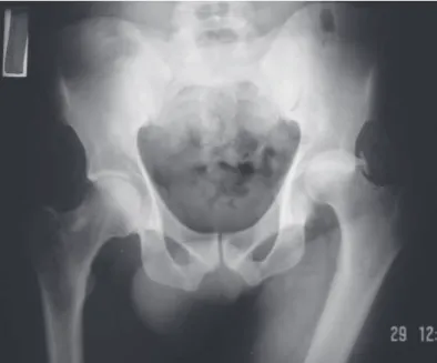
Radiological examination showed diagnostic of left hip posterior dislocation associated with fracture of posterior acetabular wall (fig-ure 1).
254e
Rev Bras Med Esporte _ Vol. 13, Nº 4 – Jul/Ago, 2007 The patient was taken to the surgical unit and was submitted toanesthesia by epidural block which allowed closed reduction of the thigh-femoral dislocation through application of Allis maneu-ver, a procedure performed in the first six hours after trauma. Af-terwards, he was tested for stability with no reoccurrence of the injury, even when the hip was placed in flexion above 100° associ-ated with adduction(8-9). Skeletal traction in distal femur was installed
and was kept until the definite therapeutic procedure was taken(9).
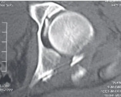
In the immediate post-surgery, the patient was submitted to a tomography (figure 2) which allowed better study of the intra-artic-ular bone fragment resultant from the posterior acetabintra-artic-ular frac-ture of the wall to determine the joint stability and to plan for the definite surgical treatment(8).
Passive mobilization of left lower extremity exercises with the aid of balance beam were initiated in the immediate post-surgery. Such mobilization became active and associated with isometric exercises as soon as the pain ceased, always according to medical prescription and under supervision.
During the infirmary follow-up the patient was kept with clutch-es and without load for eight weeks when he was authorized par-tial touch on the ground still with the aid of two clutches for four weeks. He gradually and progressively started to use load until reaching total load fifteen days after definite surgical procedure.
Physical examination six months after surgery showed that the movement arch of the left hip was normal and painless. Clinic sig-nals by complementary examinations for complementary sequelae have not been evidenced, which enabled the patient to be dis-charged for physical conditioning activity. Yet, he should avoid ex-ercises which result in excessive overload in the operated hip. He was not allowed to perform any sportive activity, regardless its nature.
DISCUSSION
Despite being rare to find posterior dislocation of the hip during soccer practice as well as having apparent less energy amount in the trauma, it does not mean that it is less serious or does not need to be seen as an orthopedic emergency to be fast and effi-ciently solved.
One of the found difficulties was the understanding about the sports gesture which had caused the injury. In the presented case, we tried to stimulate the patient to the most to describe the whole sequence of traumatic events which caused the injury, trying hence, to better understand the way it has occurred.
Since we did not get answers to these questions, we searched for explanation in the studies by Judet and Lettournel who related the position of the lower extremity at the moment of the trauma with the kind of resultant injury(10). Such analysis enabled us to
deduct that the fracture-dislocation occurred when the lower ex-tremity was in internal rotation and maximal adduction, a position which is not common in landing after jumps in sports, increased by the irregular nature of the ground(9-10). Perhaps this is the
me-chanic explanation for the injury, representing a technical mistake when performing the sports gesture.
The neurological and vascular examinations should be carefully performed in the patient’s initial physical examination when he was admitted in the emergency room, since it considerably improves prognostic(5,9). The neural injuries which may occur to patients with
posterior hip fracture-dislocation many times are not diagnosed due to intense pain(9,11). Vascular injuries are relatively rare;
howev-er, they should always be remembered and the perfusion carefully evaluated(4,9,11).
The diagnosis and technical knowledge for fast time reduction in the emergency room may make the difference concerning the future of the traumatized joint(3,9). Reductions higher than 12 hours
are usually associated with fairly unfavorable prognostics, com-promising both the sportive performance and daily activities(4,12).
The treatment principles for posterior hip dislocation should be followed in trauma with any origin(3,8). There are many maneuvers
for the reduction of posterior hip dislocation. We consider the maneuver described by Allis less traumatic and technically easy, besides being previously familiarized with it(8-9).
Once the dislocation is solved, complementary examinations which allow better research by image of the bone fragments ori-gin, enable suitable surgical planning with the purpose to give back to the joint normal conditions for its function with no sequelae(8).
These procedures become essential in athletes and should be per-formed by registered professionals who follow suitable techniques in order to provide the athletes return to their activities in the same level previous to the injury or at least very close to it(3,10-11).
Figure 2 – Tomography showing intra-articular bone fragment, derived from the acetabulum
Since we were not able to identify in the traumatic mechanism how a high energy would explain the occurrence of fracture of the posterior acetabular wall, associated pathologies which would act as predisposition for the occurrence of the injury were researched and discarded(9).
The patient was then submitted to surgery: small intra-articular bone fragments were removed and the collocation of the fragments of the main bone of the acetabular posterior wall in its original loca-tion was performed using the bone synthesis principle with com-pression bolt between the fragments and support with plaque spe-cific for acetabular fractures (figure 3).
Rev Bras Med Esporte _ Vol. 13, Nº 4 – Jul/Ago, 2007
255e
Depending on the severity of the injury as well as the treatmentnature, a change in the sport and even the practice of regular ical activity for good health, with orientation from a certified phys-ical educator, should be discussed with the patient within the re-habilitation program to which he has been submitted(3-4,12-13).
CONCLUSION
Despite being a serious and rare situation in soccer practice, traumatic posterior hip fracture-dislocation when suitably diagnosed and treated presents good results with functional recovery of the joint without sequalae.
Since it is an injury which occurs with individuals who regularly practice the sport, it is desirable that equal or close performance to what they had prior to the injury is recovered, allowing return to the sport or even adaptation to an activity compatible with this new situation, with no compromising of the traumatized joint.
All the authors declared there is not any potential conflict of inter-ests regarding this article.
REFERENCES
1. Dvorak J, Junge A. Football injuries and physical symptoms. Am J Sports Med. 2000;28:53-9.
2. Anderson S. Injuries in youth football: a subject review. Pediatrics. 2000;105: 659-61.
3. Goga IE, Gongal P. Severe soccer injuries in amateurs. Br J Sports Med. 2003;37: 498-501.
4. Giza E, Mithofer K, Matthews H, Vrahas M. Hip fracture-dislocation in football: a report of two cases and review of the literature. Br J Sports Med. 2004;38(4):e17. 5. Pallia CS, Scott RE, Chao AD. Traumatic hip dislocation in athletes. Curr Sports
Med Rep. 2002;1:338-45.
6. Junge A, Dvorak J. Soccer injuries: a review on incidence and prevention. Sports Med. 2004;34:928-38.
7. Wong P, Hong Y. Soccer injury in the lower extremities. Br J Sports Med. 2005;39: 473-82.
8. Fujiki EM, Yamaguchi E, Nicolau R, Chikude, T, Fukushima W, Oliveira HC, et al. Índice tomográfico como critério de instabilidade nas fraturas-luxações da pare-de posterior do acetábulo. Rev Bras Ortop. 2006;41(1-2):29-33.
9. Giordano V, Costa PRL, Esteves JD, Félix S Junior J, Franklin CE, Amaral NP. Luxações traumáticas do quadril em pacientes esqueleticamente maduros. Rev Bras Ortop. 2003;38(8):462-72.
10. Letournel E, Judet R. Fractures of the acetabulum. 2nd ed. Berlin:
Springer-Ver-lag; 1993.
11. Toth C, McNeil S, Feasby T. Peripheral nervous system injuries in sport and recreation: a systematic review. Sport Med. 2005;35:717-38.
12. Lequesne MG, Dang N, Lane NE. Sport practice and osteoarthritis of the limbs. Osteoarthritis Cartilage. 1997;5:75-86.
13. Anderson K, Strickland SM, Warren R. Hip and groin injuries in athtletes. Am J Sports Med. 2001;29:521-33.
14. Matsumoto K, Sumi H, Sumi Y, Shimizu K. An analysis of hip dislocations among snowboarders and skiers: a 10-year prospective study from 1992 to 2002. J Trauma. 2003;55:946-8.
15. Mohanty K, Gupta SK, Langston A. Posterior dislocation of hip in adolescents attributable to casual rugby. J Accid Emerg Med. 2000;17:429.
16. Mitchell JC, Giannoudis PV, Millner PA, Smith RM. A rare fracture-dislocation of the hip in a gymnast and review of the literature. Br J Sports Med. 1999;33:283-4.